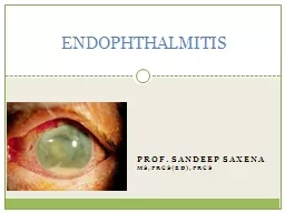
sANDEEP saxena msFRCS ed FRCS ENDOPHTHALMITIS DEFINITION An intraocular inflammation involving ocular cavities vitreous cavity and or anterior chamber and their adjacent structures ID: 439383
Download Presentation The PPT/PDF document "Prof." is the property of its rightful owner. Permission is granted to download and print the materials on this web site for personal, non-commercial use only, and to display it on your personal computer provided you do not modify the materials and that you retain all copyright notices contained in the materials. By downloading content from our website, you accept the terms of this agreement.
Slide1
Prof. sANDEEP saxena ms,FRCS(ed),FRCS
ENDOPHTHALMITISSlide2
DEFINITIONAn intraocular inflammation involving ocular cavities (vitreous cavity and/ or anterior chamber) and their adjacent structures.Slide3
CLASSIFICATIONINFECTIOUSExogenous-Acute onset -Post traumatic-Delayed onset-Bleb associated
Endogenous-
Haematogenous
spread
STERILE
Lens induced
Toxic
Post surgical
Non surgicalSlide4
CAUSATIVE ORGANISMSGram +ve: Bacteria: Bacteria: Bacteria:S.
epidermidis Propionibacterium Bacillus
Bacillus
cereus
S.
aureus
acne
S.epidermidis
StreptococciStreptococci Streptococci Streptococci
S.aureusGram –ve: Fungi: Fungi: N.meningitides
Pseudomonas
Aspergillus
Fusarium H.influenzaeH.influenzae Candida Fungi:Klebsiella spp Fusarium MucorE. coli Penicillium CandidaBacillus sppAnaerobes
Acute Post-op
Delayed Post-op
Post- Traumatic
EndogenousSlide5
POST- SURGICAL ENDOPHTHALMITISMost common form70% cases of infective endophthalmitisWorldwide incidence 0.04 - 4%Incidence in India 0.7 - 2.4%Slide6
Commonly associated with:Cataract extraction (most common)Secondary lens implantationPars plana
vitrectomy
Glaucoma
filteration
surgery
Penetrating
keratoplastySlide7
RISK FACTORSPRE- OPERATIVE RISK FACTORS:BlepharitisConjunctivitisLacrimal drainage system infection Contact lenses wear
Contaminated eyedropsSlide8
INTRA-OP RISK FACTORS:Clear corneal incisionTemporal incisionPosterior capsule ruptureVitreous incarceration in woundProlonged procedure timeContaminated irrigation solutionsCombined proceduresSlide9
POST- OPERATIVE RISK FACTORS:Inadequately buried suturesWound leak on the first dayDelaying post-op topical antibiotics until the day after surgerySlide10
CLINICAL PRESENTATION
Acute onset
Delayed onset
Within 6 weeks
After 6 weeksSlide11
ACUTE POST-OP ENDOPHTHALMITISMost common organism - Coagulase negative Staphylococcus species (S.epidermidis)Hyperacute infections - Pseudomonas
aeruginosa and Bacillus species.
Source of infection- lid flora
-
conjunctival
flora
Entry occurs at the time of surgerySlide12
DELAYED- ONSET ENDOPHTHALMITISLow virulence organisms:Propionibacterium acneStaphylococcus epidermidis
FungiRelease of organisms sequestered within the capsular bag-
saccular
endophthalmitisSlide13
CLINICAL FEATURESSYMPTOMS:Blurred vision (94%)Red eye (82%)
Pain (74%)Slide14
CLINICAL FEATURESSIGNS:Decreased visual acuityLid edema, conjunctival chemosis, congestion and discharge
Corneal edemaKeratic
precipitates (delayed-onset)
Fibrinous
exudates and
hypopyon
in ACSlide15
SIGNSRelative afferent pupillary defectLoss of red reflex, impaired fundal view, vitritis
Scattered retinal haemorrhages
,
periphlebitis
Capsular plaque (
Propionibacterium
acnes
endophthalmitis
)Slide16
BLEB- ASSOCIATED ENDOPHTHALMITISIncidence:Acute- 0.06-0.2% Predisposing factors:BlepharitisUse of anti- fibrotic agents (
Mitomycin- C, 5- fluorouracil)
Long term topical antibiotic use
Inferior or nasally placed bleb
Bleb leak
Pathogens:
Streptococcus
H.influenzae
Staphylococcus
Delayed- 0.2-18%Slide17
POST- TRAUMATIC ENDOPHTHALMITISOccurs following penetrating trauma (7%)Intraocular foreign body increases the risk (30%)Common organisms inolved:Gram positive cocciBacillus
spp
Fungi (esp.
Fusarium
)
May occur anytime from days to weeks following injury
Delay in diagnosis: Post- traumatic inflammation
vs
infectionSlide18
ENDOGENOUS ENDOPHTHALMITISHaematogenous spread of micro-organisms from a site external to the eyePredisposing host factors:Age (children)Immune suppression MalnutritionDiabetes mellitusAlcoholism MalignancyPresents with less inflammation and pain than other forms of
endophthalmitisReduced vision and floaters in one or both eyesSlide19
DIAGNOSIS OF ENDOPHTHALMITISEarly recognition is critical.High index of suspicion to be maintained.A complete ocular and medical history is essential.Thorough ophthalmic examination performed.Slide20
OPHTHALMIC INVESTIGATIONSConjunctival swabFor pre-existing organisms in adnexae
UltrasonographyUseful in anterior segment media opacity
Confirm presence of variable echoes in vitreous
Retained lens remnants in posterior segment
Intraocular foreign body in post- traumatic cases
Retinal or
choroidal
detachment
Provide a baseline to compareSlide21
IDENTIFICATION OF PATHOGENSAqueous tap:0.1-0.2 ml of aqueous is aspirated via a limbal paracentesis using a 25-G needleVitreous tap:0.2-0.4 ml is aspirated from mid-vitreous cavity using a 23-G needle
Distance from limbus-
3mm for
aphakic
eye
3.5mm for
pseudophakic
eye
4mm for phakic eyeSlide22
Samples are subjected to:Gram stainingGiemsa stainingKOH mountCulture on-
Blood agarChocolate agar
Sabouraud
dextrose agar
Thioglycollate
broth
Anaerobic medium
Polymerase chain reactionSlide23
Reasons for negative culture results:Fastidious organismsInsufficient samplingSterile endophthalmitisRepeat cultures may be needed:When clinical response is not good
Presence of contaminants in mediaPresence of fungus- especially likely to be missed initiallySlide24
SYSTEMIC INVESTIGATIONSComplete haemogramBlood sugar (predisposition in diabetics)Blood and urine cultures (endogenous endophthalmitis)
Cultures from other sites (catheter tips, skin wounds, abscesses and joints)Slide25
TREATMENTAntibioticsSteroidsTopical mydriaticsVitrectomy
IOL managementEvisceration
MEDICAL
SURGICALSlide26
INTRAVITREAL ANTIBIOTICSGram positive:Vancomycin (1.0 mg in 0.1 ml)Broad spectrumBoth coagulase positive and coagulase negative
cocciGram negative:
Ceftazidime
(2.25 mg in 0.1 ml)
No retinal toxicity
Amikacin
(0.4 mg in 0.1 ml)
Retinotoxic
Alternative to ceftazidime
in penicillin allergyGentamicinSlide27
OTHER MODESTopical antibiotics:Fortified cefazoline (5%) OR Fortified vancomycin (5%) PLUSFortified tobramycin
(1.3%)Given half hourly alternatelySystemic antibiotics:
Clindamycin
1g iv 8
hrly
Ceftazidime
2g iv 8
hrly
Ciprofloxacin 750 mg P.O. bid
Moxifloxacin 400 mg P.O. odSlide28
STEROIDSControl inflammation mediated damageBut no influence on visual outcomeINTRAVITREAL:Dexamethasone (0.4 mg in 0.1 ml)Triamcinolone (long acting) can also be usedSUBCONJUNCTIVAL:
Dexamethasone (6mg in 0.25 ml)TOPICAL:
Prednisolone
1% 2
hrly
Dexamethasone
0.1%
SYSTEMIC:
Prednisolone
1mg/kg OD (started after 12-24 hrs)Slide29
FUNGAL INFECTIONIntravitreal Amphotericin B (5µg in 0.1 ml)Newer agents- Voriconazole (200µg in 0.1 ml) and CaspofunginTopical Natamycin (5%) and Itraconazole (1%)Systemic therapy-
Fluconazole (150mg od)Steroids are contraindicatedSlide30
SURGICAL MANAGEMENTVITRECTOMY:Advantages of early vitrectomy:Clearing of ocular mediaReduction of bacterial loadRemoval of bacterial productsRemoval of vitreous scaffolding- which may cause retinal detachmentSlide31
Disadvantages:Iatrogenic retinal holes and detachmentsChoroidal haemorrhageRetinal detachment - difficult to treat in vitrectomized
eyes Slide32
COMPLICATIONS RELATED TO IOLFibrin exudates on IOL- removed with a needle or forcepsExudates trapped between the posterior capsule and IOL - Posterior capsulotomyFungal endophthalmitis and sequestered organisms in the capsular bag (P.acnes) - en bloc removal of IOL and capsular bagSlide33
MANAGEMENT PROTOCOLAssess visual acuity
Only PL+
Early VIT +
i
/
vit
Antibiotics
HM or better
I/
vit
Antibiotics
Improves
Does not improve
Repeat
i/vit antibioticsVitrectomy
Repeat culture
Watch for 48 hoursSlide34
EMPIRICAL MEDICAL THERAPY OF ENDOPHTHALMITIS (as per EVS 1996)ACUTE ONSET POST CATARACT EXTRACTIONINTRAVITREALVancomycin
Ceftazidime OR amikacin
Dexamethasone
(optional)
SUBCONJUNCTIVAL
Vancomycin
Ceftazidime or
Amikacin (if B-lactam allergy) Dexamethasone Slide35
TOPICALVancomycin hydrochlorideAmikacinAtropine sulphatePrednisolone
acetate 1%ORAL
Prednisone 30mg twice daily for 5 to 10 days (optional)Slide36
POST- TRAUMATICSame as that for Post- cataract Sx with:Intravitreal Clindamycin (450 micrograms)Systemic antibioticsBLEBITISTopicals
are sufficient:Vancomycin
hydrochloride
Amikacin
Atropine
sulphate
Prednisolone acetate 1%
BLEB- ASSOCIATED ENDOPHTHALMITISSame as that for Post- cataract Sx with systemic antibioticsSlide37
FOLLOW-UP AND OUTCOMESigns of improvement:AC reactionHypopyonFundal glowFinal outcome:Duration of infectionVirulence of organism
(EVS- Final outcomes)53% patients visual acuity >6/12
75% patients visual acuity >6/30
89% patients visual acuity >6/240Slide38
PROPHYLAXIS5% povidone iodine - 3 minutesTreatment of pre-existing infectionsProphylactic antibiotics:Pre-operative topical fluoroquinolones
Intracameral cefuroxime (1 mg in 0.1 ml)
Post-operative sub-
conjunctival
antibiotics
Systemic 4
th
generation
fluoroquinolones
Early resuturing of wound leaks