/
The anti-inflammatory properties of lipids extracted from Omani camel milk The anti-inflammatory properties of lipids extracted from Omani camel milk

The anti-inflammatory properties of lipids extracted from Omani camel milk - PowerPoint Presentation

myesha-ticknor
myesha-ticknor . @myesha-ticknor
Follow
348 views
Uploaded On 2019-11-08

The anti-inflammatory properties of lipids extracted from Omani camel milk - PPT Presentation

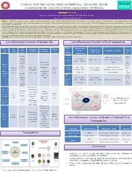
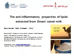
The antiinflammatory properties of lipids extracted from Omani camel milk Raya Hamdan Salim AlNasseri MSc Morris Keith Director of study Cardiff school of Health Sciences Cardiff Metropolitan University Wales UK ID: 764844

camel lipids cells milk lipids camel milk cells dthp gbsa expression acid inflammatory figure induced significantly secretion significant macrophages

Share:

Link:

Embed:

Download Presentation from below link

Download Presentation The PPT/PDF document "The anti-inflammatory properties of lipi..." is the property of its rightful owner. Permission is granted to download and print the materials on this web site for personal, non-commercial use only, and to display it on your personal computer provided you do not modify the materials and that you retain all copyright notices contained in the materials. By downloading content from our website, you accept the terms of this agreement.


Presentation Transcript

The anti-inflammatory properties of lipids extracted from Omani camel milk Raya Hamdan Salim Al-Nasseri , M.Sc * Morris Keith , Director of study, Cardiff school of Health Sciences, Cardiff Metropolitan University, Wales, UK.Kanakenian Ara , Co-Supervisor, Cardiff Metropolitan University.Boulassel Mohamed , Co-Supervisor, Sultan Qaboos University, Sultanate of Oman.*Corresponding E-mail: raal-nasseri@cardiffmet.ac.uk*Sultanate of Oman, Royal Court affairs, Directorate of veterinary services.

Overview There is anecdotal and epidemiological evidence suggesting that camel milk has potential benefits in preventing diseases such as diabetes (1, 2). In addition, there is considerable interest in the role of dietary lipids in regulating the inflammatory response in many disease where inflammation plays a prominent role.These diseases are major public health problems world-wide and include type 2 Diabetes (T2D), cardiovascular disease, atherosclerosis and Alzheimer’s disease.This study decided to focus on camel milk lipids and their possible immunoregulatory effects on human macrophages- a cell critical in the development of inflammatory diseases.Meena et al Journal of Dairy Research (2016) 83 412–419.Milhic et al Journal of Evidence-Based Complimentary and Alternative Mil (2015)

To extract and characterise camel milk lipids from pooled camel milk To Investigate if these lipids regulate Macrophage inflammatory responses using the human cellular macrophage model THP-1-a well-characterised macrophage model. To induce inflammation in this model, we used a glycated protein (gBSA). Glycated proteins are important inducers of inflammation in T2D. To determine dTHP-1 Cell Viability after treatment with Camel Milk Lipids. To Determine if any modulation of macrophage inflammation is associated with changes in macrophage polarization from proinflammatory M1 sub-type to the antiinflammatory sub-type (M2). Study Aims

RESULTSGas Chromatography – Mass Spectrometer (GC-MS) analysis.

Major Saturated fatty Acids (% of Total Fatty Acids)% Major Unsaturated fatty (% of Total Fatty Acids) %   Palmitic acid (C16:0) Myristic acid ( C14:0) Stearic acid ( C18:0)   35.285 14.4567.599 Oleic Acid (C18:1) Palmitoleic acid (C16:1)Linoleic acid (C18:2) 18.99713.658 11.283 Table 1: Total lipids were extracted from pooled camel milk (n=7), using the Bligh and Dyer Standard (BDS) method. Lipids were converted to their Fatty Acids Methyl Ester (FAME) and analysed using Gas Chromatography –Mass Spectrometer (GC-MS). Over 50% of the lipids identified were saturated, with Palmitic acid (C16:0) and Myristic acid (C14:0) being the greatest proportion. Of the unsaturated fatty acids Oleic Acid (18:1) and Palmitoleic acid were proportionally the greatest The Major Lipids Present in Omani Camel Milk Extract

RESULTS The effect of camel milk derived lipids on dTHP-1 cell viability

Figure 1: (A) Immature Monocytic THP-1 cells (No PMA) undifferentiated (B) Differentiated cells (dTHP-1) Macrophages (+PMA) incubation for 48hours. NB adherence and pseudopodia (arrow) x200 with Nikon U-200 attached to an inverted Leica microscope.Differentiation of THP-1 cells was also confirmed by the expression of the CD36 receptor.PMA- Phorbol 12Myristate 13Acetate a PKC activator THP-1 monocytes were differentiated to dTHP-1 (macrophage like cells) AB

Figure 2: dTHP-1 cells incubated with total camel lipids for 6 hours and cell viability was quantified using the Cell-Titre Blue® assay. Results are the mean (±SD) of three experiments. No significant difference in cell viability (p<0.05; one-way ANOVA) was observed with any of the lipid concentrations.The assay is based on the ability of living cells to convert a redox dye (resazurin) into a fluorescent end product. Nonviable cells rapidly lose metabolic capacity and thus do not generate a fluorescent signal.dTHP-1 Cell Viability Post-Treatment with Camel Milk Lipids

RESULTSThe effect of camel milk derived lipids on gBSA induced inflammatory cytokine secretion in dTHP-1 cells

Figure 3: gBSA induced TNFα and IL-1β secreted by dTHP-1 macrophages significantly decreased after the cells were pre-incubated for 6hrs, and in 24hrs with total camel milk lipids (* Denotes a significant reduction in secretion p<0.05; one-way ANOVA).TNF-α and IL-1β-inflammatory cytokines important in the development of T2D and its complicationsCamel milk lipids significantly reduce g-BSA induced TNF- α and IL-1β secretion in dTHP-1 cells * * *

Camel milk lipids significantly reduce gBSA induced TNF- α secretion in human primary *(PBMC) cells Figure 5: gBSA induced TNFα secretion by PBMC significantly decreased after the cells were pre-incubated for 6hrs, and in 24hrs with camel milk lipids. (* Denotes a significant reduction in secretion p<0.05; one-way ANOVA).*PBMC /Human Peripheral Blood Mononuclear Cells *

Figure 4: dTHP-1 cells incubated with respectively total, saturated and un-saturated, camel milk lipids and stimulated with gBSA. The IL-1β secreted was found to be significantly decreased by all three lipids with no significant difference in their effect (p<0.05 ANOVA, Tukey’s pairwise analysis)No significant difference in the effect of Total, Saturated and Un-saturated camel milk lipids on IL1β secretion was observed. *

CD206CD163 Figure 6: Macrophages switching to an anti-inflammatory M2 phenotype.Increased M2 expression is regarded as beneficial in preventing the development of inflammatory diseases such as T2D and atherosclerosis.CD86 Macrophages are highly plastic cells with two major subtypes- M1 and M2.THP-1 Monocytes can be polarized into M1 and M2 forms. CD86 M2b Regulate inflammation: IL-10 CD86 (M1-M2b) CD163 CD206

Given that Camel Milk Lipids significantly down regulated gBSA induced TNF-α and IL-1β secretion in dTHP-1 cellsWe hypothesised : Do these lipids impact on macrophage polarization? In particular towards the M2 phenotype ? Camel lipids

RESULTSFlow – Cytometry and Real-Time PCR

Figure 7: dTHP-1 Macrophages cells incubated for 72hrs, (M2) marker CD163 highly expressed when the lipids combined with inflammatory stimulus (gBSA). * denotes a significant result (p<0.05 one-way ANOVA)The expression of the M2 marker CD163 was up-regulated by gBSA and total lipids. *

M1 marker CD86 and the M2 markers Cd163 and CD206 gene expression. Figure 8. Camel milk derived lipids alone significantly enhanced gene expression of the M2 markers CD163 and CD206. However, the lipids on their own also enhanced expression of the traditional M1 marker CD86 in d-THP1 cells.This results is suggestive of the M2b phenotype of M2 Macrophages *

Camel milk lipids stimulate the gene expression of Interleukin-10 (IL-10)Figure 9. Camel milk derived lipids i(100µg/ml) induced significant (IL-10) expression in dTHP-1 cells and enhanced gBSA induced expression of IL-10Ouyang W1, Rutz S, Crellin NK, Valdez PA, Hymowitz SG Annu Rev Immunol. 2011;29:71-109. doi: 10.1146/annurev-immunol-031210-101312.Regulation and functions of the IL-10 family of cytokines in inflammation and disease.. *

SUMMARY The significant induction of CD86 together with an increased IL-10 is suggestive that camel milk lipids can induce a beneficial M2b sub-type in dTHP-1 cells.

Conclusion Camel milk lipids were capable of significantly in reducing pro-inflammatory cytokine (TNF-α and IL-1β) in gBSA treated dTHP-1 cells. gBSA treated primary macrophages also supported this data.Camel milk lipids enhanced certain aspects of M2 polarization however in particular the lipids enhanced C86 and IL-10 expression data that suggests that the M2b phenotype is being induced in THP-1 cells.Further investigations are being undertaken on the ability of these lipids to regulate the transcription factors NF-B and PPAR- and if IL-10 regulates CD86 expression directly in camel milk treated cells. This study presents novel evidence of a mechanism by which camel milk lipids could regulate the pathogenies of diseases such as T2D.  

Thank you