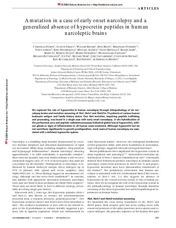
/
volume 6 number 9 september 2000 volume 6 number 9 september 2000

volume 6 number 9 september 2000 - PDF document

myesha-ticknor
myesha-ticknor . @myesha-ticknor
Follow
367 views
Uploaded On 2016-06-30

volume 6 number 9 september 2000 - PPT Presentation

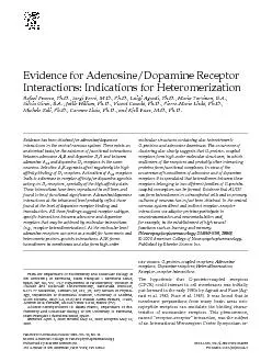
ARTICLES Most cases are sporadic and twin studies indicate a role for envi C JULIETTE ID: 383662

ARTICLES Most cases are sporadic

Share:

Link:

Embed:

Download Presentation from below link

Download Pdf The PPT/PDF document "volume 6 number 9 september 2000" is the property of its rightful owner. Permission is granted to download and print the materials on this web site for personal, non-commercial use only, and to display it on your personal computer provided you do not modify the materials and that you retain all copyright notices contained in the materials. By downloading content from our website, you accept the terms of this agreement.


Presentation Transcript

¥VOLUME 6¥NUMBER 9¥SEPTEMBER 2000 ARTICLES Most cases are sporadic, and twin studies indicate a role for envi- C, JULIETTE © 2000 Nature America Inc. Â¥ http://medicine.nature.com © 2000 Nature America Inc. Â¥ http://medicine.nature.com © 2000 Nature America Inc. Â¥ http://medicine.nature.com © 2000 Nature America Inc. Â¥ http://medicine.nature.com . We amplified and sequenced atleast 50 bp flanking each exon to identify coding alterations andmutations affecting mRNA splicing. We included all patientswith a family history or lacking HLA-DQB1*0602 from our data-morphisms were not linked or associated with narcolepsy (Table ¥VOLUME 6¥NUMBER 9¥SEPTEMBER 2000 ARTICLEScolepsy. We did not identify common polymorphisms of mutation in early onset narcolepsyof the Hcrt signal peptide. This allele was not present in 212 con-DNA from the unaffected father was unavailable to establish thisde novocinations. Notably, he first demonstrated cataplexy (expressed ashead dropping when laughing) at age 6 months. Sudden episodesthis early age. Sleep onset REM periods (SOREMPs) were first docu-mented at 2.5 years of age. Multiple sleep latency testing per- a Lack of co-localization of significant variation in size and morphology. e Allelic variance of the HCRT, HCRTR-1, and HCRTR-2 loci in narcoleptic and control caucasian subjectsDNA changeAmino acid changeDomainNarcolepsyControlNotesF+F-S+S--20CAnon-coding5Õ UTR0.00 (17)0.00 (10)0.00 (26)0.053 (19)0.00 (24)Presumed benign polymorphism47TGLeu16Argsignal peptide0.00 (17)0.00 (10)0.00 (26)0.026 (19)0.00 (106)Dominant mutation111TCsynonymousN-terminus0.32 (17)0.56 (8)0.35 (27)0.44 (16)0.36 (40)Benign polymorphism793CALeu265MetI 30.00 (18)0.00 (9)0.02 (27)0.00 (16)0.00 (45)Presumed benign polymorphism842GAArg281HisI 30.00 (18)0.00 (9)0.00 (27)0.00 (16)0.01 (45)Benign polymorphismIVS6(+6CT)non-codingintron0.06 (18)0.06 (9)0.02 (27)0.00 (16)0.06 (40)Benign polymorphism1222GAVal408IleC-terminus0.28 (18)0.50 (9)0.33 (27)0.44 (16)0.34 (46)Benign polymorphism28CTPro10SerN-terminus0.00 (17)0.00 (10)0.02 (25)0.00 (18)0.000 (90)Presumed benign polymorphism31CAPro11ThrN-terminus0.00 (17)0.10 (10)0.00 (25)0.00 (18)0.006 (90)Unlinked with phenotypeIVS1(-25AC)non-codingintron0.15 (17)0.06 (9)0.17 (24)0.18 (17)0.16 (58)Benign polymorphismIVS2(+49CT)non-codingintron0.29 (17)0.35 (10)0.18 (25)0.26 (17)0.17 (61)Benign polymorphism577TACys193SerTM IV0.00 (16)0.00 (10)0.00 (25)0.00 (17)0.01 (39)Benign polymorphism922GAVal308IleTM VI0.12 (17)0.05 (10)0.18 (25)0.24 (17)0.19 (35)Benign polymorphism942AGsynonymousTM VI0.06 (17)0.00 (10)0.00 (25)0.00 (17)0.01 (35)Benign polymorphism1202CTThr401IleC-terminus0.03 (17)0.00 (10)0.00 (25)0.00 (16)0.00 (99)Possible weakly penetrant allele Data reported are allele frequencies. The number of subjects studied in each group for each polymorphism is indicated in parentheses. F+, familial, DQB1*0602 positive F- , familial,DQB1*0602 negative S+, sporadic, DQB1*0602 positive. SÐ , sporadic, DQB1*0602 negative. DNA and amino acid changes are counted from the ATG-codon and Met-residue re-spectively. Cosegregation in multiplex families was tested whenever possible (see Notes). IVS, intervening sequence (intron), position relative to adjacent exon. 5, 5 region; TM, transmembrane domain; I, intracellular loop. ARTICLES tremely short sleep latencies and multiple SOREMPs. Examples ofcataplectic attacks in this patient triggered by laughter may beviewed at http://www.med.stanford.edu/school/psychiatry/nar-colepsy/. His symptoms are partially controlled withmethylphenidate and either imipramine, chlomipramine or flu-movements and episodic nocturnal bulimia focused on sweetsfrom the age of 5. HLA typing indicates DRB1*0402, DRB1*0701;DQB1*0202, DQB1*0302. Hcrt-1 concentrations are undetectablein lumbar CSF (concentrations 15.3 pg/mlin three unrelated controls from the same geographical area).Impaired processing and trafficking of mutant HcrtWe examined the cellular phenotypes of the wild type and mu-or the V5 epitope tag transiently transfected into Neuro-2A). The wild-type proteinpassed through the secretory pathway from the Golgi appara--Golgi network (TGN) and finally into ma-ture secretory vesicles, as seen by immunostaining withwas markedly different, with diminished localization to theGolgi, TGN and scant amounts in mature secretory granules). The mutant peptide accumulated in a membrane sys-tem with a characteristic branching tubular appearance (Fig.tion. At 12 h post-transfection, 77% of cells expressing the0.001). At this time, 5% of cells ex-pressing wild type had localization in branching tubules, ver-sus 54% of cells with the mutant construct (0At 72 hours post-transfection, 35% of wild type versus 10% for0.01). Although8% of wild-type expressing cells contained branching tubules,these were seen in 85% of cells expressing the mutant construct0Immunostaining with anti-calnexin antibodies indicated thatthe tubular network was not rough endoplasmic reticulum (ER)(data not shown). Instead, the appearance of the network wasreminiscent to that of syntaxin 17, a protein thought to localize to17 construct, the two proteins showed partial co-localization, in-bly consist of SER (Fig. 1). When wild type and mutant constructs were co-expressed, the presence of tubular networksdid not apparently affect the trafficking of the wild type (Fig. 1The V5 tagged wild type and mutant constructs were also usedform was processed at markedly decreased concentrations ()These results indicate abnormal trafficking and processing of themutant allele.Localization of Hcrt neurons in the hypothalamusWe mapped Hcrt-containing neurons in 13 control subjectshybridization. As illustrated in Fig. 2hybridizing cells were detected in the tuberal region of the hy-pothalamus. The radiolabeled cells were localized throughoutthe dorsomedial (DMH) and the ventromedial (VMH) hypothal-amic nuclei, and the tuberal lateral hypothalamic area (Fig. 2). A few radiolabeled cells were also present in the mam-millary (lateral part of the DMH) and anterior (lateral part of theparaventricular nucleus) hypothalamic areas (Fig. 2Cell distribution and signal intensity were independent of agetwo independent oligoprobes complementary to the adjacent sections produced no signal (data not shown).distributed over medium-sized cells of oval shape with maxi-lation of Hcrt-expressing cells was estimated at 15,000Ð20,000from a series of representative sections across the entire hypo-mRNA in the hypothalami of patientshybridization studies were conducted for melanin con-MCH), a peptide also expressed in theperifornical area of the human hypothalamusexpressing cells were more widely distributed than Hcrt posi-tive cells, as previously reportedbetween MCH- and Hcrt-expressing cells was suggested, espe-cially dorsal and dorsolateral to the fornix, the respective pat-terns of radiolabeling were generally distinct. We nextperformed Hcrt and MCH hybridizations on adjacent a Fig. 2Distribution of hypocretin-containing cells in the human hypothal-amus. The preprohypocretinmRNA expressing neurons are localized dis-cretely in the perifornical area. Their distribution is illustrated on schematicdiagrams of representative coronal planes through the human hypothala-mus. Each black circle represents 3Ð5 cells detected in emulsion-coated sec-tions. DHA, dorsal hypothalamic area; DMH, dorsal hypothalamic nucleus;f, fornix; H2, lenticular fasciculus; Inf, infundibular nucleus; LHA, lateral hy-pothalamic area; MM, mammillary nucleus; opt, optic tract; Pa, paraven-tricular hypothalamic nucleus; PaF, parafornical nucleus; TM, tuberomammillary nucleus; VMH, ventromedial hypothalamic nucleus. ¥VOLUME 6¥NUMBER 9¥SEPTEMBER 2000 ARTICLESsections in control and narcoleptic tissues. Sections from fourcontrols and two narcoleptic subjects were processed in paral-lel. We found no signal for Hcrt in the hypothalamus of). In contrast, MCH neu-). In control tis-sues, both peptides were highly expressed (Fig. 3expression was similar in control and narcoleptic brains. Ofnote, both narcoleptic patients and 3 of 13 controls were HLA-Absence of Hcrt peptides in the CNS of patientsWe measured the concentrations of Hcrt-1 and Hcrt-2 pep-tides in brain tissue from eight control and six narcolepticsubjects using radioimmunoassays (Fig. 4). Two of the nar-coleptic subjects and four of the controls were also used in thein situhybridization study. Peptide concentrations were mea-sured in cortex (14 subjects) and available pons samples (4subjects); these structures are known to receive hypocretinsamples, independent of their DQB1*0602 status. As previ-2001 pg/g, and Hcrt-2: 13,340 ±1231 pg/g; =2) than in thecortex (Hcrt-1: 939 ±239 pg/g; Hcrt-2: 1561 ±323 pg/g; =8).However, in the pons of two narcoleptic subjects, one ofinsituHcrt-2 were in the undetectable range ()tides were also undetectable in cortex samples, with the excep-tion of one subject with low cortical concentrations (Hcrt-1:347 pg/g and Hcrt-2: 485 pg/g) and undetectable concentra-tions in the pons. These results show that Hcrt-1 and Hcrt-2are absent in narcoleptic patients.Lack of overt immunopathology in the perifornical area The absence of hypocretin signal, together with the establishedtoimmune destruction of Hcrt-containing cells in the hypothala-(GFAP) staining of brain sections from two narcoleptic subjectsdisclosed no obvious lesions or gliosis in the perifornical areaand microglial activation is a sensitive indicator of pathologicalevents in the CNS (ref. 19). HLA-DR immunocytochemistry wasperformed in tissue from two narcoleptics and four controls. TheMCHinfornical area was moderate and none of the cases were associatedwith activated, amoeboid microglia. Microglial HLA labeling washigher in the white matter (fornix) than the gray matter (perifor-nical area), but did not differ between control and disease statusshown). These results exclude the possibility of persistent in-flammation or extensive neuronal loss in the region.in the locus. This case displays all the classical features of theDominant effects of signal peptide mutations have been fre-signal peptide of the parathyroid hormone gene causes domi-nant hypoparathyroidism. The mutant polypeptide has im-paired translocation into the ER and is poorly cleaved by signal, as observed in our hypocretin mutant. In autosomalpairing signal peptide cleavage result in the accumulation oftions cause loss of viability in stably transfected, differentiated Fig. 3Hypocretin, MCH and HLA expression studies in the hypothalamusof control and narcoleptic subjects. Preprohypocretintranscripts are de-tected in the hypothalamus of control (b) but not narcoleptic (a) subjects.MCHtranscripts are detected in the same region in both control (d) andnarcoleptic (c) sections. Immunohistochemical staining of HLA-DR disclosesnormally distributed resting microglia in both white and gray matters ofcontrol and narcoleptic subjects (e,f,2 narcoleptic subjects; g,1 control subject). , fornix. Scale bar represents 10 mm (aÐd) and 200 m (eÐg ARTICLES neuro-2A cells, but do not affect the viability of transientlyin vivoby magnetic resonance imaging studies of the. Our mutant allele has a similar, but notcumulates in the SER. The SER is not extensive in neurons;rather, it is enriched in cells that synthesize or modify largeamounts of steroids. We propose that the accumulation of themutant peptide within the SER exerts a dominant effectthrough degeneration of Hcrt neurons in vivomains possible that the cells are intact, but do not secreteconcentrations of Hcrt-1 in this patient. The small number ofThe low frequency of mutations among our 74 narcolepticsnegative and familial cases. For example, there was no mutationdetected in a three generation HLA-DQB1*0602 negative family,with normal Hcrt-1 CSF concentrations in two affected membersmilial cases had delayed, peripubertal onset, in contrast to ourpatient with the signal peptide mutation. These findings in-dicate etiologic and genetic heterogeneity in familial nar-colepsyÐcataplexy. As most human narcolepsy cases are sporadicDQB1*0602, were not associated with common informativepolymorphisms. Further studies will be neededto explore the effects of these polymorphisms on disease expres-not typically involved in predisposition to human narcolepsy.We next investigated hypocretin system function in HLA-hybridization studies demonstrated an absence oftranscripts in the hypothalamus of all patients tested. These re-sults indicate either a lack of transcription in intact cells or a previ-ous destruction of Hcrt-containing neurons. Radio-immunoassaysof Hcrt-1 and Hcrt-2 indicate an absence of both peptides in twoprojection areas, extending our finding that Hcrt-1 concentrationssult was especially striking in the pons where hypocretin projec-, indicating a total loss of neurotransmission.The lack of excitatory Hcrt projections to monoaminergic cellgroups containing Hcrtr2 receptors might be involved in generat-. The dopaminergic ventral tegmen-tal area and histaminergic tuberomammillary nucleus, twowake-promoting systems, might be critical in this modelsociated with HLA-DR2 (ref. 33). Further studies extended the as-pothesis that Hcrt neurons may be the target of an autoimmuneprocess leading to cell destruction. In situ years after disease onset). More surprisingly, however, we alsothe region. The disease process did not affect MCH positive neu-Hcrt-containing neurons are few in number (15,000Ð20,000 neu-overt lesion in our histopathological studies.control the condition. The finding that hypocretins are absent insuitable and effective treatment strategy may therefore be to sup-plement hypocretin transmission, for example, using hypocretinUnderstanding the mechanism leading to this process will beneeded before preventative or truly curative strategies can be de- a Hypocretin concentrations in the cortex and pons in narcolepticand control subjects.Cortical Hcrt-1 (0.002 and 0.003 for Hcrt-1concentrations was also tested in the pons (N2) and found to have unde- ¥VOLUME 6¥NUMBER 9¥SEPTEMBER 2000 ARTICLES118controls. For the neuropathological studies, eighteen controls (13tion conditions, refer to the Genbank entries.Hcrt primers were as follows:vestigator was blinded to transfected constructs during quantification ofvestigator was blinded to transfected constructs during quantification of3H]-Leucine and with or without the addition of caninepancreatic microsomal membranes (Promega, Madison, Wisconsin).Products were analyzed on SDS-PAGE gels with Glycine or tricine buffer.Signals were enhanced using Amplify (Amersham Pharmacia Biotech,Piscataway, New Jersey).Preparation of brain tissue.Post mortem delays were 13.46 ±1.88 h (5 to26 h) in controls and 24.6 ±15.2 h (4.5 to 98 h) in patients. Coronal slicesmogenized and cleared in three rounds of centrifugation.The supernatantroacetic acid (TFA) and purified on a C-18 Sep-Column(Phoenixtained from Oncogene Research Products (Boston, Massachusetts).tained from Oncogene Research Products (Boston, Massachusetts).35S]-dATP to a specific activity of atleast 1 108cpm/g and column purified. Corresponding sense oligo-probes were used as controls. Coronal sections were fixed in 4%paraformaldehyde for 10 min. Slides were rinsed 5 minutes in 2xSSC, im-mersed 10 min in 0.1M triethanolamine (pH 8) containing 0.25% of aceticCalifornia) or monoclonal mouseanti-GFAP(Chemicon, Temeluca, ARTICLES dpm [68 pmol]) and was found to be 58.3 ±2.5%. All reported valuesAll values are reported as means ±SEM when applica-Renswoude/ Dr. Hendrik Muller Vaderlandsch Fonds: grants GAUK 56/99, andRECEIVED 19 JUNE; ACCEPTED 14 JULY 20001.Aldrich, M.S. Narcolepsy. 2.Mignot, E. Genetic and familial aspects of narcolepsy. 3.Mignot, E., Hayduk, R., Black, J., Grumet, F.C. & Guilleminault, C. HLA4.Carlander, B., Eliaou J.F., Billiard M. Autoimmune hypothesis in narcolepsy.5.Mignot, E., Tafti, M., Dement, W.C. & Grumet, F.C. Narcolepsy and immunity.6.de Lecea, L. 7.Sakurai, T. 8.Peyron, C. 9.Lin, L. 10.Chemelli, R.M. 11.Nishino, S., Ripley, B., Overeem, S., Lammers, G.J. & Mignot, E. Hypocretin12.Hagan, J.J. 13.Sakurai, T. 14.Steegmaier, M. 15.Elias, C.F. 16.Broberger, C., De Lecea, L., Sutcliffe, J.G. & Hokfelt, T. Hypocretin/orexin- andthe rodent lateral hypothalamus: relationship to the neuropeptide Y and agouti17.Mondal, M.S. 18.Taheri, S., Mahmoodi, M., Opacka-Juffry, J., Ghatei, M.A. & Bloom, S.R.19.Schmitt, A.B. 20.Kollias, G., Douni, E., Kassiotis, G. & Kontoyiannis, D. The function of tumour21.Boivin, D.B., Montplaisir, J., Poirier, G. The effects of L-DOPA on periodic leg22.Hong, S., Hayduk, R., Lim, J. & Mignot, E. Clinical features in DQB1*0602 posi-23.Honda, Y., Asaka, A., Tanimura, M.& Furusho, T. in (ed. C. Guilleminault, E. Lugaresi)24.Yoss, R. & Daly, D. Narcolepsy in children. 25.Guilleminault, C. & Pelayo, R. Narcolepsy in prepubertal children. 26.Mignot, E. 27.Karaplis, A.C., Lim, S.K., Baba, H., Arnold, A. & Kronenberg, H.M. Inefficient28.Ito, M., Jameson, J.L. & Ito, M. Molecular basis of autosomal dominant neuro-,1897Ð1905 (1997).29.Nijenhuis, M., Zalm, R. & Burbach, J.P. Mutations in the vasopressin prohor-30.Gagliardi, P., Bernasconi, S. & Repaske, D. Autosomal dominant neurohy-31.Mignot, E. Perspectives in narcolepsy and hypocretin (orexin) research. 32.Kilduff, T.S. & Peyron, C. The hypocretin/orexin ligand-receptor system: impli-33.Juji, T., Satake, M., Honda, Y. & Doi, Y. HLA antigens in Japanese patients with34.Mignot, E. 35.Nishino, S. & Mignot, E. Pharmacological aspects of human and canine nar-36.Charnay, Y. J. Chem.37.Mai, J.K., Assheuer, J. & Paxinos, G. 38.Tafti, M.