/
Molecular Analysis of Bacterial Isolates Used as Unknowns in a Bacteriology Laboratory Molecular Analysis of Bacterial Isolates Used as Unknowns in a Bacteriology Laboratory

Molecular Analysis of Bacterial Isolates Used as Unknowns in a Bacteriology Laboratory - PowerPoint Presentation

obrien
obrien . @obrien
Follow
342 views
Uploaded On 2022-05-31

Molecular Analysis of Bacterial Isolates Used as Unknowns in a Bacteriology Laboratory - PPT Presentation

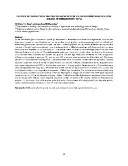
Authors Melissa Hyatt Gabriel J Swenson Richard A Long Citation Melissa Hyatt Gabriel J Swenson Richard A Long 2011 Molecular analysis of bacterial isolates used as unknowns in a bacteriology laboratory exercise ID: 912210

pcr bacterial bands gel bacterial pcr gel bands µl species analysis products dna agarose unknowns 100 ladder 515 region

Share:

Link:

Embed:

Download Presentation from below link

Download Presentation The PPT/PDF document "Molecular Analysis of Bacterial Isolates..." is the property of its rightful owner. Permission is granted to download and print the materials on this web site for personal, non-commercial use only, and to display it on your personal computer provided you do not modify the materials and that you retain all copyright notices contained in the materials. By downloading content from our website, you accept the terms of this agreement.


Presentation Transcript

Slide1

Molecular Analysis of Bacterial Isolates Used as Unknowns in a Bacteriology Laboratory Exercise

Authors: Melissa Hyatt, Gabriel J. Swenson, Richard A. Long

Citation: Melissa Hyatt, Gabriel J. Swenson, Richard A. Long. 2011. Molecular analysis of bacterial isolates used as unknowns in a bacteriology laboratory exercise.

Publication Date : November 2011

Slide2

Introduction

This figure shows an agarose gel image of PCR products to differentiate bacterial species based on variation in the genome content.  Each lane shown on the gel represents an individual bacterial unknown with one lane containing a size referencing ladder. The bands are the PCR products of the intergenic transcribed spacer (ITS), the region between the 16S and 23S ribosomal DNA genes in bacterial genomes. Multiple interval bands within a lane reflect multiple ITS regions of different bacterial species. To visualize bands, PCR products were separated on a high resolution agarose gel that was then stained with ethidium bromide, a DNA binding stain.  For analytical purposes, only dominant bands are considered; these bands were compared to previously empirically-derived bands for referencing by students.

Abbreviations used in the figure:  St (

Salmonella typhimurium

, 609 bp, 515 bp),

Bc

(

Bacillus cereus

, 229 bp),

Ea

(

Enterobacter aerogenes

, 591 bp, 443 bp),

Ec

(

Escherichia coli

, 539 bp, 436 bp),

Kp

(

Klebsiella pneumoniae

, 515 bp, 428 bp), L (Lonza DNA reference ladder, 100 bp ext. range; 3 kb, 2.5 kb, 2 kb, 1.5 kb, 1 kb, 0.8 kb, 0.6 kb, 0.4 kb, 0.1 kb),

Ml

(

Micrococcus luteus

, 515 bp),

Ms

(

Mycoplasma smegmatis,

500 bp, 400 bp),

Pv

(

Proteus vulgaris,

826 bp, 619 bp), Pa (

Pseudomonas aeruginosa

, 556 bp), and Sa (

Staphylococcus aureus,

556 bp, 515 bp).

Methods

The following bacterial isolates were obtained from the American Type Culture Collection and used as unknowns:

Bacillus cereus, Enterobacter aerogenes

,

Escherichia coli

,

Klebsiella pneumoniae

,

Micrococcus luteus

,

Mycoplasma smegmatis

,

Proteus vulgaris

, and

Pseudomonas aeruginosa

. These unknowns were incubated overnight on tryptic soy agar plates.

Staphylococcus aureus

and

Salmonella typhimurium

were incubated overnight in tryptic soy broth, pelleted, and the supernatant decanted. Either colonies or pelleted cells were suspended in 100 µl of PCR water.  Cells were lysed using 8 µl of Lyse-N-Go reagent (Pierce) and 2 µl of the cell suspension in an 0.2-ml PCR tube.  Upon completion of the Lyse-N-Go thermal cycling program, 15 µl of Apex

TaqRed

master mix amended with the ARISA primer set was added directly to each PCR tube and amplified following parameters of Cardinale et al. (2).  The primers used were ITSF (5-GTCGTAACAAGGTAGCCGTA-3) and

ITSReub

(5-GCCAAGGCATCCACC-3).

To separate PCR products, 5 µl of the sample were loaded onto a 2% high resolution agarose gel (USB Corporation) in 1X TAE; also loaded on the gel were 3 µl of Lonza ladder (100 bp ext. range DNA ladder, cat. no. 50322) for referencing. The agarose gel was run for 95 minutes at 100 V; 15% of the 1X TAE running buffer was exchanged with chilled buffer every 15 minutes.  The gel image was obtained by staining with ethidium bromide and capturing in an EpiChemi

3

darkroom using the visualization software

LabWorks

(v. 4.6; excitation 254 nm, ethidium bromide emission filter).

Discussion

Bacterial species differ in the size of the ITS region and visualization of this region though PCR and gel electrophoresis can be used to identify unknown bacterial species (1).  This relates to the applicability of PCR in economically and rapidly distinguishing between bacterial species in comparison with the classical biochemical dichotomous key commonly used in microbiology labs. For example, advanced high school or undergraduate students could perform this analysis and then compare the banding patterns, using dominant bands, to identify their unknown bacterial isolate of interest. 

References

1. 

Scheinert

, P., R.

Krausse

, U. Ullman, R.

Soller

, and G. Krupp.

1996. Molecular differentiation of bacteria by PCR amplification of the 16S-23S rRNA spacer. J. Microbiol. Methods

26:

103–117.

2.

Cardinale, M., L.

Brusetti

, P. Quatrini, S.

Borin

, A. M. Puglia, A.

Rizzi

, E.

Zanardini

, C. Sorlini, C.

Corselli

, and D.

Daffonchio

.

2004. Comparison of different primer sets for use in automated ribosomal intergenic spacer analysis of complex bacterial communities. Appl. Environ. Microbiol.

70:

6147–6156.

Slide3

Slide4