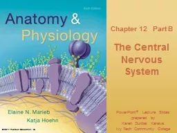
Inc 122 Cerebral Hemispheres cont Cerebral White Matter Second of the three basic regions of cerebral hemispheres Responsible for communication between cerebral areas and between cortex and lower CNS ID: 775208
Download Presentation The PPT/PDF document " © 2017 Pearson Education" is the property of its rightful owner. Permission is granted to download and print the materials on this web site for personal, non-commercial use only, and to display it on your personal computer provided you do not modify the materials and that you retain all copyright notices contained in the materials. By downloading content from our website, you accept the terms of this agreement.
Slide1
©
2017 Pearson Education
, Inc.
Slide212.2 Cerebral Hemispheres (cont.)
Cerebral White MatterSecond of the three basic regions of cerebral hemispheresResponsible for communication between cerebral areas, and between cortex and lower CNS Consists of myelinated fibers bundled into large tracts Classified according to direction they run: Association, commissural, and projection fibers
©
2017 Pearson Education
, Inc.
Slide3Cerebral White Matter (cont.)
Association fibers: horizontal running fibers that connect different parts of same hemisphereCommissural fibers: horizontal fibers that connect gray matter of two hemispheres Projection fibers: vertical fibers that connect hemispheres with lower brain or spinal cordInternal capsule: projection fibers on each side of brain stem form compact bandPasses between thalamus and some of basal nucleiCorona radiata: projection fibers that radiate through cerebral white matter to cortex
©
2017 Pearson Education
, Inc.
Slide4Figure
12.9a
White
fiber
tracts of the cerebral hemispheres.
©
2017 Pearson Education
, Inc.
Longitudinal fissure
Gray matter
White matter
Third
ventricle
Thalamus
Pons
Medulla oblongata
Superior
Frontal section
Lateral
ventricle
Basal nuclei
Decussation
(cross-over)
of pyramids
Association fibers
(within hemisphere)
Commissural fibers
(between hemispheres)
Projection
fibers
(cerebral cortex to
or from lower
area)
• Corpus
callosum
• Corona
radiata
• Internal
capsule
• Globus
pallidus
• Caudate
• Putamen
Slide5Figure
12.9b
White
fiber
tracts of the cerebral hemispheres.
©
2017 Pearson Education
, Inc.
Association fibers
Projection fibers
Commissural fibers
Parasagittal section and dissection
• Corpus
callosum
• Corona
radiata
• Internal
capsule
Gray
matter
Slide6Basal Nuclei (Ganglia)
Third of the three basic regions of cerebrumEach hemisphere’s basal nuclei include a:Caudate nucleusPutamenGlobus pallidusCaudate nucleus + putamen = striatumClosely associated with subthalamic nuclei (diencephalon) and substantia nigra (midbrain)
©
2017 Pearson Education
, Inc.
Slide7Figure
12.10a Basal nuclei.
© 2017 Pearson Education, Inc.
Striatum
Thalamus
Putamen
Tail of caudate
nucleus
Caudate
nucleus
Slide8Basal Nuclei (Ganglia) (cont.)
Functions of basal nuclei are thought to:Influence muscle movements Play role in cognition and emotionRegulate intensity of slow or stereotyped movementsFilter out incorrect/inappropriate responsesInhibit antagonistic/unnecessary movementsParkinson’s disease and Huntington’s disease are disorders of the basal nuclei
©
2017 Pearson Education
, Inc.
Slide9Figure
12.10b Basal nuclei.
© 2017 Pearson Education, Inc.
Corpus callosum
Head of caudate nucleus
Putamen
Globus
pallidus
Thalamus
Tail of caudate nucleus
Third ventricle
Cerebral cortex
Cerebral white matter
Anterior
Posterior
Anterior horn of
lateral ventricle
Inferior horn of
lateral ventricle
Slide1012.3 The Diencephalon
Consists of three paired gray-matter structures:ThalamusHypothalamusEpithalamusAll three enclose third ventricle
©
2017 Pearson Education
, Inc.
Slide11Animation – Rotatable Brain (sectioned)
© 2017 Pearson Education, Inc.
Slide12Figure
12.11a
Midsagittal
section of the brain.
©
2017 Pearson Education
, Inc.
Corpus callosum
Choroid plexus
Pineal gland
Arbor vitae (of cerebellum)
Fourth ventricle
Choroid plexus
Cerebellum
Septum
pellucidum
Hypothalamus
Optic
chiasma
Pituitary gland
Cerebral hemisphere
Mammillary body
Pons
Medulla oblongata
Spinal cord
Midbrain
Epithalamus
Fornix
Posterior
commissure
Thalamus
(encloses
third ventricle)
Cerebral
aqueduct
Corpora
quadrigemina
Interthalamic
adhesion
(intermediate
mass of thalamus)
Interventricular
foramen
Anterior
commissure
Slide13Thalamus
Bilateral egg-shaped nuclei that form superolateral walls of third ventricleMakes up 80% of diencephalonBilateral nuclei connected by interthalamic adhesion (intermediate mass)Contains several nuclei, named for locationNuclei project and receive fibers from cerebral cortex
©
2017 Pearson Education
, Inc.
Slide14Figure
12.12a
Selected structures of the diencephalon.
©
2017 Pearson Education
, Inc.
Medial
dorsal
nucleus
Lateral
dorsal
nucleus
Lateral
posterior
nucleus
Pulvinar
Medial
geniculate
body
Lateral
geniculate
body
Anterior
nuclei
Reticular
nucleus
Ventral
anterior
Ventral
lateral
Ventral
postero
-
lateral
The main thalamic nuclei.
(The reticular nuclei that
“cap” the thalamus laterally are depicted as curving
translucent structures.)
Ventral nuclei
Slide15Thalamus (cont.)
Main thalamic function is to act as relay station for information coming into cortexSorts, edits, and relays ascending input such as:Impulses from hypothalamus for regulating emotion and visceral functionImpulses from cerebellum and basal nuclei to help direct motor corticesImpulses for memory or sensory integrationOverall, it acts to mediate sensation, motor activities, cortical arousal, learning, and memory
©
2017 Pearson Education
, Inc.
Slide16Hypothalamus
Located below thalamusForms cap over brain stem and forms inferolateral walls of third ventricleContains many important nuclei such as:Mammillary bodies: paired anterior nuclei that act as olfactory relay stationsInfundibulum: stalk that connects to pituitary gland
©
2017 Pearson Education
, Inc.
Slide17Figure
12.12b
Selected structures of the diencephalon.
©
2017 Pearson Education
, Inc.
The main hypothalamic nuclei.
Anterior
commissure
Preoptic
nucleus
Anterior
hypothalamic
nucleus
Supraoptic
nucleus
Suprachiasmatic
nucleus
Optic
chiasma
Infundibulum
(stalk of the
pituitary gland)
Paraventricular
nucleus
Fornix
Dorsomedial
nucleus
Posterior
hypothalamic
nucleus
Lateral
hypothalamic
area
Ventromedial
nucleus
Arcuate
nucleus
Pituitary
gland
Mammillary
body
Slide18Hypothalamus (cont.)
The hypothalamus is the main visceral control and regulating center that is vital to homeostasis Chief homeostasis controls:Controls autonomic nervous systemExamples: blood pressure, rate and force of heartbeat, digestive tract motility, pupil sizeInitiates physical responses to emotionsPart of limbic system: perceives pleasure, fear, rage, biological rhythms, and drives (sex drive)
©
2017 Pearson Education
, Inc.
Slide19Hypothalamus (cont.)
The hypothalamus also:Regulates body temperature: sweating or shiveringRegulates hunger and satiety in response to nutrient blood levels or hormonesRegulates water balance and thirstRegulates sleep-wake cyclesSuprachiasmatic nucleus of thalamus sets our biological clock
©
2017 Pearson Education
, Inc.
Slide20Hypothalamus (cont.)
Controls endocrine system functions such as:Secretions of anterior pituitary glandProduction of posterior pituitary hormones
©
2017 Pearson Education
, Inc.
Slide21Clinical – Homeostatic Imbalance 12.4
Hypothalamic disturbances cause a number of disorders such as:Severe body wastingObesitySleep disturbancesDehydrationEmotional imbalances Implicated in failure to thrive: delay in growth or developmentOccurs when child is deprived of a warm, nurturing relationship
©
2017 Pearson Education
, Inc.
Slide22Epithalamus
Most dorsal portion of diencephalonForms roof of third ventricleContains pineal gland (body) Extends from posterior borderSecretes melatonin that helps regulate sleep-wake cycle
©
2017 Pearson Education
, Inc.
Slide23Figure
12.11a
Midsagittal
section of the brain.
©
2017 Pearson Education
, Inc.
Corpus callosum
Choroid plexus
Pineal gland
Arbor vitae (of cerebellum)
Fourth ventricle
Choroid plexus
Cerebellum
Septum
pellucidum
Hypothalamus
Optic
chiasma
Pituitary gland
Cerebral hemisphere
Mammillary body
Pons
Medulla oblongata
Spinal cord
Midbrain
Epithalamus
Fornix
Posterior
commissure
Thalamus
(encloses
third ventricle)
Cerebral
aqueduct
Corpora
quadrigemina
Interthalamic
adhesion
(intermediate
mass of thalamus)
Interventricular
foramen
Anterior
commissure
Slide24Figure
12.11b
Midsagittal
section of the
brain
(continued
).
©
2017 Pearson Education
, Inc.
Arbor vitae
Fourth ventricle
Cerebellum
Lateral ventricle
Corpus callosum
Fornix
Thalamus
Third ventricle
Hypothalamus
Optic
chiasma
Pons
Medulla oblongata
Mammillary body
Pineal gland
Midbrain
Epithalamus
Posterior
commissure
Corpora
quadrigemina
Cerebral
aqueduct
Anterior
commissure
Slide25Figure
12.13a
Three views of the brain stem (green) and the diencephalon (purple).
©
2017 Pearson Education
, Inc.
Optic
chiasma
Optic nerve (II)
Mammillary body
Oculomotor
nerve (III)
Optic tract
Trigeminal nerve (V)
Abducens
nerve (VI)
Facial nerve (VII)
Vagus
nerve (X)
Accessory nerve (XI)
Hypoglossal nerve (XII)
Ventral root of first cervical nerve
Trochlear nerve (IV)
Pons
Middle cerebellar peduncle
Pyramid
Decussation
of pyramids
Ventral view
Spinal cord
Vestibulocochlear
nerve (VIII)
Glossopharyngeal nerve (IX)
Diencephalon
• Thalamus
• Hypothalamus
Brain stem
Diencephalon
Thalamus
Hypothalamus
Midbrain
Pons
Crus
cerebri
of cerebral
peduncles (midbrain)
View
(b)
View
(c)
View
(a)
Medulla
oblongata
Slide26Figure
12.13b
Three views of the brain stem (green) and the diencephalon (purple).
©
2017 Pearson Education
, Inc.
Thalamus
Superior
colliculus
Inferior
colliculus
Trochlear nerve (IV)
Superior cerebellar peduncle
Middle cerebellar peduncle
Inferior cerebellar peduncle
Vestibulocochlear
nerve (VIII)
Olive
Optic tract
Infundibulum
Pituitary gland
Trigeminal nerve (V)
Abducens
nerve (VI)
Facial nerve (VII)
Vagus
nerve (X)
Accessory nerve (XI)
Hypoglossal nerve (XII)
P
ons
Glossopharyngeal nerve (IX)
Crus
cerebri
of cerebral
peduncles (midbrain)
Left lateral view
View
(b)
Brain stem
Diencephalon
Thalamus
Hypothalamus
Midbrain
Pons
View
(c)
View
(a)
Medulla
oblongata
Slide27Figure
12.13c
Three views of the brain stem (green) and the diencephalon (purple).
©
2017 Pearson Education
, Inc.
Pineal gland
Diencephalon
Floor of
fourth ventricle
Facial nerve (VII)
Dorsal view
Dorsal median sulcus
Choroid plexus
(fourth ventricle)
Dorsal root of
first cervical nerve
Thalamus
Midbrain
• Superior
colliculus
• Inferior
colliculus
• Trochlear
nerve (IV)
• Superior cerebellar peduncle
Pons
• Middle cerebellar peduncle
Medulla oblongata
• Inferior cerebellar peduncle
•
Vestibulocochlear
nerve (VIII)
• Glossopharyngeal nerve (IX)
•
Vagus
nerve (X)
• Accessory nerve (XI)
Corpora
quadrigemina
of
tectum
View
(a)
View
(c)
View
(b)
Brain stem
Diencephalon
Thalamus
Hypothalamus
Midbrain
Pons
Medulla
oblongata
Slide2812.4 Brain Stem
Consists of three regions: midbrain, pons, medulla oblongataSimilar in structure to spinal cord but contains nuclei embedded in white matterControls automatic behaviors necessary for survivalContains fiber tracts connecting higher and lower neural centersNuclei are associated with 10 of the 12 pairs of cranial nerves
©
2017 Pearson Education
, Inc.
Slide29Figure 12.14 Inferior view of the brain, showing the three parts of the
brain stem
: midbrain, pons, and
medulla oblongata
.
©
2017 Pearson Education
, Inc.
Frontal lobe
Optic
chiasma
Optic nerve (II)
Optic tract
Mammillary body
Pons
Cerebellum
Temporal
lobe
Spinal cord
Midbrain
Medulla
oblongata
Olfactory bulb
(synapse point of
cranial nerve I)
Slide30Midbrain
Located between diencephalon and ponsCerebral peduncles: two ventral bulges that contain pyramidal motor tractsForm pillars that hold up cerebrumCerebral aqueduct: channel running through midbrain that connects third and fourth ventricles
©
2017 Pearson Education
, Inc.
Slide31Midbrain (cont.)
Periaqueductal gray matter: nuclei that play a role in pain suppression and fight-or-flight responseAlso include nuclei that control cranial nerve III (oculomotor) and IV (trochlear)
©
2017 Pearson Education
, Inc.
Slide32Figure
12.13c
Three views of the brain stem (green) and the diencephalon (purple).
©
2017 Pearson Education
, Inc.
Pineal gland
Diencephalon
Floor of
fourth ventricle
Facial nerve (VII)
Dorsal view
Dorsal median sulcus
Choroid plexus
(fourth ventricle)
Dorsal root of
first cervical nerve
Thalamus
Midbrain
• Superior
colliculus
• Inferior
colliculus
• Trochlear
nerve (IV)
• Superior cerebellar peduncle
Pons
• Middle cerebellar peduncle
Medulla oblongata
• Inferior cerebellar peduncle
•
Vestibulocochlear
nerve (VIII)
• Glossopharyngeal nerve (IX)
•
Vagus
nerve (X)
• Accessory nerve (XI)
Corpora
quadrigemina
of
tectum
View
(a)
View
(c)
View
(b)
Brain stem
Diencephalon
Thalamus
Hypothalamus
Midbrain
Pons
Medulla
oblongata
Slide33Midbrain (cont.)
Midbrain nuclei scattered throughout white matter include:Corpora quadrigemina: paired dorsal protrusions Superior colliculi: visual reflex centers Inferior colliculi: auditory relay centersSubstantia nigra: functionally linked to basal nucleiParkinson’s disease is degeneration of this area
©
2017 Pearson Education
, Inc.
Slide34Midbrain (cont.)
Red nucleus: relay nuclei for some descending limb flexion motor pathwaysPart of reticular formation
©
2017 Pearson Education
, Inc.
Slide35Figure
12.13c
Three views of the brain stem (green) and the diencephalon (purple).
©
2017 Pearson Education
, Inc.
Pineal gland
Diencephalon
Floor of
fourth ventricle
Facial nerve (VII)
Dorsal view
Dorsal median sulcus
Choroid plexus
(fourth ventricle)
Dorsal root of
first cervical nerve
Thalamus
Midbrain
• Superior
colliculus
• Inferior
colliculus
• Trochlear
nerve (IV)
• Superior cerebellar peduncle
Pons
• Middle cerebellar peduncle
Medulla oblongata
• Inferior cerebellar peduncle
•
Vestibulocochlear
nerve (VIII)
• Glossopharyngeal nerve (IX)
•
Vagus
nerve (X)
• Accessory nerve (XI)
Corpora
quadrigemina
of
tectum
View
(a)
View
(c)
View
(b)
Brain stem
Diencephalon
Thalamus
Hypothalamus
Midbrain
Pons
Medulla
oblongata
Slide36Figure
12.15a
Cross sections through different regions of the brain stem.
©
2017 Pearson Education
, Inc.
Tectum
Fibers of
pyramidal tract
Substantia
nigra
Midbrain
Red
nucleus
Medial
lemniscus
Oculomotor
nucleus (III)
Periaqueductal gray
matter
Dorsal
Cerebral
aqueduct
Superior
colliculus
Reticular
formation
Ventral
Crus
cerebri
of cerebral
peduncle
Slide37Pons
Located between midbrain and medulla oblongata Fourth ventricle separates pons from cerebellumComposed of conduction tracts:Longitudinal fibers connect higher brain centers and spinal cordTransversal/dorsal fibers relay impulses between motor cortex and cerebellum
©
2017 Pearson Education
, Inc.
Slide38Pons (cont.)
Origin of cranial nerves V (trigeminal), VI (abducens), and VII (facial)Some nuclei play role in reticular formation, and some help maintain normal rhythm of breathing
©
2017 Pearson Education
, Inc.
Slide39Figure
12.15b
Cross sections through different regions of the brain stem.
©
2017 Pearson Education
, Inc.
Medial
lemniscus
Pontine
nuclei
Fibers of
pyramidal tract
Reticular
formation
Fourth
ventricle
Superior cerebellar
peduncle
Trigeminal
main sensory
nucleus
Trigeminal
motor
nucleus
Middle
cerebellar
peduncle
Trigeminal
nerve (V)
Pons
Slide40Medulla Oblongata
Also known as medullaBlends into spinal cord at foramen magnumContains fourth ventricleContinuation of central canal of spinal cordMedulla and pons form ventral wallContains choroid plexus: capillary-rich membrane that forms cerebral spinal fluid
©
2017 Pearson Education
, Inc.
Slide41Medulla Oblongata (cont.)
Structures of the medulla oblongataPyramids: two ventral longitudinal ridges formed by pyramidal tracts from motor cortexDecussation of the pyramids: point where pyramidal tracts cross over to opposite side of bodyOlives: swellings caused by underlying inferior olivary nuclei that relay stretch information from muscles and joints to cerebellum
©
2017 Pearson Education
, Inc.
Slide42Medulla Oblongata (cont.)
Cranial nerves VIII, IX, X, and XIIVestibular and cochlear nuclei: mediate responses that maintain equilibriumNucleus cuneatus and nucleus gracilis: relay ascending sensory information from spinal cord
©
2017 Pearson Education
, Inc.
Slide43Figure
12.11a
Midsagittal
section of the brain.
©
2017 Pearson Education
, Inc.
Corpus callosum
Choroid plexus
Pineal gland
Arbor vitae (of cerebellum)
Fourth ventricle
Choroid plexus
Cerebellum
Septum
pellucidum
Hypothalamus
Optic
chiasma
Pituitary gland
Cerebral hemisphere
Mammillary body
Pons
Medulla oblongata
Spinal cord
Midbrain
Epithalamus
Fornix
Posterior
commissure
Thalamus
(encloses
third ventricle)
Cerebral
aqueduct
Corpora
quadrigemina
Interthalamic
adhesion
(intermediate
mass of thalamus)
Interventricular
foramen
Anterior
commissure
Slide44Figure
12.13c
Three views of the brain stem (green) and the diencephalon (purple).
©
2017 Pearson Education
, Inc.
Pineal gland
Diencephalon
Floor of
fourth ventricle
Facial nerve (VII)
Dorsal view
Dorsal median sulcus
Choroid plexus
(fourth ventricle)
Dorsal root of
first cervical nerve
Thalamus
Midbrain
• Superior
colliculus
• Inferior
colliculus
• Trochlear
nerve (IV)
• Superior cerebellar peduncle
Pons
• Middle cerebellar peduncle
Medulla oblongata
• Inferior cerebellar peduncle
•
Vestibulocochlear
nerve (VIII)
• Glossopharyngeal nerve (IX)
•
Vagus
nerve (X)
• Accessory nerve (XI)
Corpora
quadrigemina
of
tectum
View
(a)
View
(c)
View
(b)
Brain stem
Diencephalon
Thalamus
Hypothalamus
Midbrain
Pons
Medulla
oblongata
Slide45Figure
12.15c
Cross sections through different regions of the brain stem.
©
2017 Pearson Education
, Inc.
Fourth
ventricle
Pyramid
Medial
lemniscus
Hypoglossal
nucleus (XII)
Medulla oblongata
Dorsal
motor
nucleus of
vagus
(X)
Inferior
cerebellar
peduncle
Lateral
nuclear
group
Medial
nuclear
group
Raphe
nucleus
Reticular formation
Choroid
plexus
Solitary
nucleus
Vestibular
nuclei
(VIII)
Cochlear
nuclei
(VIII)
Nucleus
ambiguus
Inferior
olivary
nucleus
Slide46Medulla Oblongata (cont.)
Functions of the medulla oblongataMedulla is an autonomic reflex centerMany functions overlap with hypothalamusHypothalamus relays instructions via medullaFunctional groups of medulla include:Cardiovascular centerCardiac center adjusts force and rate of heart contractionVasomotor center adjusts blood vessel diameter for blood pressure regulation
©
2017 Pearson Education
, Inc.
Slide47Medulla Oblongata (cont.)
Respiratory centersGenerate respiratory rhythmControl rate and depth of breathing (with pontine centers)Various other centers regulate:Vomiting Hiccupping Swallowing CoughingSneezing
©
2017 Pearson Education
, Inc.
Slide4812.5 Cerebellum
11% of brain massLocated dorsal to pons and medullaProcesses input from cortex, brain stem, and sensory receptors to provide precise, coordinated movements of skeletal musclesAlso plays a major role in balance
©
2017 Pearson Education
, Inc.
Slide49Cerebellar Anatomy
Cerebellar hemispheres connected by wormlike vermisFolia: transversely oriented gyriEach hemisphere has three lobesAnterior, posterior, and flocculonodular Contains thin cortex of gray matter with distinctive treelike pattern of white matter called arbor vitae
©
2017 Pearson Education
, Inc.
Slide50Cerebellar Anatomy (cont.)
Purkinje fibers originate in cortex, synapse with cerebellumCerebellar homunculi show sensory maps of entire body
©
2017 Pearson Education
, Inc.
Slide51Figure
12.16a
Cerebellum.
©
2017 Pearson Education
, Inc.
Medulla oblongata
Fourth ventricle
Arbor vitae
Folia
Anterior lobe
Choroid plexus
Pons
Cerebellar
cortex
Posterior
lobe
Slide52Figure
12.16b
Cerebellum.
©
2017 Pearson Education
, Inc.
Medulla oblongata
Flocculonodular
lobe
Arbor vitae
Cerebellar cortex
Anterior lobe
Cerebellar
peduncles
• Superior
• Middle
• Inferior
Posterior
lobe
Choroid
plexus of
fourth
ventricle
Slide53Figure
12.16c
Cerebellum.
©
2017 Pearson Education
, Inc.
Anterior lobe
Vermis
Posterior
lobe
Primary
fissure
Horizontal
fissure
Slide54Cerebellar Peduncles
All fibers in cerebellum are ipsilateral—from and to the same side of bodyThree paired fiber tracts connect cerebellum to brain stemSuperior cerebellar peduncles connect cerebellum to midbrainMiddle cerebellar peduncles connect pons to cerebellumInferior cerebellar peduncles connect medulla to cerebellum
©
2017 Pearson Education
, Inc.
Slide55Figure
12.16b
Cerebellum.
©
2017 Pearson Education
, Inc.
Medulla oblongata
Flocculonodular
lobe
Arbor vitae
Cerebellar cortex
Anterior lobe
Cerebellar
peduncles
• Superior
• Middle
• Inferior
Posterior
lobe
Choroid
plexus of
fourth
ventricle
Slide56Cerebellar Processing
Cerebellum fine-tunes motor activity as follows:1. Receives impulses from cerebral cortex of intent to initiate voluntary muscle contraction2. Receives signals from proprioceptors throughout body, as well as visual and equilibrium pathways that:Pathways continuously “inform” cerebellum of body’s position and momentumCerebellar cortex calculates the best way to smoothly coordinate muscle contractionSends “blueprint” of coordinated movement to cerebral motor cortex and brain stem nuclei
©
2017 Pearson Education
, Inc.
Slide57Cognitive Functions of Cerebellum
Neuroimaging suggests that cerebellum plays role in thinking, language, and emotionAs it does for motor processes, it may compare actual output of higher functions with expected output and adjust accordingly
©
2017 Pearson Education
, Inc.