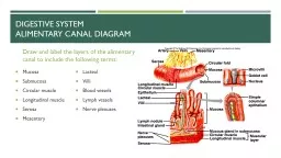
Metallic 0 Mind Mucsularis mucosae surround lamina propria and composed of Inner circular layer Outer longitudinal layer Mucosa Lined by epithelium Deep is a loose connective tissue ID: 659786
Download Presentation The PPT/PDF document "Digestive System: Alimentary Canal" is the property of its rightful owner. Permission is granted to download and print the materials on this web site for personal, non-commercial use only, and to display it on your personal computer provided you do not modify the materials and that you retain all copyright notices contained in the materials. By downloading content from our website, you accept the terms of this agreement.
Slide1
Digestive System: Alimentary Canal
Metallic
0
MindSlide2Slide3
Mucsularis
mucosae
surround lamina propria and composed of :
Inner circular layer.Outer longitudinal layer
Mucosa:Lined by epithelium.
Deep is a loose connective tissue (
Lamina
Propria
)
that houses glands and lymph vessels.
Submucosa
:
Dense irregular
fibroelastic
connvtive
tissue.
No glands
Except
in the esophagus and duodenum.
Meissner’s
submucosal
plexus:
houses postganglionic nerve cell bodies.Slide4
Muscularis
Externa
Responsible for
peristaltic activity.
Smooth muscle Except in the esophagus.Interstitial cells of
cajal are the pacemakers.Arranged helically
Composed of
Between the muscle is
auerbach’s
myenteric
plexus composed of postganglionic
parasympatheric
nerve cells.
Connective tissue envelopes the
muscularis
externa
that
may or may not
surrounded by
squamous epithelium If:
the organ is inraperitoneal it’s known as Serosa
The organ is retroperitoneal it’s known as
adventitiaSlide5
Mucousa
:
Epithelium:
Stratified
squamous
nonkeratinized epithelium.
Lamina
propria
:
Unremarkable.
Houses
esophageal cardiac glands
in two clusters:
Near the pharynx.
Near its juncture with stomach.
Muscularis
Mucosae
:
Single layer of longitudinal smooth muscle.Slide6
Submusosa:
fibroelastic
connective tissue.
Houses
esophageal glands proper.
Muscularis
externa
Inner circular and outer longitudinal layers.
They have both skeletal and smooth.
In the upper third of esophagus it’s mostly skeletal
In the middle third it’s both skeletal and smooth
In the lowest third it’s only smooth
The esophagus it covered by and
adventitia
until it pierces the diaphragm after that it’s covered by
serosa
.Slide7
Stomach
Rugae
:
Longitudinal folds (transverse in the
anturm
) of mucosa and
submucosa
.
Disappear in distended stomach.
Gastric Pits:
Formed by epithelial lining.
Gastric glands
empty in the bottom of each gastric pit.Slide8
Fundic Mucosa
Epithelium :
Simple columnar epithelium.
surface lining cells : manufacture
mucin
.
No goblet cells.
Lamina
propria
:
Connective tissue.
Occupied by
Fundic
(
oxyntic
) Glands.
Fundic
Glands:
short pits.
Simple or branched.
Have 3 regions:
Isthmus.
Neck.
Base. Slide9
Mucous neck cells:
In the neck region.
Secrete mucous to lubricate gastric content.
Golgi apparatus , RER are present.
Mitochondria in the basal region of the cell.
Apical cytoplasm has
secretory
granules.
Parietal (
oxyntic
) Cells:
at the periphery of the gland.
Produce (HCL) and intrinsic factors.
Basally located nuclei.
Acidophilic
Apical membrane
invaginates
to form
intracellular
canaliculi
lined by
microvilli
.
Cytoplasm in the
canaliculi
has round and tubular vesicles
tubulovesicular
system.
Rich in mitochondria.
Regenerative (stem) cells:
In the neck region.
Proliferate to replace all of the specialized cells
Chief (
zymogenic
) cells:
In the base region.
Manufacture
pepsinogen
,
renin
, gastric lipase
Exhibit rich RER,
golgi
apparatus, apical granules.
Basophilic.
DNES cells (APUD or
enteroendocrine
cells)
In the base region.
Manufacture:
endocrine,
paracrine
,
neurocrine
hormones.
Well-developed RER and
golgi
apparatus and numerous mitochondria.
Basal granules.
If:
The cell reach the lumen of the gut called (
the open type)
The cell doesn’t reach the lumen called (
the closed type)
Cell Types
Region
Surface lining cells
Isthums
Mucous neck cells, regenerative cells, parietal cells
Neck
Chief cells,
DNES cells
BaseSlide10
Submucosa
of the stomach:
Dense, irregular
collagenous
connective tissue.
Muscularis
Externa:Three layers:
Innermost Oblique muscle layer:
Not well defined
except
in the cardiac region.
Middle Circular muscle layer:
Especially pronounced where it forms the pyloric sphincter.
Outer longitudinal muscle layer
Serosa
:
Thin loose
subserous
connective tissue.
Covered by wet smooth wet simple
squamous
epithelium.Slide11
Small Intestine
3 types of modification are present in the small intestine to increase the surface area:
Plicae
Circulares
(valves of
Kerckring)Transverse folds of
submucosa
and mucosa.
Permanent.
Inncrease
surface area by factor 2 to 3
Villi
:
Protrusions of the lamina
propria
.
Epithelially
covered.
The core composed of :
Capillary loops
Lymphatic channel (lacteal)
Few smooth muscle fibers.Loose connective tissue rich in lymphoid cells
Numbers are greater in the duodenum.Increase the surface area 10 times
Microvilli
:
Increase by factor of 20
Invaginations of the epithelium into the lamina
propria
between
villi
form glands (
crypts of
Lieberkühn
)Slide12
Intestinal mucosa
in regions where lymphoid nodules abut the epithelium.
M cells
replace simple columnar epithelial lining of the small intestine.
Function:
presents antigenSlide13
Lamina
Propria
Loose connective tissue.
Crypts of
Lieberkuhn
:Tubular (or branched) gland.Open into the
intervillus space.
Paneth
cells:
In the bottom of the crypts.
Acidophilic
Apical granules.
Manufacture
lysozyme
.Slide14
Muscularis
mucoasae
:
Inner circular layer.Outer longitudinal layer
Submucosa:
Irregular fibroelastic connective tissue.Submucosa of the
duodenum
houses glands known as
Brunner’s Glands.
Muscularis
Externa:
Inner circular layer.Outer longitudinal layer.
Second and third part of the duodenum have
adventitia.
Everything else has
serosa
.Slide15
Large Intestine
Colon Histology:
No
villi
.
Have crypts of
lieberkühn.Absent Paneth cells.
Lamina
propria
,
muscularis
mucosa,
submuosa resemble
small intestine.
Muscularis externa:
have unusual outer longitudinal muscle layer.
The muscle gathered in three narrow ribbons known as
taenie
coli
Serosa
has a fat-filled pouches called
appendices
epiploicae.Slide16
Appendix
Vermiform appendix
Mucosa:
Simple columnar epithelium consisting of
surface absorptive , goblet cells, M cells.
Lamina
propria
: loose connective tissue with lymph nodules and crypts of
liebekühn
Same
muscularis
mucosae as colon.
Same
submucosa also it have lymphoid nodules and fatty infiltration.
Same
muscularis
externa
.
Covered by
serosaSlide17
Please Remember
Meissner’s
plexus and
Auerbach
plexus present in all tissues of alimentary canal.
Goblet cells starts at the duodenum and
increases gradually