/
Treatment of dental and skeletal bimaxillary protrusion in patient wit Treatment of dental and skeletal bimaxillary protrusion in patient wit

Treatment of dental and skeletal bimaxillary protrusion in patient wit - PDF document

olivia-moreira
olivia-moreira . @olivia-moreira
Follow
398 views
Uploaded On 2016-07-15

Treatment of dental and skeletal bimaxillary protrusion in patient wit - PPT Presentation

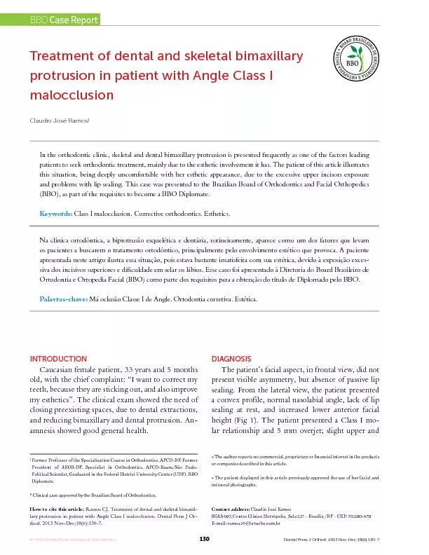
130 INTRODUCTION Caucasian female patient 33 years and 5 months old with the chief complaint 147I want to correct my teeth because they are sticking out and also improve my esthetics148 Th ID: 405475

130 INTRODUCTION Caucasian female patient

Share:

Link:

Embed:

Download Presentation from below link

Download Pdf The PPT/PDF document "Treatment of dental and skeletal bimaxil..." is the property of its rightful owner. Permission is granted to download and print the materials on this web site for personal, non-commercial use only, and to display it on your personal computer provided you do not modify the materials and that you retain all copyright notices contained in the materials. By downloading content from our website, you accept the terms of this agreement.


Presentation Transcript

130 Treatment of dental and skeletal bimaxillary protrusion in patient with Angle Class I » The author reports no commercial, proprietary or nancial interest in the products » The patient displayed in this article previously approved the use of her facial and intraoral photographs.Contact address: Claudio José RamosSGAS 607, Centro Clínico Metrópolis, Sala 217 – Brasília /DF – CEP: 70.200-670E-mail: ramos.16@brturbo.com.br INTRODUCTION Caucasian female patient, 33 years and 5 months old, with the chief complaint: “I want to correct my teeth, because they are sticking out, and also improve my esthetics”. The clinical exam showed the need of closing preexisting spaces, due to dental extractions, and reducing bimaxillary and dental protrusion. Anamnesis showed good general health.AGThe patient’s facial aspect, in frontal view, did not present visible asymmetry, but absence of passive lip sealing. From the lateral view, the patient presented a convex profile, normal nasolabial angle, lack of lip sealing at rest, and increased lower anterior facial height (Fig 1). The patient presented a Class I molar relationship and 5 mm overjet; slight upper and Former Professor of the Specialization Course in Orthodontics, APCD-DF. Former President of ABOR-DF. Specialist in Orthodontics, APCD-Bauru/São Paulo. Political Scientist, Graduated in the Federal District University Center (UDF). BBO Clinical case approved by the Brazilian Board of Orthodontics.How to cite this articleRamos CJ. Treatment of dental and skeletal bimaxil In the orthodontic clinic, skeletal and dental bimaxillary protrusion is presented frequently as one of the factors leading patients to seek orthodontic treatment, mainly due to the esthetic involvement it has. The patient of this article illustrates this situation, being deeply uncomfortable with her esthetic appearance, due to the excessive upper incisors exposure and problems with lip sealing. This case was presented to the Brazilian Board of Orthodontics and Facial Orthopedics (BBO), as part of the requisites to become a BBO Diplomate. Keywords:Class I malocclusion. Corrective orthodontics. Esthetics. Na clínica ortodôntica, a biprotrusão esquelética e dentária, rotineiramente, aparece como um dos fatores que levam os pacientes a buscarem o tratamento ortodôntico, principalmente pelo envolvimento estético que provoca. A paciente apresentada neste artigo ilustra essa situação, pois estava bastante insatisfeita com sua estética, devido à exposição excessiva dos incisivos superiores e diculdade em selar os lábios. Esse caso foi apresentado à Diretoria do Board Brasileiro de Ortodontia e Ortopedia Facial (BBO) como parte dos requisitos para a obtenção do título de Diplomado pelo BBO. Palavras-chave:Má oclusão Classe I de Angle. Ortodontia corretiva. Estética. © 2013 Dental Press Journal of Orthodontics Dental Press J Orthod. 2013 Nov-Dec;18(6): BBO Case Report 131 Inskeletal terms, according to the lateral cephalometric radiograph and respective tracing (Fig5), it was observed that the patient presented unbalanced skeletal bases, characterized by angles SNA90°, SNB83° and ANB7°, with a dolichofacial pattern, and presenting protruded upper incisors, with slight lingual tipping, and protruded lower incisors with buccal tipping. The functional analysis of mandibular movements revealed absence of anterior guides.lower crowding; moderate Curve of Spee; absence of teeth #36 and #37; posterior cross bite on the right side, with buccal cross bite between teeth #15 and #45; lower midline deviated 1 mm to the left; and upper and lower incisors with incisal wear (Fig 2). The periapical and panoramic radiographs demonstrated root shortening of the upper right central incisor, absence of teeth #36 and #47, lower molar mesialization and lack of parallelism between roots (Figs3 and4). Figure 1 132 Figure 2 Figure 3Figure 4 PLADue to compromised facial esthetics and dental and skeletal bimaxillary protrusion, the treatment plan had the following objectives: Maintain canine occlusion; align and level the teeth; eliminate posterior cross bites; reduce overbite and overjet; eliminate crowding on both arches; level the Curve of Spee, close the spaces due to extraction of teeth #36 and #47, and also achieve lower molar root parallelism.For this purpose, extractions of the rst upper and right lower premolars were necessary, besides the retraction of upper and lower anterior teeth, to reduce bimaxillary protrusion and correct the lower midline. All this very carefully, in order to avoid root shortening of tooth #11.The treatment also aimed at obtaining lateral and protrusion guides, and reduction of facial convexity. The nasolabial angle would be preserved, as well as 133 Figure 5 - Initial lateral cephalometric radiograph () and cephalometric tracing ( passive lip sealing would be expected and decrease of the lower anterior facial height, in order to reach smile harmony.ROThe treatment was performed with the Straight wire technique, using metallic orthodontic brackets in both arches, according to Roth’s prescription (0.022x0.028-in), with a removable transpalatal bar (stainless steel archwire, 0.032-in) as upper arch anchorage, and a lip bumper as lower arch anchorage. Leveling and alignment were carried with a sequence of NiTi round archwires, NiTi rectangular archwires 0.017 x 0.025-in and stainless steel rectangular archwires 0.019 x 0.025, with loops for retraction of anterior teeth and 5/16-in elastics to assist in the nishing.Bands were placed on the rst and second upper molars, as well as on the rst, second and third lower molars, with a transpalatal bar on the rst upper molars and lip bumper on lower molars.The placement of the xed orthodontic appliance was completed with the direct bonding of brackets on the remaining upper and lower teeth, except on teeth #14, #24 and #44 — which would be extracted, with the objective of eliminating crowding and allow the correction of the dental bimaxillary protrusion, by means of retracting the anterior teeth.1,3,4 Alignment and leveling were carried with 0.012, 0.014, 0.016 and 0.018-in NiTi archwires; and 0.017 x 0.025-in NiTi rectangular, and stainless steel 0.019 x 0.025-in archwires. Then, the retraction of upper and lower anterior teeth, and the correction of the lower midline were performed. Theextraction of tooth #18 was suggested because it would be without its antagonist due to the mesial movement of tooth #48. For intercuspation, 5/16-in intermaxillary elastics were used.A stainless steel 0.028-in fixed lower retainer was bonded to teeth #33 to #45, and to teeth #35 to #37. On the upper arch, a wraparound removable plate was used.The evaluation of the nal records shows that there was 1° reduction for SNA and SNB angles, a slight decrease in the vertical direction (Table1). The canine guidance was kept, there was overjet reduction and overbite and Curve of Spee correction. All teeth were aligned and leveled, and the posterior cross bites eliminated; the crowding B A 134 Figure 6on both arches was corrected; Curve of Spee was leveled. Thespaces for teeth #36 and #47, and also the extraction spaces for teeth #14, #24 and #44 were properly closed; upper and lower anterior retraction was satisfactory.The lower midline correction and anterior guides were obtained. However, lip sealing was not completely passive, due to the patient’s vertical growth pattern. The nasolabial angle was preserved, despite the decrease of incisor protrusion, which promoted smile harmony (Figs 6 to 10). The treatment lasted 35 months.CONThe treatment of dental skeletal bimaxillary protrusion was successful, once the main objectives proposed to the patient were achieved, resulting in good occlusion and pleasant facial esthetics (Fig 6). A good parallelism was obtained between roots, especially for lower molars. The pre-existing spaces before treatment and those caused by the extraction of teeth #14, #24 and #44 were properly closed, with upper and lower anterior retraction (Fig 7). 135 Figure 7 Figure 8Figure 9 136 Figure 10 - Final lateral cephalometric radiograph () and cephalometric tracing ( Figure 11 - Total superimposition of initial (black) and nal (red) cephalometric tracings, registered on SN line.Figure 12 - Partial superimposition of the maxilla (A) and the mandible (B), of initial (black) and nal (red) cephalometric tracings, evidencing tooth movements. In facial terms, passive lip sealing, initially expected, was not completely obtained, mainly due to increased anterior facial height. Nevertheless, greater smile harmony was obtained, which fully met the patient’s expectations, who always collaborated regarding oral hygiene and clinical recommendations.The level of root resorptions was acceptable, except for the upper right central incisor, in which the root shortening increased signicantly,7,9 but without any mobility observed in regular evaluation of each treatment phase (Fig 8). The patient was pleased with the treatment result, since it improved her facial esthetics B A 137 Almeida FM, Siqueira VCV. O efeito da exodontia dos primeiros pré-molares sobre a AFAI. Rev Dental Press Ortod Ortop Facial. 2004;9(6):48-62.Bills DA, Handelman CS, BeGole EA. Bimaxillary dentoalveolar protrusion: traits and orthodontic correction. Angle Orthod. 2005;75(3):333-9.3.Bravo LA. Soft tissue facial prole changes after orthodontic treatment with four premolars extracted. Angle Orthod. 1994;64(1):31-42.4.Erdinc AE, Nanda RS, Dandajena TC. Prole changes of patients treated with and without premolar extractions. Am J Orthod Dentofacial Orthop. 2007;132(3):324-31. 5.Kim TK, Kim JT, Mah J, Yang WS, Baek SH. First or second premolar extraction eects on facial vertical dimension. Angle Orthod. 6.Maltagliati LA, Montes LAP. Análise dos fatores que motivam os pacientes adultos a buscarem o tratamento ortodôntico. Rev Dental Press Ortod Ortop Facial. 2007;12(6):54-60.7.Mohandesan H, Ravanmehr H, Valaei N. A radiographic analysis of external apical root resorption of maxillary incisors during active orthodontic treatment. Eur J Orthod. 2007;29(2):134-9. 8.Oliveira GF, Almeida MR, Almeida RR, Ramos AL. Alterações dentoesqueléticas e do perl facial em pacientes tratados ortodonticamente com extração de quatro primeiros pré-molares. RevDental Press Ortod Ortop Facial. 2008;13(2):105-14. 9.Simplício H. Avaliação da reabsorção radicular apical em incisivos submetidos à retração [tese]. Araraquara (SP): Universidade Estadual substantially, with the reduction of dental and skeletal bimaxillary protrusion. Aer removing the appliances and placing new retainers, the patient was referred to dental whitening and composite restoration of the incisal borders of the upper anterior teeth. Thus, aer the recommended procedures, an even more pleasant smile was obtained, which contributed signicantly for the patient’s complete satisfaction. A/B di.Skeletal patternFacial angle Facial angle FMA(Tweed)24°ental patternIMPA(Tweed) .NA (degrees)14° 1(Steiner)4 mm7 mm3 mm4 .NB (degrees) 1(Steiner)4 mm12 mm9 mm3 11 - Interincisal angle (Downs)130°117°126°9 -APo (mm)(Ricketts)Lower lip – S line Table 1 - Cephalometric measures. © 2013 Dental Press Journal of Orthodontics Dental Press J Orthod. 2013 Nov-Dec;18(6): Ramos CJBBO Case Report Treatment of dental and skeletal bimaxillary protrusion in patient with Angle Class I malocclusion © 2013 Dental Press Journal of Orthodontics Dental Press J Orthod. 2013 Nov-Dec;18(6):130-7