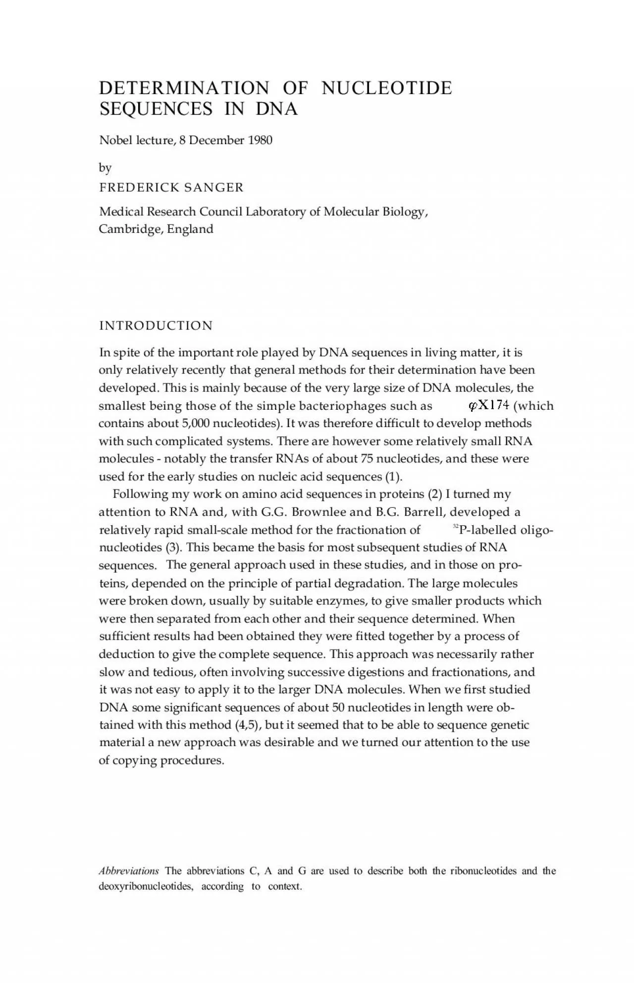
/
DETERMINATION OF NUCLEOTIDESEQUENCES IN DNANobel lecture 8 December 1 DETERMINATION OF NUCLEOTIDESEQUENCES IN DNANobel lecture 8 December 1

DETERMINATION OF NUCLEOTIDESEQUENCES IN DNANobel lecture 8 December 1 - PDF document

oneill
oneill . @oneill
Follow
344 views
Uploaded On 2022-08-22

DETERMINATION OF NUCLEOTIDESEQUENCES IN DNANobel lecture 8 December 1 - PPT Presentation

which 432Chemistry 1980COPYING PROCEDURESIn the RNA field these procedures had been pioneered by C Weissmann andhis colleagues 6 in their studies on the RNA sequence of the bacteriophage contain ID: 939818

sequence dna genes codons dna sequence codons genes method code 1980 fig coding mitochondria trnas mtdna reading codon protein

Share:

Link:

Embed:

Download Presentation from below link

Download Pdf The PPT/PDF document "DETERMINATION OF NUCLEOTIDESEQUENCES IN ..." is the property of its rightful owner. Permission is granted to download and print the materials on this web site for personal, non-commercial use only, and to display it on your personal computer provided you do not modify the materials and that you retain all copyright notices contained in the materials. By downloading content from our website, you accept the terms of this agreement.


Presentation Transcript

DETERMINATION OF NUCLEOTIDESEQUENCES IN DNANobel lecture, 8 December 1980byFREDERICK SANGERMedical Research Council Laboratory of Molecular Biology,Cambridge, EnglandINTRODUCTION (which 432Chemistry 1980COPYING PROCEDURESIn the RNA field these procedures had been pioneered by C. Weissmann andhis colleagues (6) in their studies on the RNA sequence of the bacteriophage contains a replicase that will synthesize a complementary copy of thesingle-stranded RNA chain, starting from its 3’ end. These workers devisedelegant procedures involving pulse-labelling with radioactively labelled nucleo-tides, from which sequences could be deduced.For DNA sequences we have used the enzyme DNA polymerase, whichcopies single-stranded DNA as shown in Fig. 1. The enzyme requires a primer,which is a single-stranded oligonucleotide having a sequence that is comple-mentary to, and therefore able to hybridize with, a region on the DNA beingsequenced (the template). Mononucleotide residues are added sequentially tothe 3’ end of the primer from the corresponding deoxynucleoside triphosphates,making a complementary copy of the template DNA. By using triphosphatescontaining32P in the a position the newly synthesized DNA can be labelled. Inthe early experiments synthetic oligonucleotides were used as primers, but afterFig. 1. Specificity requirements for DNA polymerase. F. Sanger433the discovery of restriction enzymes it was more convenient to use fragmentsresulting from their action as they were much more easily obtained.The copying procedure was used initially to prepare a short specific region oflabelled DNA which could then be subjected to p

artial digestion procedures.One of the difficulties of sequencing DNA was to find specific methods forbreaking it down into small fragments. No suitable enzymes were known thatwould recognise only one nucelotide. However, Berg, Fancher & Chamberlin(7) had shown earlier that under certain conditions it was possible to incorpo-rate ribonucleotides, in place of the normal deoxyribonucleotides, into DNAchains with DNA polymerase. Thus, for instance, if copying were carried outusing riboCTP and the other three deoxynucleosidc triphosphates, a chaincould be built up in which the C residues were in the ribo form. Bondsinvolving ribonucleotides could be broken by alkali under conditions wherethose involving the deoxynucleotides were not, so that a specific splitting at Cresidues could be obtained. Using this method we were able to extend oursequencing studies to some extent (8). However extensive fractionations andanalyses were still required.THE ‘PLUS AND MINUS’ METHODIn the course of these experiments we needed to prepare DNA copies of highspecific radioactivity, and in order to do this the highly labelled substrates hadto be present in low concentrations. Thus if was used for label-ling its concentration was much lower than that of the other three triphos-phates and frequently when we analysed the newly synthesized DNA chains wefound that they terminated at a position immediately before that at which an Ashould have been incorporated. Consequently a mixture of products was pro-duced all having the same 5’ end (the 5’ end of the primer) and terminating atthe 3’ end at the position of the A residues. If these products cou

ld befractionated on a system that separated only on the basis of chain length, thepattern of their distribution on fractionation would be proportional to thedistribution of the A’s along the DNA chain. And this, together with thedistribution of the other three mononucleotidcs, is the information required forsequence determination. Initial experiments carried out with J.E. Donelsonsuggested that this approach could be the basis for a more rapid method, and itwas found that good fractionations according to size could be obtained byionophoresis on acrylamide gels.The method described above met with only limited success but we were ableto develop two modified techniques that depended on the same general princi-ple and these provided a much more rapid and simpler method of DNAsequence determination than anything we had used before (9). This, which isknown as the “plus and minus” technique, was used to determine the almostcomplete sequence of the DNA of bacteriophage 174 which contains 5,386nucleotides (10). Chemistry 1980434THE ‘DIDEOXY’ METHODMore recently we have developed another similar method which uses specificchain-terminating analogues of the normal deoxynucleoside triphosphates(11). This method is both quicker and more accurate than the plus and minustechnique. It was used to complete the sequence of (12), to determinethe sequence of a related bacteriophage, G4 (13), and has now been applied tomammalian mitochondrial DNA.The analogues most widely used are the dideoxynucleoside triphosphates(Fig. 2). They are the same as the normal deoxynucleoside triphosphates butlack the 3’ hydroxyl group. They can be incor

porated into a growing DNAchain by DNA polymerase but act as terminators because, once they areincorporated, the chain contains no 3’ hydroxyl group and so no other nucelo-tide can be added.The principle of the method is summarised in Fig. 3. Primer and templateare denatured to separate the two strands of the primer, which is usually arestriction enzyme fragment, and then annealed to form the primer-templatecomplex. The mixture is then divided into four samples. One (the T sample) isincubated with DNA polymerase in the presence of a mixture of ddTTP(dideoxy thymidine triphosphate) and a low concentration of TTP, togetherwith the other three deoxynucleoside triphosphates (one of which is labelledFig. 2. Diagram showing chain termination with dideoxythymidine triphosphate (ddTTP). Thetop line shows the DNA polymerase-catalysed reaction of the normal deoxynucleoside triphosphate(TTP) with the 3’ terminal nucleotide of the primer: the bottom line the corresponding reactionwith ddTTP. F. Sanger435Fig. 3. Principle of the chain-terminating method.with 32P) at normal concentration. As the DNA chains are built up on the 3’end of the primer the position of the T’s will be tilled, in most cases by thenormal substrate T and extended further, but occasionally by ddT and termi-nated. Thus at the end of incubation there remains a mixture of chainsterminating with T at their 3’ end but all having the same 5’ end (the 5’ end ofthe primer). Similar incubations are carried out in the presence of each of theother three dideoxy derivatives, giving mixtures terminating at the positions ofC, A and G respectively, and the four mixtures are

fractionated in parallel byelectrophoresis on acrylamide gel under denaturing conditions. This systemseparates the chains according to size, the small ones moving quickly and thelarge ones slowly. As all the chains in the T mixture end at T the relativeposition of the T’s in the chain will define the relative sizes of the chains, andtherefore their relative positions on the gel after fractionation. The actualsequence can then simply be read off from an autoradiograph of the gel (Fig. 4).The method is comparatively rapid and accurate and sequences of up to about300 nucleotides from the 3’ end of the primer can usually be determined. 436Fig. 4. Autoradiograph of a DNA sequencing gel. The origin is at thr top and migration of the DNAchains, according to size, is downwards. The gel on the left has been run for 2 5 hr and that on theright for 5 hr with the same polymerisation mixtures. Considerably longer sequences have been read off but these are usually lessreliable.One problem with the method is that it requires single-stranded DNA astemplate. This is the natural form of the DNA in the bacteriophages and G4, but most DNA is double-stranded and it is frequently difficult toseparate the two strands. One way of overcoming this was devised by A.J.H.Smith (14). If the double-stranded linear DNA is treated with exonuclease III(a double-strand specific 3’ exonuclease) each chain is degraded from its 3’end, as shown in Fig. 5, giving rise to a structure that is largely single-strandedand can be used as template for DNA polymerase with suitable small primers.This method is particularly suitable for usewith fragments cloned in

plasmidvectors and has been used extensively in the work on human mitochondrialDNA.CLONING IN SINGLE-STRANDED BACTERIOPHAGEAnother method of preparing suitable template DNA that is being more widelyused is to clone fragments in a single-stranded bacteriophage vector (15-17).This approach is summarised in Fig. 6. Various vectors have been described.We have used a derivative of bacteriophage M 13 developed by Gronenborn &Messing (16) which contains an insert of the gene with anEcoRI restriction enzyme site in it. The presence of in a plaquecan be readily detected by using a suitable colour-forming substrate (X-gal).The presence of an insert in the EcoRI site destroys the gene,giving rise to a colourless plaque.Besides being a simple and general method of preparing single-strandedDNA this approach has other advantages. One is that it is possible to usethesame primer on all clones. Heidccker et al. (18) prepared a 96-nucleotide longrestriction fragment derived from a position in the Ml3 vector flanking theEcoRI site (see Fig. 6). This can be used to prime into, and thus determine, asequence of about 200 nucleotides in the inserted DNA. Smaller syntheticprimers have now been prepared (19,20)which allow longer sequences to bedetermined. The approach that WC have used is to prepare clones at randomfrom restriction enzyme digests and determine the sequence with the flankingprimer. Computer programmrs (21) are then used to store, overlap and ar-range the data. 438Chemistry 1980Another important advantage of the cloning technique is that it is a veryefficient and rapid method of fractionating fragments of DNA. In all se

quenc-ing techniques, both for proteins and nucleic acids, fractionation has been animportant step and major progress has usually been dependent on the develop-ment of new fractionation methods. With the new rapid methods for DNAsequencing fractionation is still important and as the sequencing procedureitself is becoming more rapid more of the work has involved fractionation of therestriction enzyme fragments by electrophoresis on acrylamide. This becomesincreasingly difficult as larger DNA molecules are studied and may involveseveral successive fractionations before pure fragments are obtained. In theFig. 6. Diagram illustrating the cloning of DNA fragments in the single-stranded bacteriophagevectorM13mp2 (16) and sequencing the insert with a flanking primer. F. Sanger439new method these fractionations are replaced by a cloning procedure. Themixture is spread on an agar plate and grown. Each clone represents theprogeny of a single molecule and is therefore pure, irrespective of how complexthe original mixture was. It is particularly suitable for studying large DNAs. Infact there is no theoretical limit to the size of DNA that could be sequenced bythis method.We have applied the method to fragments from mitochondrial DNA (22,23)and to bacteriophage l DNA. Initially new data can be accumulated veryquickly (under ideal conditions at about 500-1,000 nucleotides a day). How-ever at later stages much of the data produced will be in regions that havealready been sequenced, and progress then appears to be much slower. Never-theless we find that most new clones studied give some useful data, either forcorrecting or confirmin

g old sequences. Thus in the work with bacteriophage DNA we have about 90% of the molecule identified in sequences and most ofthe new clones we study contribute some new information. In most studies onDNA one is concerned with identifying the reading frames for protein genes,and to do this the sequence must be correct. Errors can readily occur in suchextensive sequences and confirmation is always necessary. We usually considerit necessary to determine the sequence of each region on both strands of theDNA.Although in theory it would be possible to complete a sequence determina-tion solely by the random approach, it is probably better to use a more specificmethod to determine the final remaining nucleotidcs in a sequencing study.Various methods are possible (22,24), but all arc slow compared with therandom cloning approach.BACTERIOPHAGE DNAThe first DNA to be completely sequenced by the copying procedures was frombacteriophage tides long, which codes for ten genes. The most unexpected finding from thiswork was the presence of ‘overlapping’ genes. Previous genetic studies hadsuggested that genes were arranged in a linear order along the DNA chains,each gene being encoded by a unique region of the DNA. The sequencingstudies indicated however that there were regions of the DNA that werecoding for two genes. This is made possible by the nature of the genetic code.Since a sequence of three nucleotides (a codon) codes for one amino acid, eachregion of DNA can theoretically code for three different amino acid sequences,depending on where translation starts. This is illustrated in Fig. 7. The readingframe or phase in whic

h translation takes place is defined by the position of theinitiating ATG codon, following which nucleotides are simply read off three ata time. In there is an initiating ATG within the gene coding for the Dprotein, but in a different reading frame. Consequently this initiates an entirely 440different sequence of amino acids, which is that of the E protein. Fig. 8 showsthe genetic map of The E gene is completely contained within the D geneand the B gene within the A.Further studies (25) on the related bacteriophage, G4, revealed the presenceof a previously unidentified gene, which was called K. This overlaps both the Aand C genes, and there is a sequence of four nuclrotides that codes for part ofall three proteins, A, C and K, using all of its three possible reading frames.It is uncertain whether overlapping genes are a general phenomenon orwhether they arc confined to viruses, whose survival may depend on their rateof replication and therefore on the size of the DNA: with the overlapping genesmore genetic information can be concentrated in a given sized DNA.Further details of the sequence of bacteriophage DNA have beenpublished elsewhere (10,12).MAMMALIAN MITOCHONDRIAL DNAMitochondria contain a small double-stranded DNA (mtDNA) which codes fortwo ribosomal RNAs (rRNAs), 22-23 transfer RNAs (tRNAs) and about 10-13 proteins which appear to be components of the inner mitochondrial mem-brane and are somewhat hydrophobic. Other proteins of the mitochondria areencoded by the nucleus of the cell and specifically transported to the mitochon-dria. Using the above methods we have determined the nucleotide sequence ofhum

an mtDNA (23) and almost the complete sequence of bovine mtDNA. Thesequence revealed a number of unexpected features which indicated that thetranscription and translation machinery of mitochondria is rather differentfrom that of other biological systems. The genetic code in mitochondriaHitherto it has been believed that the genetic code was universal. No differ-cnces were found in the E. coli, yeast or mammalian systems that had beenstudied. Our initial sequence studies were on human mtDNA. No amino acidsequence of the proteins that were encoded by human mtDNA were known.However Steffans & Buse (26) had determined the sequence of subunit II ofcytochrome oxidase (COII) from bovine mitochondria, and Barrell, Bankier &Drouin (27) found that a region of the human mtDNA that they were studyinghad a sequence that would code for a protein homologous to this amino acidsequence - indicating that it most probably was the gene for the human COII.Surprisingly the DNA sequence contained TGA triplets in the reading frame ofthe homologous protein. According to the normal genetic code TGA is atermination codon and if it occurs in the reading frame of a protein thepolypetidc chain is terminated at that position. It was noted that in thepositions where TGA occured in the human mtDNA sequence, tryptophan wasfound in the bovine protein sequence. The only possible conclusion seemed tobe that in mitochondria TGA was not a termination codon but was coding fortryptophan. It was similarly concluded that ATA, which normally codes forisoleucine, was coding for methionine. As these studies were based on aRNA 442Chemistry 1980comparison of a hum

an DNA with a bovine protein, the possibility that thedifferences were due to some species variation, although unlikely, could not becompletely excluded. For a conclusive determination of the mitochondrial codeit was necessary to compare the DNA sequence of a gene with the amino acidsequence of the protein it was coding for. This was done by Young & Anderson(28) who isolated the bovine mtDNA, determined the sequence of its COIIgene and confirmed the above differences.Fig. 9 shows the human and bovine mitochondrial genetic code and thefrequency of use of the different codons in human mitochondria. All codons areused with the exception of UUA and UAG, which are terminators, and AGAand AGG, which normally code for arginine. This suggests that AGA and AGare probably also termination codons in mitochondria. Further support for thisis that no tRNA recognizing the codons has been found (see below) and thatsome of the unidentified reading frames found in the DNA sequence appear toend with these codons.In parallel with our studies on mammalian mtDNA, Tzagoloff and hiscolleagues (29,30) were studying yeast mtDNA. They also found changes in thegenetic code, but surprisingly they are not all the same as those found inmammalian mitochondria. These differences are summarised in Table 1.Fig. 9. The human mitochondrial genetic code, showing the coding properties of the tRNAs (boxedcodons) and the total number of codons used in the whole gcnome shown in Fig. 10. (Onemethionine tRNA has been detected, but as there is some uncertainty about the number presentand their coding properties, these codons are not boxed.) F. Sanger443Table 1,

Coding changes in mitochondriaAmino acid codedTermTransfer RNAsTransfer RNAs have a characteristic base-pairing structure which can bedrawn in the form of the ‘cloverleaf’ model. By examining the DNA sequencefor cloverleaf structures and using a computer programme (31) it was possibleto identify genes coding for the mt-tRNAs.Besides the cloverleaf structure, normal cytoplasmic tRNAs have a numberof so-called ‘invariable’ features which are believed to be important to theirbiological function. Most of the mammalian mt-tRNAs are anomalous in thatsome of these invariable features are missing. The most bizarre is one of theserine tRNA in which a complete loop of the cloverleaf structure is missing(32,33). Nevertheless it functions as a tRNA.In normal cytoplasmic systems at least 32 tRNAs are required to code for allthe amino acids. This is related to the ‘wobble’ effect. Codon-anticodon rela-tionships in the first and second positions of the codons are defined by thenormal base-pairing rules, but in the third position G can pair with U. Theresult of this is that one tRNA can recognise two codons. There are many casesin the genetic code where all four codons starting with the same two nucleotidescode for the same amino acid. These are known as ‘family boxes’. The situationfor the alanine family box is shown in Table 2, indicating that with the normalwobble system two tRNAs are required to code for the four alanine codons.Table 2. Coding properties of alanine tRNAsCodonGCUGCCAnticodon(wobble)GGCAnticodon(mitochondria)UGCCAUGCGCG 444Chemistry 1980Only 22 tRNA genes could be found in mammalian mtDNA, and for all thefamily b

oxes there was only one, which had a T in the position correspondingto the third position of the codon (34). It seems very unlikely that none of theother predicated tRNAs would have been detected and the most feasibleexplanation is that in mitochondria one tRNA can recognise all four codons ina family box and that a U in the first position of the anticodon can pair with U,C, A or G in the third position of the codon. Clearly in boxes in which two ofthe codons code for one amino acid and two for a different one, there must betwo different tRNAs and the wobble effect still applies. Such tRNAs are found,as expected, in the mitochondrial genes. The coding properties of the mt-tRNAs are shown in Fig. 9. Similar conclusions have been reached by Heck-man et al. (35) and by Bonitz et al. (36),working respectively on neurosporaand yeast mitochondria.Distribution of protein genesMitochondrial DNA was known to code for three of the subunits of cytochromeoxidase, probably three subunits of the ATPase complex, cytochrome b, and anumber of other unidentified proteins. In order to identify the protein-codinggenes, the DNA was searched for reading frames; i.e. long stretches of DNAcontaining no termination codons in one of the phases and thus being capableof coding for long polypeptide chains. Such reading frames should start with aninitiation codon, which in normal systems is nearly always ATG, and end witha termination codon. Fig. 10 summarises the distribution of the reading frames* Indicates that termination codons arc created by polyadenylation of the mRNA on the DNA and these are believed to be the genes coding for the protein

s. Thegene for COII was identified from the amino acid sequence as described above,for subunit I of the cytochrome oxidase from amino acid sequence studies onthe bovine protein by J. E. Walker (personal communication), and COIII,cytochrome b and, probably, ATPase 6 were identified by comparison with theDNA sequences of the corresponding genes in yeast mitochondria. Tzagoloffand his colleagues were able to identify these genes in yeast by genetic methods.It has not yet been possible to assign proteins to the other reading frames.One unusual feature of the mtDNA is that it has a very compact structure.The reading frames coding for the proteins and the rRNA genes appear to beflanked by the tRNA genes with no, or very few, intervening nucleotides. Thissuggests a relatively simple model for transcription, in which a large RNA iscopied from the DNA and the tRNAs are cut out by a processing enzyme, andthis same processing leads to the production of the rRNAs and the messengerRNAs (mRNAs), most of which will be monocistronic. Strong support for thismodel comes from the work of Attardi (37,38) who has identified the RNAsequences at the 5’ and 3’ ends of the mRNAs, thus locating them on the DNAsequence. One consequence of this arrangement is that the initiation codon isat, or very near, the 5’ end of the mRNAs. This suggests that there must be adifferent mechanism of initiation from that found in other systems. In bacteriathere is usually a ribosomal binding site before the initiating ATG codon,whereas in eucaryotic systems the ‘cap’ structure on the 5’ end of the mRNAappears to have a similar function and the first ATG follow

ing the cap acts asinitiator. It seems that mitochondria may have a more simple, and perhapsmore primitive, system with the translation machinery recognising simply the5’ end of the mRNA. Another unique feature of mitochondria is that ATA andpossibly ATT can act as initiator codons as well as ATG.On the basis of the above model, some of the mRNAs will not containtermination codons at their 3’ ends after the tRNAs are cut out. However theyhave T or TA at the 3’ end. The mRNAs are generally found polyadenylated attheir 3’ ends and this process will necessarily give rise to the codon TAA toterminate those protein reading frames.The compact structure of the mammalian mitochondrial genome is inmarked contrast to that of yeast, which is about five times as large and yetcodes for only about the same number of proteins and RNAs. The genes arcseparated by long AT-rich stretches of DNA with no obvious biological func-tion. There are also insertion sequences within some of the genes, whereas theseappear to be absent from mammalian mtDNA. 446Chemistry 1980REFERENCESI. Holley, R. W., Les Prix Nobel, p. 183 (1968)2. Sanger, F., Les Prix Nobel, p. 134 (1958)3. Sanger, F., Brownlee, G. G. & Barrell, B. G.,J. Mol. Biol. 13, 373 (1965)4. Robertson, H. D., Barrell, B. G., Weith, H. L. &onelson, J. E., Nature, New Biol. 2441, 38(1973)5. Ziff, E. B., Sedat, J, W. & Galibert, F., Nature, New Biol. 241, 34 (1973)6. Billeter, M. A., Dahlbcrg, J. E., Goodman, H. M., Hindley, J. & Weissmann, C., Nature 224,1083 (1969)7. Berg, P., Fancher, H. & Chamberlin, M., Symp.“Informational Macromolecules”, pp 467-483, Academic Press: New York & London (

1963)8. Sanger, F., Donelson, J. E., Coulson, A. R., Kössel, H. & Fischer, D., Proc. Natl. Acad. Sci.USA 70, 1209 (1973)9. Sanger, F. & Coulson, A. R. J. Mol. Biol. 94, 441 (1975)10. Sanger, F., Air, G. M., Barrell, B. G., Brown, N. L., Coulson, A. R. Fiddes, J. C., Hutchison,C. A., Slocombe, P. M. & Smith, M., Nature 26.5, 687 (1977)11. Sanger, F., Nicklen, S. & Coulson, A. R., Proc. Natl. Acad. Sci. USA 74, 5463 (1977)12. Sanger, F., Coulson, A. R., Friedmann, T., Air, G. M., Barrell, B. G., Brown, N. L., Fiddes, J.C., Hutchison, C. A., Slocombe, P. M. & Smith, M., J. Mol. Biol. 125, 225 (1978)13. Godson, G. N., Barrell, B. G., Staden R. & Fiddes, J. C., Nature 276, 236 (1978)14. Smith, A. J. H., Nucl. Acids Res. 6, 831 (1979)15. Barnes, W. M., Gene 5. 127 (1979)16. Gronenborn, B. & Messing, J., Nature 272, 375 (1978)17. Herrmann, R., Neugebauer, K., Schaller, H. & Zentgraf, H., In “The Singlestranded DNAPhages” (Eds. Denhardt, D. T., Dressier, D. & Ray, D. S.) pp 473-476, Cold Spring HarborLaboratory, New York (1978)18. Heidecker, G., Messing, J. & Gronenborn, B., Gene IO, 69 (1980)19. Anderson, S., Gait, M. J., Mayol, L. & Young, I. G., Nucl. Acids. Res. 8, 1731 (1980)20. Gait, M. J., Unpublished results (1980)21. Staden, R., Nucl. Acids Res. 8, 3673 (1980)22. Sanger, F., Coulson, A. R., Barrell, B. G., Smith, A. J. H. & Roe, B. A., J. Mol. Biol. 143, 161(1980)23. Barrell, B. G., Anderson, S., Bankier, A. T., de Bruijn, M. H. L., Chen, E., Coulson, A. R.,Drouin, J., Eperon, I. C., Nierlich, D. P., Roe, B. A., Sanger, F., Schreier, P. H., Smith, A. J.H., Staden, R. & Young, I. G., 31st Mosbach Colloq.“Biological C

hemistry of organelleFormation”, (Eds. Bücher, T., Sebald, W. & Weiss, H.) pp 11 -25, Springer-Verlag, Berlin(1980)24. Winter, G. & Fields, S., Nucl. Acids Res. 8, 1965 (1980)25. Shaw, D. C., Walker, J. E., Northrop, F. D., Barrell, B. G., Godson, G. N. & Fiddes, J. C.,Nature 272, 510 (1978)26. Steffans, G. J. & Bose, G., Hoppe-Seyler’s Z. Physiol. Chem. 360, 613 (1979)27. Barrell, B. G., Bankier, A. T. & Drouin, J., Nature 282, 189 (1979)28. Young, I. G. & Anderson, S., Gene, 12, 257 (1980)29. Macino, G., Coruzzi, G., Nobrega, F. G., Li, M. & Tzagoloff, A., Proc. Natl. Acad. Sci. USA76, 3784 (1979)30. Macino, G. & Tzagoloff, A., Proc. Natl. Acad. Sci. USA 76, 131 (1979)31. Staden, R., Nucl. Acids Res. 8, 817 (1980)32. de Bruijn, M. H. L., Schreier, P. H., Eperon, I. C., Barrell, B. G., Chen, E. Y., Armstrong, P.W., Wong, J. F. H. & Roe, B. A., Nucl. Acids Res. 8, 5213 (1980)33. Arcari, P. & Brownlee, G. G., Nucl. Acids. Res. 8,5207 (1980)34. Barrell, B. G., Anderson, S., Bankier, A. T., de Bruijn, M. H. L., Chen, E., Coulson, A. R.,Drouin, J., Eperon, I. C., Nierlich, D. P., Roe, B. A., Sanger, F., Schreier, P. H., Smith, A. J.H., Staden, R. & Young, I. G., Proc. Natl. Acad. Sci. USA 77, 3164 (1980) F. Sanger44735. Heckman, J. E., Samoff, J., Alzner-De Weerd, B., Yin, S. & RajBhandary, U. L., Proc. Natl.Acad. Sci. USA 77, 3159 (1980)36. Bonitz, S. G., Berlani, R., Coruzzi, G., Li, M., Macino. G., Nobrega, F. G.. Nobrcga, M. P.,Thalenfeld, B. E. & Tzagoloff, A., Proc. Natl. Acad. Sci. USA, 77, 3167 (1980)37. Montoya, J., Ojala, D. & Attardi, G., Nature 290,465 (1981)38. Ojala, D., Montoya, J. & Attardi, G., Nature 29