/
Bovine viral diarrhea (BVD) Bovine viral diarrhea (BVD)

Bovine viral diarrhea (BVD) - PowerPoint Presentation

pamela
pamela . @pamela
Follow
27 views
Uploaded On 2024-02-09

Bovine viral diarrhea (BVD) - PPT Presentation

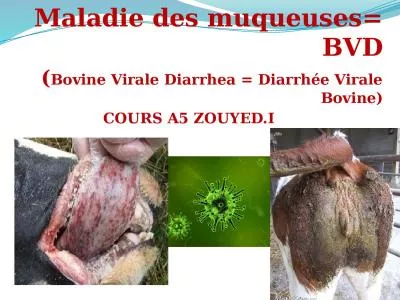
is most common in young cattle 624 mo old The clinical presentation can range from inapparent or subclinical infection to acute and severe enteric disease to the highly fatal mucosal disease complex characterized by profuse enteritis in association with typical mucosal lesions BVD mus ID: 1045325

disease bvdv infected cattle bvdv disease cattle infected persistently infection mucosal bvd clinical lesions virus acute diarrhea fetal viral

Share:

Link:

Embed:

Download Presentation from below link

Download Presentation The PPT/PDF document "Bovine viral diarrhea (BVD)" is the property of its rightful owner. Permission is granted to download and print the materials on this web site for personal, non-commercial use only, and to display it on your personal computer provided you do not modify the materials and that you retain all copyright notices contained in the materials. By downloading content from our website, you accept the terms of this agreement.


Presentation Transcript

1.

2. Bovine viral diarrhea (BVD) is most common in young cattle (6–24 mo old). The clinical presentation can range from inapparent or subclinical infection to acute and severe enteric disease to the highly fatal mucosal disease complex characterized by profuse enteritis in association with typical mucosal lesions. BVD must be distinguished from other viral diseases that produce diarrhea and mucosal lesions.

3. Bovine viral diarrhea virus (BVDV), the causal agent of BVD and mucosal disease complex, is classified in the genus Pestivirus in the family Flaviviridae. Although cattle are the primary host for BVDV, several reports suggest most even-toed ungulates are also susceptible.Noncytopathic BVDV is the predominant viral biotype in nature, whereas cytopathic BVDV is relatively rare and of little epidemiologic relevance.

4. Etiology and EpidemiologyThe prevalence of antiviral antibody in cattle varies greatly among countries and geographic regions because of differing cattle housing practices, population densities, vaccination practices, and implementation of different control or eradication programs. Prevalence of antiviral antibody may be >90% if vaccination is practiced commonly in a geographic region. Although cattle of all ages are susceptible, most cases of overt clinical disease are seen in cattle between 6 mo and 2 yr old.

5. Cattle persistently infected with noncytopathic BVDV serve as a natural reservoir for virus. Persistent infection develops when noncytopathic BVDV is transmitted transplacentally during the first 4 mo of fetal development. The calf is born infected with virus, remains infected for life, and usually is immunotolerant to the resident noncytopathic virus. Transplacental infection that occurs later in gestation can result in abortion, congenital malformations, or birth of healthy calves that have antibody against BVDV.

6. Persistently infected cattle can shed large amounts of BVDV in their secretions and excretions and readily transmit virus to susceptible herdmates. Clinical disease and reproductive failure often are seen after healthy cattle come in contact with a persistently infected animal. Although persistently infected cattle are important in transmission of BVDV, the virus also may be spread by biting insects, fomites, semen, biologic products, and other animals, including swine, sheep, goats, camelids, and possibly wild ruminants.

7. Clinical Findings and LesionsDisease induced by BVDV varies in severity, duration, and organ systems involved. Biphasic fever (~104°F [40°C]), depression, decreased milk production, transient inappetence, rapid respiration, excessive nasal secretion, excessive lacrimation, and diarrhea are typical signs of acute clinical BVD. Clinical signs of disease usually are seen 6–12 days after infection and last 1–3 days. Transient leukopenia may be seen with onset of signs of disease. Recovery is rapid and coincides with production of viral neutralizing antibody. Gross lesions seldom are seen in cases of mild disease. Lymphoid tissue is a primary target for replication of BVDV, which may lead to immunosuppression and enhanced severity of intercurrent infections.

8.

9. Some isolates of BVDV (BVD type 2) have been associated with severe clinical disease that manifests as;high fever 41°–42°C, oral ulcerations, eruptive lesions of the coronary band and interdigital cleft, diarrhea, dehydration, leukopenia, thrombocytopenia.

10. In thrombocytopenic cattle, petechial hemorrhages may be seen in the;conjunctiva, sclera, nictitating membrane of the eyes,on mucosal surfaces of the mouth and vulva. Prolonged bleeding from injection sites also occurs. Swollen lymph nodes,erosions and ulcerations of the GI tract, petechial and ecchymotic hemorrhages on the serosal surfaces of the viscera, and extensive lymphoid depletion are associated with severe forms of acute BVD. (The duration of overt disease may be 3–7 days. High morbidity with a mortality of ≥25% is common.)

11. In pregnant cattle, BVDV may cross the placental barrier and infect the fetus. The consequences of fetal infection usually are seen several weeks to months after infection of the dam and depend on the stage of fetal development and on the strain of BVDV. Infection of the dam near the time of fertilization may result in reduced conception rates. Infection during the first 4 mo of fetal development may lead to embryonic resorption, abortion, growth retardation, or persistent infection. Congenital malformations of the eye and CNS result from fetal infections that occur between months 4–6 of development. Fetal mummification, premature birth, stillbirth, and birth of weak calves also are seen after fetal infection.

12.  Young calf persistently infected with BVD (right) compared to similarly-aged normal herd mate.BVD virus is most important when it infects susceptible breeding cattle during early pregnancy causing foetal death/abortion, and birth defects.

13. Persistently infected calves may appear healthy and normal in size, or they may show stunted growth and be prone to respiratory or enteric ailments. They often have a short life span, and death before 2 yr of age is common.Persistently infected cows always give birth to persistently infected calves, but most calves sired by a persistently infected bull will not be infected with virus in utero.

14. Mucosal disease is an uncommon but highly fatal form of BVD occurring in persistently infected cattle and can have an acute or chronic presentation. Mucosal disease is induced when persistently infected cattle become superinfected with cytopathic BVDV. The origin of the cytopathic BVDV is usually internal, resulting from a mutation of the resident persistent, noncytopathic BVDV. Acute mucosal disease is characterized by;fever, leukopenia,dysenteric diarrhea,inappetence, dehydration,erosive lesions of the nares and mouth,and death within a few days of onset. At necropsy, erosions and ulcerations may be found throughout the GI tract. The mucosa over Peyer’s patches may be hemorrhagic and necrotic. Extensive necrosis of lymphoid tissues, especially gut-associated lymphoid tissue, is seen on microscopic examination.

15. Poorly grown persistently-infected BVDv calf.  This calf has chronic pneumonia and ringworm infection.Chronic pneumonia secondary to persistent BVDv infection (PI calf).

16. Clinical signs of chronic mucosal disease may last several weeks to months and are less severe than those of acute mucosal disease. Intermittent diarrhea and gradual wasting are common. Coronitis and eruptive lesions on the skin of the interdigital cleft cause lameness in some cattle. Lesions found at necropsy are less pronounced than, but similar to, those seen in acute mucosal disease. Often, the only gross lesions seen are focal ulcerations in the mucosa of the cecum, proximal colon, or rectum, and the mucosa over Peyer’s patches of the small intestine may appear sunken.

17. Diagnosis:BVD is diagnosed tentatively from disease history, clinical signs, and gross and microscopic lesions. Diagnostic laboratory support is required when clinical signs and gross lesions are minimal. Laboratory support also is required in some outbreaks of mucosal disease or clinically severe acute BVD, because either disease may appear similar to rinderpest or malignant catarrhal fever.Laboratory tests for BVDV include isolation of virus or viral antigen in clinical specimens and tissues, and assays that detect anti-BVDV antibody in serum or milk.  At necropsy, tissues of choice for viral isolation include spleen, lymph node, and ulcerated segments of the GI tract.

18.

19.

20.

21.

22.

23.

24.