/
3 : Organelles and their structures 3 : Organelles and their structures

3 : Organelles and their structures - PowerPoint Presentation

pamella-moone
pamella-moone . @pamella-moone
Follow
398 views
Uploaded On 2016-07-24

3 : Organelles and their structures - PPT Presentation

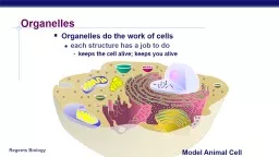
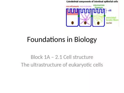
Lesson Objectives Describe the structure of an animal and prokaryotic cell AO1 C Starter RECAP from GCSE Draw amp label a typical plant and animal cell Cells All cells have an internal structure known as the ID: 418497

cell cells apparatus membrane cells cell membrane apparatus golgi ribosomes protein material controls structure lipids enzymes organelles nucleus eukaryotic

Share:

Link:

Embed:

Download Presentation from below link

Download Presentation The PPT/PDF document "3 : Organelles and their structures" is the property of its rightful owner. Permission is granted to download and print the materials on this web site for personal, non-commercial use only, and to display it on your personal computer provided you do not modify the materials and that you retain all copyright notices contained in the materials. By downloading content from our website, you accept the terms of this agreement.


Presentation Transcript

Slide1

3: Organelles and their structures

Lesson Objectives:

Describe the structure of an animal and prokaryotic cell.

(AO1:

C)

Slide2

StarterRECAP from GCSE

Draw & label a typical

plant

and animal cellSlide3

CellsAll cells have an internal structure known as the

ultra structure

.

2 types of cell

Eukaryotic

Prokaryotic

Eukaryotic cells have a distinct nucleus and possess membrane bound organelles.Slide4

Eukaryotic CellsSlide5

The NucleusContains genetic material and controls cell activity. Made up of:

The nuclear envelope

– double membrane that controls material entry and exit

Nuclear pores

– allows large molecules like mRNA out of the nucleus

Chromatin –

made of proteins and DNA. Controls the cell activity

Nucleolus

– makes ribosomesSlide6

The NucleusNucleus functions:

Produce mRNA for

protein

synthesis

Retain genetic material in the form of DNA or chromosomes

Manufacture ribosomal RNA and ribosomesSlide7

MitochondriaMitochondria are the main organelles involved in

respiration

so

are responsible for the production of the energy carrier molecule ATP

.Slide8

MitochondriaDouble membrane

– outer controls flow of materials in/out

Cristae

– extensions of inner membrane. Increased SA for attachment of

enzymes

Matrix

– Semi rigid. Contains protein, lipid and trace DNA. Also the site of the Krebs cycleSlide9

Endoplasmic ReticulumSystem of sheet-like membranes throughout the cytoplasm

Continuous with the

outer

nuclear membraneSlide10

Endoplasmic Reticulum2 types of endoplasmic reticulum

Rough ER

ribosomes on surface

Large SA for protein synthesis

Pathway to transport proteins out of cell

Smooth ER

no

ribosomesSynthesises, stores and transports lipidsSynthesises, stores and transports carbohydratesSlide11
Slide12

Golgi ApparatusStructure is similar to S-ER, just more compact.

Flattened stacks of membranes with rounded structures called

vesicles.

Proteins and lipids produced by R-ER pass through the

golgi

apparatus to me modified.

Once sorted, proteins and lipids are transported in vesicles that fuse with the cell surface membrane to release their contentsSlide13

Golgi ApparatusSlide14

Golgi ApparatusFunctions:

Adds carbohydrate to protein to make

glycoproteins

Produces secretory enzymes

Secretes carbohydrates

Transports and modifies lipids

Forms

lysosomesSlide15

LysosomesFormed from Golgi Apparatus and contain enzymes. They isolate these potentially harmful enzymes from the rest of the cell

.

Breakdown material ingested by phagocytic cells, such as white blood cells

Release enzymes to the outside of the cell

Digest worn out

organelles

Break down cells when they have diedSlide16

LysosomesSlide17

RibosomesFound in cytoplasm or on R-ER

Have 2 sub-units containing RNA and protein

Important in protein synthesis

2 types:80S type – eukaryotic cells about 25nm diameter

70S

type – prokaryotic cells (smaller)Slide18

Microvilli

Finger-like projections of epithelial cells to increase surface area for absorptionSlide19