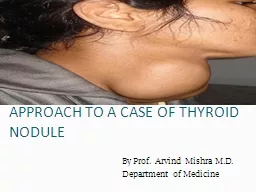
By Prof Arvind Mishra MD Department of Medicine Causes of Thyroid Nodularity Benign Follicular Adenomas Multinodular goiter Hashimotos thyroiditis Cysts colloid simple hemorrhagic ID: 351319
Download Presentation The PPT/PDF document "APPROACH TO A CASE OF THYROID NODULE" is the property of its rightful owner. Permission is granted to download and print the materials on this web site for personal, non-commercial use only, and to display it on your personal computer provided you do not modify the materials and that you retain all copyright notices contained in the materials. By downloading content from our website, you accept the terms of this agreement.
Slide1
APPROACH TO A CASE OF THYROID NODULE
By Prof. Arvind Mishra M.D.Department of MedicineSlide2
Causes of Thyroid Nodularity
BenignFollicular AdenomasMultinodular goiterHashimoto’s thyroiditisCysts (colloid, simple, hemorrhagic)Slide3
MalignantPapillary CarcinomaFollicular Carcinoma
Medullary CarcinomaAnaplastic and poorly differentiated carcinomaPrimary lymphoma of the thyroidMetastatic carcinomaSlide4
Clinical features
Most thyroid nodules are asymptomaticNodules that produce thyroid hormones in excess -palpitation -anxiety -clammy skin
-increased appetite
-weight loss
-heat intolerance
Nodules can press adjacent structures in neck causing
-hoarseness of voice(recurrent laryngeal N compression)
-
dysphagia
-
dyspnoea
-pain in neck Slide5
Nodules sometimes found in
hashimoto’s thyroiditis, which may cause symptoms of an underactive thyroid gland -dry skin -face swelling -intolerance to cold -weight gain
-decreased appetite
-hair lossSlide6
INVESTIGATIONS
ULTRASONOGRAPHYFNACTHYROID SCANSlide7
Ultrasonography
Most sensitive test to detect lesions in the thyroidIndicated in all patients who have a nodular thyroid, with a palpable solitary nodule or a multinodular goiter,be evaluated by USNot as screening test in general population
7Slide8
USG findings
NumberSizeExtracapsular growthCystic lesionsCervical LN
8Slide9
Findings suggestive of malignancy:
–Presence of halo–Irregular border–Presence of cystic components–Presence of calcifications–Heterogeneous echo pattern–Extrathyroidal extensionSlide10
Radionuclide Scanning
Used to identify whether a nodule is functioning or not. Functioning nodules are nearly always benign Approximately 90 percent of nodules are nonfunctioning5 percent of nonfunctioning nodules are malignant However even with suppressed level of serum
TSH patient can have both functioning and non functioning
nodules.Thus
, even suppressed level of serum TSH may obviate the need for biopsy.Slide11
Usually either Technetium(Tc
99) or Radioiodine(I123) used.Normal follicular cells will trap both but only radioiodine is added to tyrosine and stored in the colloid space
Both
benign and almost all malignant
neoplastic
tissue concentrate both radioisotopes less than normal thyroid
tissueSlide12
Cold Nodules
CystNon-functioning AdenomaMalignancySlide13Slide14
Hot Nodules
Functioning AdenomaThyroiditisMultinodular goiterSlide15Slide16
Limitations of Thyroid scan
Two dimensional scanning techniqueInability to measure the size of a nodule accuratelyMissed malignant thyroid nodulesSlide17
Other imaging tech
CT and MRI not as routine. Can asses size, retrosternal extension, position and relation to the surrounding structure.
17Slide18
18
Images of a large, asymmetric
multinodular
goiter. (A) Chest radiography shows marked
tracheal deviation
to the right (arrow). (B) Chest CT confirmed the presence of a large
substernal
goiter
on the left to the level of tracheal bifurcation.Slide19
USG guided FNAC
Indicated if:Palpation-guided FNAC non-diagnosticComplex (solid/cystic) nodulePalpable small nodule (<1.5 cm)Impalpable noduleAbnormal cervical nodesNodule with suspicious US features
FNAC results are:70% Benign, 10% Malignant or suspicious of malignancy, and 20% Unsatisfactory
19Slide20Slide21
Malignant (+ve) cytology
Commonest is PTC(Papillary thyroid carcinoma):Characteristics cytological feature- psammoma bodies, orphan annie eye nuclei (cleaved nuclei)Others include:
Medullary
thyroid carcinoma(
amyloid
deposits,intracytoplasmic
calcitonin
),
anaplastic
carcinoma(cellular
anaplasia
) and high-grade metastatic cancers
21Slide22
Suspicious cytology in FNAC
Diagnosis cannot be madeInculdes: Follicular neoplasms, Atypical PTC, or Lymphoma
22Slide23
FNAC Limitations
The absence of malignant cells in an acellular or hypocellular specimen does not exclude malignancy
Inability
to reliably distinguish a benign follicular
adenoma
from a
follicular malignant
tumour
Slide24
TSH
To detect early or subtle thyroid dysfunction.If TSH levels abnormal, free T3 & T4 should be measured to confirm the diagnosisTPOAb
Thyroid
peroxidase
antibody
Though characteristically observed in hypothyroidism, can also be seen in patients of hyperthyroidism and
subacute
thyroiditisSlide25
Serum Tg
Correlates with iodine intake and the size of the thyroid gland rather than with the nature or function of the noduleSeldom used in nodule diagnosis Extremely elevated levels of Tg
may suggest thyroid metastasis.
Serum
Calcitonin
Good marker for
medullary
carcinoma and correlates well with tumor burdenSlide26Slide27
MCQsSlide28
1)Thyroid carcinoma associated with
hypocalcemia isa)Follicular CAb)Medullary CAc)Anaplastic CA
d)Papillary CASlide29
2)Lab investigation of patient shows low T3, low T4 and
highTSH.Diagnosisa)Primary hypothyroidismb)Grave’s diseasec)Hypothalamic failured)Pituitary failureSlide30
3)Excess iodine intake causes hypothyroidism.It
is known asa)Wolff chaikoff effectb)Jod basedow effectc)
Thyrotoxicosis
factitia
d)De
quervain’s
thyroiditisSlide31
4)FNAC can not differentiate between follicular adenoma and carcinoma because it can not clearly shows
a)Vascular invasion b)Extracapsular extensionc)a+b
d)Nuclear
pleomorphismSlide32
5)Subclinical hypothyroidism stands for biochemical evidence of hypothyroidism without any clinical
features.Cut off TSH values area)<5mU/Lb)<8mU/Lc)Normald)<10mU/L