/
DISORDERS OF THE GASTROINTESTINAL SYSTEM DISORDERS OF THE GASTROINTESTINAL SYSTEM

DISORDERS OF THE GASTROINTESTINAL SYSTEM - PowerPoint Presentation

pamella-moone
pamella-moone . @pamella-moone
Follow
342 views
Uploaded On 2019-11-20

DISORDERS OF THE GASTROINTESTINAL SYSTEM - PPT Presentation

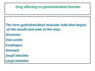
DISORDERS OF THE GASTROINTESTINAL SYSTEM DR ADIBE MAXWELL CLINICAL PHARMACY 2017 Anatomy amp Physiology of the Gastrointestinal System GI tract breakdown absorption and elimination ID: 766050

amp pain bowel treatment pain amp treatment bowel 2010 day disease 2011 diet symptoms care small abdominal inflammation mfmer

Share:

Link:

Embed:

Download Presentation from below link

Download Presentation The PPT/PDF document "DISORDERS OF THE GASTROINTESTINAL SYSTEM" is the property of its rightful owner. Permission is granted to download and print the materials on this web site for personal, non-commercial use only, and to display it on your personal computer provided you do not modify the materials and that you retain all copyright notices contained in the materials. By downloading content from our website, you accept the terms of this agreement.


Presentation Transcript

DISORDERS OF THE GASTROINTESTINAL SYSTEM DR ADIBE MAXWELL CLINICAL PHARMACY 2017

Anatomy & Physiology of the Gastrointestinal System GI tract: breakdown, absorption and elimination Upper potionthe mouthesophagusstomachLower portionsmall intestinelarge intestinerectumanus. (Day et al., 2010)

DIGESTIVE SYSTEM FUNCTIONS : ingest food DIGESTION:break it down into small molecules ABSORPTION:absorb nutrient moleculesELIMINATION:eliminate nondigested wastesASSESSORY ORGANS :pancreas, liver, gallbladder

Disorders of the upper GI system Disorders affecting Ingestion ANOREXIA: lack of appetite, could be from emotional or physical factorslab tests may be done to assess nutritional status Medical treatment: supplements may be ordered, TPN or enteral feedingsInterventions: oral hygiene, clean room, determine cause of nausea and treat, include family and friends(socialization), respect likes and dislikes, education

STOMATITIS Inflammation of the oral mucosa (mouth) Causes: trauma, organisms, irritants, nutritional deficiency, diseases, chemotherapyS/S: swelling, pain, ulcerations, excessive salivation, halitosis, sore mouthTreatment:pain relief, removal of causative factor, oral hygiene, medications, soft bland diet

GINGIVITIS Inflammation of the gums Causes : poor oral hygiene, poorly fitting dentures, nutritional deficiencyS/S: red, swollen, bleeding gums, painfulTreatment: dental hygiene, prevention of complications

Interventions: Stomatitis and Gingivitis Assess mouth condition Administer medicationsMouth careSoft bland diet, no spicy foodsObserve for complicationsTeach importance of mouth and gum care

HERPES SIMPLEX TYPE 1 Infection affecting the lips and mucous membranes of the mouth Causes : Herpes simplex virusS/S: Vesicles on the mouth, nose or lips, malaise, edema of surrounding areaTreatment: Antiviral medication(Zovirax), analgesics, symptomatic reliefInterventions: Administer meds, keep lesions dry, provide symptomatic relief

LEUKOPLAKIA Abnormal thickening and whitening of the epithelium of the mucous membranes of the cheeks and tongue Causes: Chronic irritation S/S: Thickened white or reddish lesions on the mucous membrane, lesions can not be rubbed off

Treatment : May be surgically removed or treated with chemotherapy, meticulous oral hygiene Interventions : Assess mouth frequently, assist with oral hygiene, discuss removal of sources of irritation

ORAL CANCER Malignant lesions may develop on the lips, oral cavity, tongue and pharynx. Generally squamous cell carcinomas Causes : high alcohol consumption, tobacco use, external irritantsS/S: Leukoplakia, swelling, edema, numbness, painDiagnosis: biopsy

Treatment: Surgery Radiation or chemotherapydepends on the size and location and the lesionInterventions: consult MD for special mouth care, monitor respiratory status, administer pain med, assess ability to swallow and talk, assess for infection at incision site, education

ESOPHAGITIS Inflammation or irritation of the esophagus Causes : Reflux of stomach contents, irritants, fungal infections, trauma, malignancy, intubationS/S: heartburn, pain, dysphagiaTreatment: treat underlying causeInterventions: soft bland diet, administer meds, observe for complications

ESOPHAGEAL VARICIES Tortuous, distended vessels of the esophagus may rupture and bleed causes: Portal hypertension caused by cirrhosis of the liverS/S Hematemesis, hemorrhage from UGI, black tarry stools, pain, shock

Treatment : Iced saline lavage Medications( Vasopressin, antibiotics, analgesics)Surgeries: ligation, injection sclerotherapyBlood transfusions

Interventions: administer meds provide pre/post op care administer blood transfusionsmonitor tube placementassess vital signs, bleeding

CANCER OF THE ESOPHAGUS Prognosis is very poor, diagnosed at late stages Causes- no known cause, predisposing factors; irritation, poor oral hygiene S/S- progressive dysphagia, painful swallowing, weight loss, vomiting, hoarseness, coughing, iron deficiency, anemia, occult bleeding or hemmorage

Treatment of CA of Esophagus Palliative treatment is common Radiation, chemotherapy surgery:EsophagectomyEsophagogastrostomyEsophagoenterostomyGastrostomy

Interventions Maintain NG tube after surgery Assess for signs of hemorrahage Monitor respiratory statusmonitor adequacy of nutritional intake ( high protein, high calorie diet)assess ability to swallowallow patient to ventilate feelings

DISORDERS OF DIGESTION AND ABSORPTION N/V Hiatal Hernia GastritisPeptic UlcerStomach Cancer

NAUSEA AND VOMITING Nausea: unpleasant sensation usually preceding vomiting, may have abdominal pain, pallor, sweating, clammy skin Causes: irritating food, infection, radiation, drugs, hormonal changes, surgery, inner ear disorders, distention of the GI tract

Vomiting: forceful expulsions of stomach contents through the mouth. Occurs when vomiting reflex in the brain is stimulated. Projectile vomiting- is forceful ejection of stomach contents. Regurgitation- gentle ejection of stomach contents without nausea or retching

Complications and Treatment May lead to dehydration, metabolic alkalosis, aspiration Treatment: Antiemetics( Phenergan, Dramamine, Scopolamine patch Reglan), IV fluids, NG tube, TPNIntervention: through assessment, keep patient comfortable, offer liquids, position on side, suction setup in the room

HIATAL HERNIA Protrusion of the lower esophagus and stomach upward through the diaphragm into the chest SLIDING-gastroesophageal junction above the hiatus ROLLING( paraesophageal)-junction in place portion of stomach rolls up through diaphramCauses; weakness in the lower esophageal sphincter, related to increased abdominal pressure, long term bedrest, trauma

Signs and Symptoms Feelings of fullness dysphagia eruptionregurgitationheartburnComplications: Ulcerations, bleeding, aspiration seen in 50% of people over 60.

Treatment for Hiatal Hernia Drug therapy H2 receptor antagonists:Tagamet,Zantac, Pepsid- reduce stomach secretionsAntacids- neutralize stomach acidsReglan, Propulsid- increase stomach emptying diet therapy- decrease caffeine fatty foods, alcohol( reduce LES tone), acidic and spicy foods

SURGERY Nissen Fundoplication Angelclik prothesisCARE: assessment, pain relief, watch for aspiration, nutrition, education

GASTRITIS Inflammation of the lining of the stomach ACUTE: excessive intake of food or alcohol. Food poisoning, chemical irritation CHRONIC: repeated episodes of acute, H Pylori

Signs/Symptoms and Complications Nausea, vomiting, feeling of fullness, pain in stomach, indigestion. With chronic may have only mild indigestion changes in stomach lining with decrease in acid and intrinsic factor ( high risk for pernicious anemia)

Treatment Treat symptoms, and fluid replacement Medications: antacids, H2 receptor blockers, B 12 injections, corticosteroids analgesics, antibiotics if H Pylori bland diet, frequent meals Eliminate the causesurgical interventionBEST DIAGNOSIS IS GASTROSOPY & BIOPSY

CARE Good HX and review of present S/S pain relief, adequate nutrition, hydration, stress management, education

PEPTIC ULCER Loss of tissue from the lining of the digestive tract. May be acute or chronic. Classified as gastric or duodental (stress- develop 24-48hr. After event) CAUSES: drugs, stress, heavy alcohol and tobacco use, infection (H .pylori bacteria) Conditions that cause high gastric acid concentration

Peptic Ulcer comparison Gastric Ulcers burning pain 1-2 hrs. after meals, upper left abd/back,relieved by food N/V, anorexia, wt lossShallow/ gastric secretions deceasedOlder men, working class, bld type A, under stressDuodenal Ulcersburning/ cramping pain 2-4hrs. P meal, beneath xiphoid and back, relieved by antacids/foodincreased gastric acid Young men, all social classes, bld type O, chronic illnesses

PEPTIC ULCER COMPLICATIONS HEMORRHAGE PERFORATION PYLORIC OBSTRUCTION

TREATMENT Drug therapy Antacids H2 RECEPTOR BLOCKERSANTICHOLINERGICS-Pro-Banthine, Robinul, BentylSUCRALFATE- CarafateAntibiotics –Flagyl, tetracycline, Biaxintreatment goals- relieve symptoms, promote healing, prevent complications and recurrence

Interventions Three meals a day – decreases acid productiondecrease foods that stimulate acid secretions and cause discomforttreat pain with rest, diet and drug therapyeducate on stress management and relaxation

Surgical options for gastric ulcers To decrease acid secretion: vagotomy pyloroplastygastroenterostomyantrectomysubtotal gastrectomy Billroth IBillroth II

care after gastric surgery No signs of complications Gastric dilation ObstructionPerforationMaintenance of NG tube:Suctiondo not irrigate or reposition tubetype of drainage

Adequate nutrition: NPO gradually advance from clear liquids to full liquids then solid foods Assess for N/V, abdominal distention Size of meals changes depending on type of surgeryGastric surgeries can have serious effects on absorption of vit. B12, folic acid, iron, calcium, vit, D

education Reinforce diet teach signs of complicatons Avoid risk factors

STOMACH CANCER Rare ( 25,000/yr.), common in males, African American, over 70 and low socioeconomic status. 60% decrease in past 40 yrs. No S/S in early stagesLate stages S/S: N/V, ascities, liver enlargement, abd. MassMets to bone and lung10% survival rate after 5 yrs.

Risk factors : pernicious anemia, chronic gastritis, cigarette smoking, diet high in starch, salt, salted meat, pickled foods, nitrates Treatment : surgery/ chemotherapy/ radiationsubtotal gastrectomy, total gastrectomy

Inflammatory Bowel Disease IBD refers to two chronic inflammatory GI disorders: Crohn’s disease and Ulcerative Colitis, both cause inflammation and ulcerations of the intestine.Two Types:Crohn’s: usually affects the intestines. Ulcerative Colitis: usually affects the large intestine. Statistics: 200,000 Canadians have IBD 15-30 years of age at highest risk. gender nonspecific. (Day et al., 2010)

Inflammatory Bowel Disease: Complications Ulcers : chronic inflammation can lead to open sores within the digestive tract. Fistulas: when an ulcer forms and extends completely through the intestinal wall. Anal fissures: crack or cleft in the anus or skin where infection occurs. Malnutrition: difficulties eating and absorbing nutrients. Other problems: Arthritis Kidney and gallstones Inflammation of eyes and skin (Day et al., 2010)

Crohn’s Disease: Etiology & Pathophysiology Chronic disorder that causes inflammation of the GI tract, most commonly affecting the small intestine. Transmural; affecting all layers of the mucosa. Begins with edema and thickening of the mucosa. Ulcers appear on the inflamed mucosa, causing fistulas and fissures. Scaring, thickening and narrowing of the GI tract. Statistics: Usually diagnosed in adolescents Prevalence has risen in the past 30 years. seen more in smokers (MFMER, 2011 ; Day et al., 2010; CCRC, 2008; CCFC, 2008 ; Mahan & Escott-Stumop , 2004;)

Crohn’s Disease: Clinical Manifestations Persistent diarrhea Loss of appetite & weight loss May have rectal bleeding Cramping abdominal pain Steatorrhea Fatigue Fever ComplicationsBowel obstructionSores of ulcers FistulasMalnutrition (CSIR, 2012; CCFC, 2008)

Crohn’s Disease: Diagnosis Health history: Onset, associated symptoms, pain, stool, & rectal bleeding. Blood tests: anemia or infection and certain antibodies Fecal occult blood test: looking rectal bleeding. Stool sample : presence of white blood cells. Colonoscopy : visualize and collect biopsy. Flexible sigmoidoscopy : examine sigmoid colon. Barium enema : evaluate large intestine with x-ray. X-ray : rule out toxic megacolon. CT scan : assess for complications and amount of infection. MRI : diagnosis and management. Capsule endoscopy : all other diagnostics are negative. Double – balloon endoscopy : still questioning diagnosis. Small bowel imaging : locate narrowing or inflammation. (CSIR, 2012; MFMER, 2011; CCFC, 2008)

Colonoscopy A procedure used to see inside the colon and rectum Used to investigate intestinal signs and symptoms. Preparation:Bowel prep to empty the bowel. No solid food the day before Laxative or enema kit Adjust medications Postoperative: Hour to recover Blood with first BM When to seek medical care Severe abdominal pain, fever, dizziness, weakness, bloody BM’S http://www.youtube.com/watch?v=rSXTIzqWc7s (NIDDK, 2011; MFMER, 2011)

Flexible Sigmoidoscopy A procedure used to evaluate the part of the large intestine and investigate signs and symptoms. Preparation: No solid foods NPO after midnight Laxative or enema kit Adjust medications What to expect: Usually does not require sedation or pain medication. May feel abdominal cramping or urge to push. Ability to take biopsies . Takes about 15 minutes. (NIDDK, 2011; MFMER, 2011)

Barium Enema A special X-ray used to detect changes or abnormalities in the large colon and part of the small intestine. Single-column: allows visualization of silhouette , shape and condition of colon. Air-contrast: Air expansion improves the quality of X-ray images. During exam: No sedation necessary. Side lying position. May manipulate the colon manually. Enema tube is inserted with a barium bag. After exam: May expel additional barium and air with BM. Drink plenty of fluids, laxative may be required. (NIDDK, 2011; MFMER, 2011)

Capsule Endoscopy A procedure that uses a tiny wireless camera to take pictures of your digestive tract. Preparation Stop eating for 12 hours before. Stop or delay certain medications. Plan to take it easy for the day. During the test Wear a recorder with a special belt. Avoid strenuous activity. May or may not be able to go back to work. After the procedure Contact doctor if capsule not eliminated within two weeks Complete after 8 hours, camera eliminated within hours After 2 hours may resume clear liquids. (MFMER, 2011)

Crohn’s Disease: Treatment Anti-inflammatory drugs: 5-Aminosalicylates Corticosteriods Immune system suppressors:Methotrexate Cyclosporine Antibiotics:Metronidazole Ciprofloxacin Anti- diarrheals : Metamucil Citrucel Pain relievers: Acetaminophen (MFMER, 2011 ; CCFC, 2008)

Crohn’s Disease: Medical Nutrition Therapy Diet changes: Limit dairy products Low fat foods Limit fiber Avoid problem foodsEat small meals Drink plenty of fluidsMultivitamins Enteral and Paraenteral NutritionAllows for bowel rest Reduces inflammation short term Used pre-op and when medications fail (MFMER, 2011)

Crohn’s Disease: Non-Pharmacologic & Alternative Therapies Stress Can worsen or precipitate flare ups. Exercise Reduces stress Relieves depression Normalizes bowel function Relaxation OtherProbioticsFish oil Acupuncture (MFMER, 2011)

Crohn’s Disease: Considerations Teaching & Education Stress management techniques Medication therapiesDiet management & exercise Diagnostic testing and procedures SupportUnderstanding the diseaseBody image Collaborate Dietitian Gastroenterologist & Surgeon Smoking cessation programs

Ulcerative Colitis: Etiology & Pathophysiology Affects the superficial mucosal layer resulting in inflamed mucosa with small ulcers that cause bleeding. Classifications: Extensive colitis – extends to the hepatic flexure. Proctosigmoidsitis - extends to the rectosigmoid junction. Left-sided colitis – extends to the splenic flexure. Pancolitis – extends from the rectum to the ceum and involves the entire colon. Proctitis - confined in the rectum. (Day et al., 2010, Sephton , 2009)

Ulcerative Colitis: Clinical Manifestations Symptoms: Diarrhea LLQ abdominal pain Intermittent tenesmusRectal bleedingPallorAnemiaFatigue Classifications: Mild Severe Fluminant (Day et al., 2010, Sephton , 2009)

Ulcerative Colitis: Diagnosis CBC – ESR, C-reactive protien, WBC, LFT, AlbuminSeries of 3 stools sent to microbiology, C & S, and for c. difficile . X-ray - assess for toxic megacolon and perforation. Sigmoidscopy & Colonoscopy – assess extent and severity of the disease. CT, MRI & Ultrasound – identify abscesses and peritoneal involvement. assessments : Tachycardia Hypotension Tachycardia Pallor Fever Bowel sounds Distention Tenderness (Day et al., 2010, Sephton , 2009)

Ulcerative Colitis: Treatment Medical management: ASA - Corticosteriods - Immunosupressive drugs –Methotrexate – Anti-TNF therapy - (Day et al., 2010, Sephton , 2009)

Ulcerative Colitis: Medical Nutrition Therapy Diet modification: Low residue High protein diet Initially include excess fibre Smaller frequent mealsExacerbations due to: Increase sucrose intake Lack of fruit and vegetable intakeLow intake of dietary fibre Altered omega 3 fatty acid ratiosOverall poor quality diet (Day et al., 2010, Sephton , 2009)

Ulcerative Colitis: Considerations Teach & Educate Early recognition Monitor hydration and keep food journal Stool chart Weight monitoring Support Emotional support Collaborative Care Infection control nurseDietitianGastroenterologist & surgeon (Day et al., 2010, Sephton , 2009)

Ostomies’s A stoma ( ostomy ) is an artificial, surgically created opening into the abdominal wall to allow exit of feces and urine. Colostomy: formed through colon (large bowel) Pass flatus & soft formed feces. Permanent end: removal of anus, anal canal, rectum and some of the distal colon.Loop: formed in transverse colon, 2 ends are brought to surface. Ileostomy: formed in ileum (small bowel) Pass flatus & loose porridge-like stool. Permanent end: removal of entire colon. Loop : creation of stoma after anastomosis. (Burch, 2011 ; Day et al., 2010)

Inflammatory Bowel Disease: Diagnosis Diarrhea related to the inflammatory process Acute pain related to increased peristalsis and GI inflammation. Fluid volume deficit related to anorexia, nausea and diarrheaImbalanced nutrition: less than body requirements related to dietary restrictions, nausea, and malabsorptionActivity intolerance related to fatigueAnxiety Ineffective coping related to repeated episodes of diarrheaRisk for impaired skin integrity related to malnutrition and diarrheaKnowledge deficit

Irritable Bowel Syndrome Common disorder based on a presentation of signs and symptoms. - intermittent to continuous, mild to severe. - Abnormal pattern in bowel elimination including constipation, diarrhea or both. - abdominal pain, feeling of fullness, gas, or bloating. Clients have a normal bowel structure with no inflammation.Abnormal function of motility or peristalsis due to: - neuroendocrine disorders - vascular disturbances - metabolic disturbances - infection - irritation (Day et al., 2010)

Irritable Bowel Syndrome: Diagnosis No definitive diagnosis, a symptom based diagnosis once other structural disorders have been ruled out. Symptoms must be present for a minimum of 3 days a month for 3 consecutive months. Procedures: looking for a spasm, distention or mucus accumulation in the intestine. stool studies x-rays & contrast x-raysbarium enemacolonoscopy (Day et al., 2010)

Irritable Bowel Syndrome Treatment There is no medical treatment, although there are medications used to treat symptoms such as: anticholinergics antidiarrheals bowel aidssome cases, antibiotics Nutritional Management: restrict foods then gradually increase. avoid large meals increase fibre. avoid foods that stimulate the bowel - caffeine - spicy foods - fried foods - alcohol - carbonated drinks (Day et al., 2010)

Irritable Bowel Syndrome: Considerations Support: tests involved & psychological support. Educate: alcohol and smoking cessation avoiding triggering foods eating regular small mealsCollaborative Care: Dietitians Gastroenterologist & surgeons (Day et al., 2010)

Diverticulitis Diverticulum : a “saclike herniation of the lining of the bowel that extends through a defect in the muscle layer.” (Day et al., 2010, pg. 1167). Diverticulosis: when multiple diverticuli exists. Diverticulitis: results when food and bacteria retained in a diverticumulum produce infection and inflammation that can impede drainage and lead to perforation or abscess formation.” The cause is unknown. Low fiber and high fat diet may cause sac formation. Symptoms include: acute onset of mild to severe pain in the lower left quadrant, accompanied by nausea, vomiting, fever, and chills. (Day et al., 2010)

Diverticulitis: Diagnosis CBC: shows an elevated WBC. Colonoscopy: shows extent of disease and biopsy is completed.CT: confirms diagnosis. (Day et al., 2010)

Diverticulitis: Treatment Nutritional Management high fiber low fat symptomatic: clear liquid diet Medical Managementantibiotics laxativesstool softeners hospitalization of immunocompromised.surgical considerations Complications perforation peritonitis hemorrhage obstruction (Day et al., 2010)

Diverticulitis: Considerations Education and Teaching Understanding the disease Avoid high fat foodsIncrease fiber intake Drink plenty of fluids How to manage attacks When to seek health careSurgical nursing considerations: Preoperative teachingPostoperative teaching Self image (Day et al., 2010)

Celiac Disease: Gluten-sensitivity enteropathy Is a autoimmune medical condition in which damage to the mucosa layer of the small intestines occurs following ingestion of a substance called gluten. Statistics: 1 in 200 Canadians; 330,000 Canadians in total Increased risk with genetic predisposition Often misdiagnosed Is more common in Caucasians More frequent in women Rates have nearly doubled in last 25 years More commonly diagnosed in children; 73,000 in total50% of clients have few or no obvious symptoms (CSIR, 2012; Canadian Digestive Health Foundation, 2012; CCA, 2011; MFMER, 2011; Day et al., 2010; PubMed Health, 2010; Mahan & Escott-Stumop , 2004).

Celiac Disease: Etiology/Pathophysiology When gluten in ingested it creates a systemic immune and inflammatory response that damages and flattens the intestinal villi. This causes malabsorption difficulties of essential macro and micronutrients. Affects primarily the proximal and midpoints of the small intestine, and possibly the distal portions. It takes only one molecule of gluten to trigger the destructive mucosal response. (Canadian Society of Intestinal Research, 2012; MFMER, 2011; Mahan & Escott-Stumop , 2004)

Celiac Disease: Clinical Manifestations Common Symptoms: Chronic diarrhea; steatorrhea and malodorous stools Constipation Weight loss or poor weight gainDelayed puberty/ missed menstrual periods Breathlessness Fatigue Abdominal cramping and bloating Irritability or apathy Easily bruised Muscle cramps and joint pain Lactose intoleranceNausea and vomiting (Canadian Digestive Health Foundation, 2012; Canadian Society of Intestinal Research, 2012; Canadian Celiac Association, 2011; PudMed Health, 2010; Mahan & Escott-Stumop , 2004)

Complications of Celiac Disease Malnutrition Malabsorption Growth delay Osteoporosis Calcium & Vitamin D deficiency Lactose IntoleranceAbdominal painDiarrhea CancerIntestinal lymphomaBowel cancer Neurological SeizuresPeripheral neuropathy (MFMER, 2011)

Celiac Disease: Diagnosis Screening Blood tests Endoscopy Gold standard Internal mucosa biopsyCapsule endoscopy:Examines entire small intestine Both biopsy and blood test results may be difficult to interpret if client has been on a gluten free diet. (CSIR, 2012; Canadian Digestive Health Foundation, 2012; MFMER, 2011; Canadian Celiac Association, 2011; PubMed , 2010. Mahan & Escott-Stumop , 2004).

Endoscopy A procedure used to visually examine the upper digestive system with a tiny camera. Preparation Fast 8 hours beforeStop taken medications During Lie down on backside. Receive a sedative IV. Tube inserted through the mouth, feel some pressureAfter procedure Stay for an hour to recover and will need transportationMay experience mild uncomfortable signs and symptoms (MFMER, 2012)

Celiac Disease: Treatment Medical Therapy: Corticosteroids ( ie: prednisone) Azathioprine Cyclosporine Anti-inflammatory Gluten free diet is the first line of treatment, it may take months or years for the intestinal mucosa to heal. (PubMed , 2010; Mahan & Escott-Stumop , 2004).

Celiac Disease: Medical Nutrition Therapy

Celiac Disease: Considerations Teach & educate: How to read food labels Avoid gluten containing products Vitamin and mineral supplementationSupport: Financial considerations Collaboration:Dietitian Community Resources

Celiac Disease Relieving pain Maintaining normal elimination patterns Maintaining fluid intakeMaintaining optimal nutritionPromoting restReducing anxietyEnhancing coping measuresPreventing skin breakdownMonitoring and managing potential complications

DISORDERS AFFECTING ABSORPTION AND ELIMINATION

MALABSORPTION CONDITION WHEN ONE OR MORE NUTRIENTS ARE NOT DIGESTED OR ABSORBED multiple causes lactase deficiencysprue: celiac/tropicaltreatment/care: depends on typelactase- hold milk productsceliac sprue- hold gluten productstropical sprue- antibiotics, folic acid

DIRRHEA The passage of loose liquid stools with increased frequency, associated with cramping, abd, pain Causes ; (many), foods, allergies, infections, stress, fecal impaction, tube feedings, medicationsComplications- usually temporary/ can be dehydration, malnutrition

Treatment/care Treatment; GI rest, antidiarrheal drugs( Lomotil, Imodium, Kaolin, Aluminum hydroxide)Care: help determine cause, assessVS, weight, skin turgor, abdominal destention, perianal irritation, skin integrity

CONSTIPATION HARD DRY INFREQUENT STOOLS PASSED WITH DIFFICULTY Causes : (many),inactivity, ignored urge, drugs,age related changesComplications: straining (Valsalva maneuver) and fecal impaction

Treatment/care Laxatives, suppositorys, enemas for prompt results stool softeners, increase fluids,dietary fiber Nursing care: assessment, monitor fluids and diet, education, check for impaction

INTESTINAL OBSTRUCTION Exists when there is obstruction in the normal flow of intestinal contents through the intestinal tract Mechanical- Pressure on the intestinal wall Paralytic- Intestinal musculature unable to propel contents along the bowelMay be partial or complete

Intestinal obstruction causes SMALL BOWEL: adhesions most common intussusceptionvolvulusparalytic ilieusabdominal hernia

LARGE BOWEL: carcinoma diverticulitis inflammatory bowel disordersvolvulus

Small Bowel vs Large Bowel Small: abdominal pain vomitingpass blood and mucous, no stool, no gasover time signs of dehydrationLarge:symptoms develop slowlyconstipationdistended abdomencrampy lower abdominal painfecal vomiting

Management of bowel obstruction Small decompression is strangulated then surgeryLargesurgical resection with formation of colostomycare: same as gastric surgery, management of NG tube

APPENDICITIS Inflammation of the appendix appendix has no known function in the body opening becomes obstructedobstruction interferes with the drainage of secretions from the appendix

Signs and symptoms Generalized epigastric pain at first that shifts to the RLQ pain at McBurney’s pointelevated temp, N/V, elevated WBC’s ( over 10,000)

Treatment/care NPO surgical removal IV’s and antibioticsice pack to the abd.LAXATIVES IS CONTRAINDICATEDCare:pain relief, fluid balanceabsence of infection, effective breathing

PERITONITIS Inflammation of the peritoneum Causes; chemicalbacterial contaminationS/S pain, rebound tenderness, rigidity, distention, fever, tachcardia, tachypnea,N/V

Treatment/care NG tube, IV fluids, antibiotics, analgisics , surgery if indicatedcare;Assessment- VS, pain, abd distention, I/O, monitor cardiac output

ABDOMINAL HERNIA A protrusion of the intestine through a weakness in the abdominal wall reducible irreducibleInguinal, umbilical, femoral, incisionalS/S: smooth lump in the abdomen, usually not painful. If incarcerated, severe pain present

Treatment /care Treatment : Herniorrhaphy, Hernioplastycare;absence of strangulation, monitor activitygeneral surgery interventions with surgery

Questions/Comments