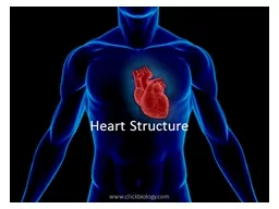
PPT-Heart Structure Heart – located in mediastinum within the thoracic cavity

Author : pamella-moone | Published Date : 2018-02-25

The Pericardium membrane surrounding heart Serous Pericardium 1 Parietal outer layer 2 Visceral inner layer part of epicardium Layers of Heart Wall Epicardium

Presentation Embed Code

Download Presentation

Download Presentation The PPT/PDF document "Heart Structure Heart – located in med..." is the property of its rightful owner. Permission is granted to download and print the materials on this website for personal, non-commercial use only, and to display it on your personal computer provided you do not modify the materials and that you retain all copyright notices contained in the materials. By downloading content from our website, you accept the terms of this agreement.

Heart Structure Heart – located in mediastinum within the thoracic cavity: Transcript


The Pericardium membrane surrounding heart Serous Pericardium 1 Parietal outer layer 2 Visceral inner layer part of epicardium Layers of Heart Wall Epicardium outermost layer. Hamot. Manisha. . Shende. , MD, FACS. Chief, Thoracic Surgery . Thoracic Surgery at . Hamot. , historically, has been “traditional” access surgery with large incisions.. Patients in this area would often travel long distances to access “Minimally Invasive” , advanced Thoracic Oncologic Surgery as well as specialized thoracic surgery.. LISA PIROPATO, PT, DPT, ATC. NORTHEAST INDIANA SPORTS MEDICINE SYMPOSIUM. MARCH 25, 2017 . Learning objectives. Explain . the etiologies of thoracic outlet syndrome. . Differentiate . between . the types of vascular and neurologic thoracic outlet syndrome. . COI Disclosure. The author has no conflict of interest to disclose with respect to this presentation.. Presenting author: . ●●. . ●●. 第. 70. 回日本胸部外科学会定期学術集会. The 70th Annual Scientific Meeting of the Japanese Association for Thoracic Surgery. Human Body. Overview of . Anatomy and Physiology. Anatomy – the study of the structure of body parts and their relationships to one another. Gross or macroscopic. Microscopic. Developmental. Physiology – the study of the function of the body’s structural machinery. Tony . Serino, Ph.D.. .. Mediastinum: Topic Objectives. Be able to identify and describe the contents of the three major divisions of the mediastinum.. Be able to identify normal and pathological variations of the aortic arch and congenital heart defects.. Assistant Professor/School of Medicine. The Hashemite University. Anatomy. The . mediastinum. is anatomically defined as the space between the two lungs; it is demarcated by the thoracic inlet superiorly, the diaphragm inferiorly, the sternum anteriorly, and by the spine and paravertebral thoracic sulci posteriorly. . Key terms. Having to do with the sinus. Sinus . Air-filled cavity within a bone that is lined with mucous membrane that makes mucus. Functions of the sinuses. Make the bones of the skull lighter. Help produce sound by giving resonance to the voice. ligamentum. . flavum. As the ligaments become ossified, it causes narrowing of thoracic canal and eventually compression of spinal cord.. Fluoride is an important factor in bone . mineralisation. . However. Dome shaped . Musculoaponeurotic. structure. Separates thorax from abdomen . Forms roof of abdominal cavity & floor of thoracic cavity. Parts of thoracic diaphragm. Thoracic surface. : convex on right & left side but depressed in centre . secretory. products into the oral cavity, the salivary glands. In the lab you will also have the opportunity the examine one other specialized epithelial area, the lip. The . oesophagus. is the first part of the alimentary canal. Its . Dr.Berlina. Terrence Mary . . Assistant Professor . . Department of Anatomy . . Mediastinum. is the middle space left in the thoracic cavity in between the lungs.. Boundaries. Anteriorly. 2 Revise the current Body Part value for Thoracic Aorta to differentiate Descending Thoracic Aorta from Ascending Thoracic Aorta/Aortic ArchRequest There are significant clinical differences in proce PATIENT EDUCATION | INFORMATIONSERIESwww.thoracic. least 3-6 months. Your healthcare provider may advise clot. Some people at high risk of blood clots may stay on blood thinner inde�nitely. Anggraheny S – FK UWKS. Indikasi Foto Thorax :. Persiapan preoperatif utk keperluan anestesi. Batuk berdahak, sesak. Post operatif, follow up. Curiga keganasan. Posisi . : PA , AP, lateral & Oblique (LAO,RAO).

Download Document

Here is the link to download the presentation.
"Heart Structure Heart – located in mediastinum within the thoracic cavity"The content belongs to its owner. You may download and print it for personal use, without modification, and keep all copyright notices. By downloading, you agree to these terms.

Related Documents