/
Immunofluorescence and Immunofluorescence and

Immunofluorescence and - PowerPoint Presentation

pamella-moone
pamella-moone . @pamella-moone
Follow
610 views
Uploaded On 2015-10-27

Immunofluorescence and - PPT Presentation

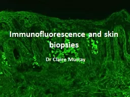
s kin biopsies Dr Claire Murray For IMF Normal Skin Perilesional skin Lesion For histology Procedure for biopsy Ellipse incisional biopsy helps preserve an intact blister Punch biopsies are more likely to disrupt the roof ID: 174048

igg skin light imf skin igg imf light bullous lesions pemphigus deposition linear inflammatory lesional split tissue igm antibodies

Share:

Link:

Embed:

Download Presentation from below link

Download Presentation The PPT/PDF document "Immunofluorescence and" is the property of its rightful owner. Permission is granted to download and print the materials on this web site for personal, non-commercial use only, and to display it on your personal computer provided you do not modify the materials and that you retain all copyright notices contained in the materials. By downloading content from our website, you accept the terms of this agreement.


Presentation Transcript

Slide1

Immunofluorescence and skin biopsies

Dr

Claire MurraySlide2

For IMF

Normal Skin

Perilesional

skin

Lesion

For histology

Procedure for biopsy

Ellipse incisional biopsy helps preserve an intact blister. Punch biopsies are more likely to disrupt the roofSlide3

Direct ImmunoflorescencePerformed on lesional or perilesional tissue from skin, mucosa or conjunctiva

Detects in vivo deposition of:

Immunoglobulins

(IgA,

IgG, IgM)Complement proteins (C1, C3)FibrinogenUsed forAutoimmune blistering disorders

Connective tissue disease (SLE, DM)VasculitisSlide4

Direct ImmunoflorescenceUses single primary antibody chemically linked to a fluorophoreFluorophore

= a

fluorsecent

chemical compound that can re-emit light upon light excitation.Antibodies are directly applied to the

lesional/perilesional tissueSlide5

Indirect ImmunofluorescencePatient’s serum is tested for antibodies directed towards a defined antigenA double layer techniquePrimary antibody (within serum) binds to the target antigen on tissue (monkey

oesophagus

or similar)

Secondary antibody carrying the fluorophore

binds to the primary antibody as fluorescent labelMultiple secondary antibodies can bind to single primary antibody providing signal amplificationSlide6

Where to take the biopsy for DIMF?Blistering disorders – perilesional skin

Perilesional

skin = normal skin immediately adjacent to a lesion

Immune deposits are degraded in inflamed or blistered skin which can result in false negative DIF

avoid an ulcer or an area where the epidermis is disruptedAvoid active lesionsConnective tissue diseases – lesional

skinFor SLE lupus band test x 2 biopsies of lesional and non-sun exposed normal skin (buttock or inner thigh)Avoid old lesions and facial lesionsVasculitis – lesional skinSlide7

How to transport the biopsy material?Rinse biopsy in salinePlace in saline soaked gauzeSend unfixed in Michels

transport medium

does not fix the tissue

maintains

isotonticity and pH of the tissue for around 2 daysstabilises proteins in tissues to allow preservation of antigenicity and use of immunofluroescence.Keep in fridge overnight (do not freeze in uncontrolled manner)Slide8

Fluorescent Microscopy

T

issue sample acts as light source

Microscope emits high intensity,

excitation

lightFluorophores illuminated by the excitation light (UV light)Flurophores emit longer lower energy wavelength light (fluorescent light)Fluorescent light is separated from surrounding radiation by filtersFilters only allow light with same wavelength as fluorescing material throughThe low energy light can be seen against a dark backgroundSlide9

Slides stored in fridge to reduce degradation of immunofluorescencePhotobleaching (fading) of slides occurs when over exposed to the high intensity light

Photobleaching

can be reduced by reducing the

insity of the light or the duration of time the tissue is exposed to the lightSlide10

Bullous pemphigoidMost common subepidermal autoimmune bullous disorderTypically affects elderly

Common sites are lower abdomen, groins, legs and armsSlide11

Bullous pemphigoidUnilocular, subepidermal blister

Roof attenuated or normal in early lesions. May become necrotic in large or older lesions

Blister contents: fibrin, inflammatory cellsSlide12

Bullous pemphigoidInflammatory (cell rich) blisterPredominant eosinophils

Variable neutrophils and lymphocytes

Non-inflammatory (cell poor) blister

Sparse inflammatory cells

Can be appearance in very early lesionsSlide13

Bullous pemphigoidFestooning of dermal papillae = preservation of outline dermal papillaeSevere dermal oedema

Perivascular

eosinophils

and histiocytes

Eosinophilic spongiosis in adjacent epidermisSlide14

Bullous pemphigoidHomogenous, linear deposition of IgG

and/or C3 along the

dermo

-epidermal junctionSlide15

Differential Diagnosis of BP

Bullous

Pemphigoid

Epidermolysis Bullosa

Bullous SLEDirect IMFLinear IgG and C3Linear IgG and C3Linear IgG and C3Indirect IMLIgG antibodies75 – 80%

IgG antibodies 25 – 50%IgG antibodies 60%Salt-split skinRoofFloorFloorSlide16

Split skin immunofluorescenceA modified indirect IMF techniqueNormal skin is split to create artificial blister cavitySplit achieved by immersing in saline

serum applied to split skin

Antibodies localised to roof or floor of blister

IgG

localised to roof in bullous pemphigoidSlide17

Epidermolysis BullosaGroup of non-inflammatory skin disorders characterised by development of blisters following minor traumaAutosomal dominant or recessive inheritancePresentation varies depending on the class of the diseaseSlide18

Epidermolysis Bullosa AcquisitaNon-inherited variantOnset in mid-life

Development of non-inflammatory bullae after minor trauma

Extensor surfaces of limbs most affectedSlide19

Epidermolysis Bullosa AcquisitaSub-epidermal bulla with fibrin

Scanty inflammatory cells

Intact roof of blister

Variable inflammatory infiltrate in dermisPAS stain demonstrates the level of split within the BM with most in the roofSlide20

Epidermolysis Bullosa Acquisita

Direct IMF: intense deposition of

IgG

and faint C3 along

dermoepidermal junctionSlide21

Epidermolysis Bullosa Acquisita

Salt-split skin IMF: antibodies bind to the floor of the blisterSlide22

Lupus Band Test for Lupus erythematosusDirect IMF performed on lesional and non-lesional

sun-protected skin

Band-like deposition of

IgG,

IgM & C3 at dermoepidermal junction in lesional skinEpidermal nuclear

IgG in small percentageSlide23

Lupus Band Test for Lupus erythematosusFalse positive lupus band test in 30% of sun-exposed skin biopsies from unaffected patientsNegative IMF can occur in early lesions, treated lesions, lesions from the trunk, when in remission.Slide24

Bullous SLEA rare variant of SLESubepidermal blistersNeutrophils in the papillary dermisLymphocytes around vessels in the superficial plexus

Linear or mixed linear/granular deposition of

IgG

and less commonly IgA and/or IgM

along dermoepidermal junctionSlide25

Bullous SLELinear or mixed linear/granular deposition of IgG and less commonly IgA and/or IgM along

dermoepidermal

junction

Salt-split IDIMF shows deposition along floor of blisterSlide26

Porphyria Cutanea TardaRare inherited or acquired disease (liver diseaseDefect in enzyme

uroprophyrinogen

decarboxylase

involved in synthesis of haem

pathway resulting in accumulation of porphyrinsBlisters arise on sun-exposed sitesSlide27

Porphyria Cutanea TardaSubepidermal blisterCell-poor

Festooning of dermal papillae

Deposition of hyaline material in BM and around dermal vessels

‘caterpillar bodies’ – hyaline material within epidermis that stains with PASSlide28

Porphyria Cutanea Tarda: DIMFIgG,

IgM

and C3 outline vessels in the papillary dermis ‘doughnut’ distribution

Linear deposition of

IgG, IgM and C3 at the dermoepidermal junction.Slide29

Dermatitis HerpetiformisAssociated with coeliac diseaseAffect all agesLesions on posterior scalp, back, buttocks, backs of arms and legs

Intensely

pruritis

, widespread, papulovesicular

erruptionSlide30

Dermatitis HerpetiformisNeutrophilic abscesses within the dermal papillae in early lesionsMultiloculated subepidermal bullae develop

Intense

neutrophilic

inflammatory infiltrate within the blister cavitySlide31

Dermatitis HerpetiformisPerilesional skin should be sampledGranular deposits of IgA seen in papillary dermisGranular-linear pattern may also be seenSlide32

PemphigusPemphigus vulgarisPemphigus vegetansPemphigus foliaceous

Paraneoplastic

pemphigus

IgA pemphigusSlide33

Pemphigus VulgarisMost common (80% cases)Middle age onsetBegins in mouth (50%)Spreads to involve the skin within weeks/months

Bullae and large and

flacid

and rupture easilyAutoantibodies to

desmoglein 3Slide34

Pemphigus VulgarisSuprabasal bullaeAcantholysisDermal papillae project into cavity like villi

‘Tombstone’ pattern – layer of basal cells remain attached to dermisSlide35

Pemphigus VulgarisAcantholytic cells round, eosinophilic & pyknotic nuclei

Occasional

eosinophils

& neutrophilsDermal perivascular infiltrate composed of

eoinophils and neutrophilsSlide36

Pemphigus IMF (perilesional skin)Intercellular deposition of IgG and C3Individual keratinocytes outlined like chicken wire

Serum antibodies can be demonstrated with indirect IMF using monkey

oesophagusSlide37

Lichen Planus‘Sawtooth’ epidermal hyperplasiaWedge shaped

hypergranulosis

Civatte

and colloid bodiesBasal vacuolar degeneration

Band-like lymphocytic infiltrate in papillary dermisSlide38

Lichen planus IMFHelps exclude SLE and other bullous disease in difficult casesDirect IMF highlights colloid bodies in papillary dermis

Colloid bodies can stain for

IgM

and C3Irregular

band of fibrinogen along basal layerSlide39

Lichen planus pemphigoidesLP associated with pemphgoid like blisters

More in common in men, 4-5

th

decadeUsually preceded by typical LP

Blisters more common on extremitiesSlide40

Lichen planus pemphigoidesLichenoid lesions are typicalBullous lesions show subepidermal blister

Cell rich or poor variants both occurSlide41

Lichen planus pemphigoides IMF

Direct IMF

Linear deposition of

IgG

and C3 along dermoepidermal

junctionSalt-split skin indirect IMFSerum contains IgG basement membrane antibody in 50-60%IgG labels roof of blisterSlide42

Leukocytoclastic VasculitisBiopsy lesional skinEarly lesions < 6 hours old more commonly positive

Deposition of fibrinogen, C3 and

IgM

all seen in vessel wallsSlide43

Henoch-Schonlien Purpura (leukocytoclastic Vasculitis)

Represents 10% of all cutaneous vasculitis

Purpuric

rash on lower legsHistology indistinguishable from other LCV

Deposition of IgA in vessel walls in involved and uninvolved skin