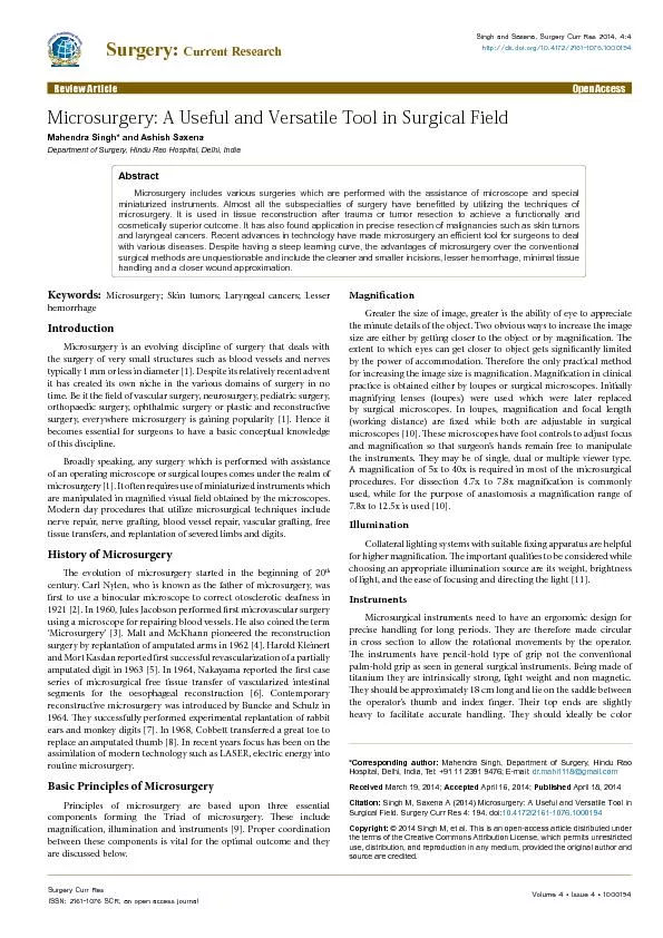
/
Mahendra Singh* and Ashish SaxenaDepartment of Surgery, Hindu Rao Hosp Mahendra Singh* and Ashish SaxenaDepartment of Surgery, Hindu Rao Hosp

Mahendra Singh* and Ashish SaxenaDepartment of Surgery, Hindu Rao Hosp - PDF document

pamella-moone
pamella-moone . @pamella-moone
Follow
405 views
Uploaded On 2016-06-26

Mahendra Singh* and Ashish SaxenaDepartment of Surgery, Hindu Rao Hosp - PPT Presentation

Corresponding author Mahendra Singh Department of Surgery Hindu Rao Hospital Delhi India Tel 91 11 2391 9476 Email drmahi1118gmailcom ID: 378591

*Corresponding author: Mahendra Singh Department

Share:

Link:

Embed:

Download Presentation from below link

Download Pdf The PPT/PDF document "Mahendra Singh* and Ashish SaxenaDepartm..." is the property of its rightful owner. Permission is granted to download and print the materials on this web site for personal, non-commercial use only, and to display it on your personal computer provided you do not modify the materials and that you retain all copyright notices contained in the materials. By downloading content from our website, you accept the terms of this agreement.


Presentation Transcript

(2014) Microsurgery: A Useful and Versatile Tool in Surgical Field. Surgery Curr Res10.4172/2161- coated to avoid the metallic glare. Needle holder should be equipped with a precise working lock that should not exceed a locking force of 50g as high locking forces generate tremor and low locking forces reduce feeling for movement. e instruments include forceps, needle holders, scissors, vascular clamps, irrigators and vessel dilators [12].It is advisable to have the hands and forearms supported by the table or other xed items. Specially designed suture material of gauges 9-0 to 12-0 and needles with a diameter of re used [13]. e color of suture material is very important since non colored material is invisible under magnication. Hence very dark-tinted sutures are used.Fundamental Techniques in MicrosurgeryFree tissue transfer/apFree tissue transfer is a surgical reconstructive procedure using microsurgery. In this a region of donor tissue is selected which can be isolated upon a feeding artery and vein. Free tissue aps can be categorized into isolated and composite types. Isolated aps consist of skin, fascia, nerve, muscle or bone taken individually, whereas composite ap is a combination of more than one tissue type. ey include myo-cutaneous, osteo-cutaneous, or innervated myo-cutaneous aps [14,15].e ap thus harvested from donor site is now transferred to recipient site requiring reconstruction and vessels of the free ap are anastomosed to vessels of the recipient site by microsurgical methods.Replantatione reattachment of a completely detached body part is known as replantation. Most commonly replanted organs are ngers and thumbs but the ear, scalp, nose, face, arm and penis replantation have also been reported. Replantation involves restoration of the blood ow, musculo-skeletal system and the nerves as required [15]. Functional demands of amputated specimen are paramount in replantation. According to modern concept the survival alone of replanted part is not sucient. e functional utility of organ also has to be taken into account and has to be weighed against the patient’s ability to tolerate long rehabilitation process, both on physical and psychological levels. Similarly it is now understood that an amputated organ can be transferred to a new site in order to enhance functional outcome of surgery. For example, a nger or a great toe can be transposed in position of a non-salvageable amputated thumb to obtain a more functional hand [8]. Such types of procedures are known as “Spare part surgery” [3].TransplantationMicrosurgery represents the link between surgery and transplant immunological research. First microsurgical experiments in transplantaion (porto-caval anastomosis in the rat) were performed by Dr. Sun Lee a pioneer in the eld of microsurgery [16]. Microsurgical techniques have played a crucial role in the eld of transplantation research because it allowed use of rodent models. It was also important to develop new techniques of transplantation that would be later performed in humans. In addition, it allows reconstruction of small arteries in clinical organ transplantation.Various Procedures in MicrosurgeryMicrosurgical reconstructive technique can be either orthoplastic or oncoplastic. In orthoplastic procedures, various free aps are used to reconstruct or replace the damaged structures such as face, digit and limbs. e oncoplastic technique is utilized in the case of malignancy where reconstruction is required aer wide excision of growth. For example, Deep Inferior Epigastric Perforator (DIEP) ap is used in breast malignancies. Otolaryngologists perform microsurgery on structures of inner ear or vocal cords [2]. Ophthalmologists perform cataract surgery, corneal transplants, and treatment of conditions like glaucoma with assistance of microsurgery. Urologists and gynecologists reverse vasectomies and tubal ligations to restore fertility using microsurgical techniques. Orthopaedic surgeons perform lumbar microsurgery for delicate procedures like removal of diseased portion of bone or inter-vertebral discs [17]. Microsurgery can play a life saving role in the eld of neurosurgery where tumors can be removed and vascular anomalies in brain can be corrected.Recent Advances in MicrosurgeryTOMS-ree Dimensional On-Screen Microsurgery SystemIt facilitates three dimensional visualization of (micro) surgical eld on a video monitor, without need to visualize physically through the microscope [18]. It provides a clear stereoscopic view with accurate sense of depth perception.HDTV single camera 3D systemis involves a high denition television attached to the microscope which enables three dimensional visualization and documentation as well [19].Supermicrosurgerye highly challenging task of anastomosis of very small vessels ()s now possible with the assistance of modern optical equipments [20]. is is known as supermicrosurgery. It has been used in the anastomosis of perforator aps.Trans oral Laser Microsurgery (TLM)It is a recent type of minimally invasive surgery for oral cancers, especially in laryngeal cancer. It employs CO laser via endoscopic approach. TLM follows an incisional resection technique under microscopic guidance. e dissection proceeds gradually bit by bit allowing the operator to precisely follow the anatomical interface between tumor and normal tissue. It thus facilitates a greater preservation of normal tissue structure and function [21].Transanal Endoscopic Microsurgery (TEMS)TEMS is a useful micro invasive technique to resect certain large or sessile adenomas of rectum. It is essentially a form of laparoscopic surgery performed in a much more conned space [22].Electromicrosurgeryis modality is used in cosmetic procedures like the destruction of feeding vessels in a skin tag, warts, and spider naevi. In it under the guidance of a magnifying lamp, the tip of the nest smallest probe possible, about the size of an eyelash is introduced along the line of a capillary and a tiny energy current is discharged [23].Mohs micrographic surgeryThis is a highly specialized surgical technique to treat certain types of skin cancers.In it, the skin cancer is removed a thin layer at a time with a very small margin of healthy skin around it. The resected specimen is immediately evaluated under microscope to find out positive margins. The corresponding area of apparently healthy skin is then resected. The entire process is repeated until no residual tumor is left behind [24]. Singh and Saxena, Surgery Curr Res 2014, 4:4 Review Article Volume 4  Issue 4  1000194Surgery Curr ResISSN: 2161-1076 SCR, an open access journal Volume 4  Issue 4  1000194Surgery Curr ResISSN: 2161-1076 SCR, an open access journal (2014) Microsurgery: A Useful and Versatile Tool in Surgical Field. Surgery Curr Res10.4172/2161- Limitations of Microsurgery Microsurgery is technically a demanding procedure and has a steep learning curve. Establishment of the set-up is costly. Other diculties include a restricted area of vision, loss of depth perception and loss of visual reference points [25].ConclusionMicrosurgery has become an indispensible tool in virtually all elds of surgery and holds a promising future. It oers several advantages over the conventional surgery such as cleaner and smaller incisions, lesser hemorrhage, minimal tissue handling and a closer wound approximation. ese factors also render the post operative period less painful and in turn lead to a faster recovery and a more satisfying outcome. Although one has to be extremely vigilant in patient selection, pre-operative planning, per-operative nesse and post operative monitoring, the rewards gained in the form of excellent results by far outweigh the eorts employed. 1. Daniel RK (1979) Microsurgery: through the looking glass. N Engl J Med 300: 2. Mudry A (2000) The history of the microscope for use in ear surgery. Am J Otol 3. Tyagi S, Kumar S (2010) Microsurgery: An important tool for reconstructivesurgery- a clinical review. Int J Pharm Bio Sci. 4. Malt Ra, Mckhann C (1964) Replantation of Severed Arms. JAMA 189: 716- 5. kleinert He, Kasdan M (1963) Restoration of Blood Flow in Upper ExtremityInjuries. J Trauma 3: 461-476. 6. Nakayama K, Yamamoto K, Tamiya T, Makino H, Odaka M, et al. (1964)Experience with Free Autografts of the Bowel with a New Venous Anastomosis 7. Buncke HJ Jr, Schulz WP (1966) Total ear reimplantation in the rabbit utilising 8. Cobbett JR (1969) Free digital transfer. Report of a case of transfer of a great 9. Kim S, Pecora G, Rubinstein RA (2001) Comparison of traditional andmicrosurgery in endodontics. In: Color Atlas of Microsurgery in Endodontics. WB Saunders Company, Philadelphia, USA. 10. Schoef� H, Lazzeri D, Schnelzer R, Froschauer SM, Huemer GM (2013) Optical magni�cation should be mandatory for microsurgery: scienti�c basis and clinical data contributing to quality assurance. Arch Plast Surg 40: 104-108. 11. Gu M, Bao H, Kang H (2014) Fibre-optical microendoscopy. J Microsc 254: 12. Chang WC, Sretavan DW (2007) Microtechnology in medicine: the emergence 13. Furka I, Brath E, Nemeth N, Miko I (2006) Learning microsurgical suturing and 14. Shenaq SM, Klebuc MJ, Vargo D (1::5) Free-tissue transfer with the aid of loupe magni�cation: experience with 251 procedures. Plast Reconstr Surg :5: 15. Morrison WA, McCombe D (2007) Digital replantation. Hand Clin 23: 1-12. 16. Lee SH, Fisher B (1961) Portacaval shunt in the rat. Surgery 50: 668-672. 17. Wang M, Zhou Y, Wang J, Zhang Z, Li C (2012) A 10-year follow-up study onlong-term clinical outcomes of lumbar microendoscopic discectomy. J NeurolSurg A Cent Eur Neurosurg 73: 195-198. 18. Franken RJ, Gupta SC, Banis JC Jr, Thomas SV, Derr JW, et al. (1995)Microsurgery without a microscope: laboratory evaluation of a three- 19. Ryo M, Schigeaki K (2001) HDTV single camera 3D system and its application in microsurgery. Sterioscopic display and virtual reality systems. Proc SPIE 20. Koshima I, Nanba Y, Tsutsui T, Takahashi Y (2003) Medial plantar perforator�aps with supermicrosurgery. Clin Plast Surg 30: 447-455, vii. 21. Pearson BW, Salassa JR (2003) Transoral laser microresection for cancer ofthe larynx involving the anterior commissure. Laryngoscope 113: 1104-1112. 22. Platell C, Denholm E, Makin G (2004) Ef�cacy of transanal endoscopic microsurgery in the management of rectal polyps. J Gastroenterol Hepatol 19: 23. Groot DW, Johnson PA (1987) Lasers and advanced dermatologicalinstrumentation. Australas J Dermatol 28: 77-85. 24. Schell AE, Russell MA, Park SS (2013) Suggested excisional margins forcutaneous malignant lesions based on Mohs micrographic surgery. JAMA 25. Ghousia A, Prabhuji MLV, Lavanya R (2012) Microsurgery: A Clinical Philosophy Mahendra Singh* and Ashish SaxenaDepartment of Surgery, Hindu Rao Hospital, Delhi, India *Corresponding author: Mahendra Singh, Department of Surgery, Hindu RaoHospital, Delhi, India, Tel: +91 11 2391 9476; E-mail: dr.mahi1118@gmail.com © 2014 , et al This is an open-access article distributed under the terms of the Creative Commons Attribution License, which permits unrestricted use, distribution, and reproduction in any medium, provided the original author and Keywords: Microsurgery; Skin tumors; Laryngeal cancers; LesserhemorrhageIntroductionMicrosurgery is an evolving discipline of surgery that deals with the surgery of very small structures such as blood vessels and nerves typically 1 mm or less in diameter [1]. Despite its relatively recent advent it has created its own niche in the various domains of surgery in no time. Be it the eld of vascular surgery, neurosurgery, pediatric surgery, orthopaedic surgery, ophthalmic surgery or plastic and reconstructive surgery, everywhere microsurgery is gaining popularity [1]. Hence it becomes essential for surgeons to have a basic conceptual knowledge of this discipline.Broadly speaking, any surgery which is performed with assistance of an operating microscope or surgical loupes comes under the realm of microsurgery [1]. It oen requires use of miniaturized instruments which are manipulated in magnied visual eld obtained by the microscopes. Modern day procedures that utilize microsurgical techniques include nerve repair, nerve graing, blood vessel repair, vascular graing, free tissue transfers, and replantation of severed limbs and digits.History of Microsurgerye evolution of microsurgery started in the beginning of 20century.Carl Nylen, who is known as the father of microsurgery, was rst to use a binocular microscope to correct otosclerotic deafness in 1921 [2]. In 1960, Jules Jacobson performed rst microvascular surgery using a microscope for repairing blood vessels. He also coined the term ‘Microsurgery’ [3]. Malt and McKhann pioneered the reconstruction surgery by replantation of amputated arms in 1962 [4]. Harold Kleinert and Mort Kasdan reported rst successful revascularization of a partially amputated digit in 1963 [5]. In 1964, Nakayama reported the rst case series of microsurgical free tissue transfer of vascularized intestinal segments for the oesophageal reconstruction [6]. Contemporary reconstructive microsurgery was introduced by Buncke and Schulz in 1964. ey successfully performed experimental replantation of rabbit ears and monkey digits [7]. In 1968, Cobbett transferred a great toe to replace an amputated thumb [8]. In recent years focus has been on the assimilation of modern technology such as LASER, electric energy into routine microsurgery.Basic Principles of MicrosurgeryPrinciples of microsurgery are based upon three essential components forming the Triad of microsurgery. ese include magnication, illumination and instruments [9]. Proper coordination between these components is vital for the optimal outcome and they are discussed below. MagnicationGreater the size of image, greater is the ability of eye to appreciate the minute details of the object. Two obvious ways to increase the image size are either by getting closer to the object or by magnication. e extent to which eyes can get closer to object gets signicantly limited by the power of accommodation. erefore the only practical method for increasing the image size is magnication. Magnication in clinical practice is obtained either by loupes or surgical microscopes. Initially magnifying lenses (loupes) were used which were later replaced by surgical microscopes. In loupes, magnication and focal length (working distance) are xed while both are adjustable in surgical microscopes [10]. ese microscopes have foot controls to adjust focus and magnication so that surgeon’s hands remain free to manipulate the instruments. ey may be of single, dual or multiple viewer type. A magnication of 5x to 40x is required in most of the microsurgical procedures. For dissection 4.7x to 7.8x magnication is commonly used, while for the purpose of anastomosis a magnication range of 7.8x to 12.5x is used [10].IlluminationCollateral lighting systems with suitable xing apparatus are helpful for higher magnication. e important qualities to be considered while choosing an appropriate illumination source are its weight, brightness of light, and the ease of focusing and directing the light [11]. InstrumentsMicrosurgical instruments need to have an ergonomic design for precise handling for long periods. ey are therefore made circular in cross section to allow the rotational movements by the operator. e instruments have pencil-hold type of grip not the conventional palm-hold grip as seen in general surgical instruments. Being made of titanium they are intrinsically strong, light weight and non magnetic. ey should be approximately 18 cm long and lie on the saddle between the operator’s thumb and index nger. eir top ends are slightly heavy to facilitate accurate handling. ey should ideally be color Microsurgery includes various surgeries which are performed with the assistance of microscope and special miniaturized instruments. Almost all the subspecialties of surgery have bene�tted by utilizing the techniques of microsurgery. It is used in tissue reconstruction after trauma or tumor resection to achieve a functionally and cosmetically superior outcome. It has also found application in precise resection of malignancies such as skin tumors and laryngeal cancers. Recent advances in technology have made microsurgery an ef�cient tool for surgeons to deal with various diseases. Despite having a steep learning curve, the advantages of microsurgery over the conventional surgical methods are unquestionable and include the cleaner and smaller incisions, lesser hemorrhage, minimal tissue Surgery: Current ResearchISSN: 2161-1076 Current Research