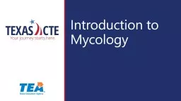
/
Medical mycology  First Medical mycology  First

Medical mycology First - PowerPoint Presentation

pamella-moone
pamella-moone . @pamella-moone
Follow
382 views
Uploaded On 2018-12-26

Medical mycology First - PPT Presentation

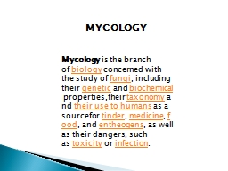
lecture   Introductions Terms Mycology Mykes mushrom fungi It is drive from a Latin word Kingdom myceteae or mycota Fungus is singular term where fungi are pleural term ID: 746105

cell fungi hyphae species fungi cell species hyphae yeasts wall kingdom mold gene eukaryotic bacteria molds spores membrane lsu

Share:

Link:

Embed:

Download Presentation from below link

Download Presentation The PPT/PDF document "Medical mycology First" is the property of its rightful owner. Permission is granted to download and print the materials on this web site for personal, non-commercial use only, and to display it on your personal computer provided you do not modify the materials and that you retain all copyright notices contained in the materials. By downloading content from our website, you accept the terms of this agreement.


Presentation Transcript

Slide1

Medical mycology

First

lecture


Introductions

Terms:

Mycology =

Mykes

=

mushrom

= fungi (It is drive from a Latin word)

Kingdom:

myceteae

or

mycota

(Fungus) is singular term where (fungi) are pleural term.

Mycology: the science of fungi.


Fungi are a group of heterotrophic

eucaryotes

that are widely distributed in nature.

They are classified into separate kingdom distinct from that of plants or animals. Fungi are considered to be saprophytes because they obtain their nutrients from dead organic matter.

 Slide2

Mycology

Divison : Mycota Subdivision Mycotina Class: mycetes Subclass :mycetidae Order: ales Family: aceae  

Mykes

=mushroom=fungi

Logos+study

of=study of fungi

Kingdom

mycetes

(=K. fungi)Slide3

Molecular identification

DNa were discovered by Friedrich Miescher. In 1869 1919, Phoebus Levene identified the component of nucleic acid and coind the terms nucleotides for the individuate bases Adenine, guanine Pyramiding thymine and cytocine. Addinin will pair with thymidine and Guanine with cytosine. The PCR technique is amplification of a part of the DNA which is present in a low concentration. The principle of molecular phylogeny is that species are understood to have arise from mother species (their accentors) during the process of as speciation.

The two closely related species from two closely related organisms should have more unique polymorphism. As a results of mutation or recombination.

So when you compare the same

piesce

ofDNA

sequences between different species so it is possible to infer between different species based on the polymorphism that are present.

One of the most used region for the Dan is the regions of DNA is the ribosomal DNA

Operone

which is consist of the small subunits (SSU), 18s

rRNA

genus the 5.8S

rRNA

gene AND THE LARG SUBUNITES (

lsu

) GET INFORMATION FROM THE THESIS Slide4

THIS OPERON IS PRESENT IN THE FUGNAL

genom as a large number of copies (50-100).

The internal transcribed spacer (ITS1 and ITS2 are found between SSU and the 5.8S

rRNA

gene. And the between the 5.8S r RNA gene and LSU respectively. The SSU and LSU evolve relatively slowly and are frequently used (50-100).

The

ssu

and LSU evolve relatively slowly and are frequently used to study higher orders relationship foe examples between families or order. Where as the ITS1 and Its2 and 5.8S

rRNA

gene region (often abbreviation to ITS 9s frequently used for species level phylogeny because of the high evolutionary rate if the internal transcribed spacers.

The IGS is found at the end of LSU and start of the SSU sequence and sometimes used for the inter or intra species variation studies

Some other species which his involve other gene sequences such as B

tubuline

and or hailstone H3. To

sovel

species and species complexity.

To use more than one gene by using

multilocus

sequencing to comply with the genealogical concordance

phylogeniatic

species recognition

The kingdom fungi is they form a kingdom which is part from plant kingdom. And animals and closer to the later. Slide5

Structure of fungi

Unicellular yeasts True yeses cell retain individuality Yeasts like cell attached to each other side by side forming pseudohyphaeFilamentous mold hyphae-hyphae-septum Septate hayphaeNonspetate hyphae coneocytic hyphaeInrerwoven hyphae

=Mycelium

Dimorphic:have

to forms depending on changes in the environment factor like temp, medium and cultures Vs host

Monomorphic

only one forms regardless of environment.

Slide6

Characterization distinguishing features

All eukaryotic Organism Heterotrophic-don’t have chlorphyll (Acrophylous)Saprobic Symbiotic Parastic The cell is surrounded by rigid cell wall made up of chitin and complex charbohydrates (chitonsan, mannan,glucan, galacotmannan)Have simple structure most of them is microscopic Reproduced by spore formation sexually or asexually

The ITS sequencing Slide7
Slide8

Kingdoms

Animilia: (eukaryotic cells) these eukaryotes are multi-cellular organisms include worms, arthropods, and animals.Plantae: (eukaryotic cells)Protoctista: eukarotic single cell like protozoa and algae Monera: (prokaryotic) single cell like bacteria, actinomycetes and blue-green algae Fungi: eukaryotic non-motile include yeasts, mold, and mushroom. Slide9

Main Characteristic of Fungi:

Features distinguish them from bacteria:Fungi ( Eukaryotic)

Bacteria

(Prokaryotic)

Nucleus

True nucleus

Prenucleus

Nuclear

membrane

define nuclear membrane

2

no well define nuclear membrane (the nuclear material is diffused in the cytoplasm)

Cytoplasmic structures

membrane bound organelles (Endoplasmic reticulum , golgi apparatus…etc)

no membrane bound organelles

size

1 um in size or wider it is larger than bacteria

1 um in size or less, smaller than fungi.

Cell wall

Composed of chitin and other polysaccharides

Composed of peptidoglycan

Replication

Mitosis and Meiosis

Simple binary fission Slide10

Features Distinguish Them From Plant:

Fungi

Planta

Nutrition

Hetrotrophic (achlorophllous)

Saprobic (saprophytic)

Symbiotic

Parasitic

Autotrophic

(photosynthesis)Slide11

Features Distinguish Them From

Animilia:Fungi

Animailia

Cell wall

Rigid cell wall mad up of chitin and other polysaccharides (manna, glucan)

No cell wall Slide12

Fungi are

Fungi are saprophytic, aerobic, nucleated, achlorophyllous organisms, which typically reproduce sexually and\or asexually. These organism usually filamentous-branched somatic structures are surrounded by true cell wall. They comprise two groups of organisms known as yeasts and molds.   Structure of fungi:  Yeasts: Unicellular, produced in two phase either budding yeasts

cells or elongated cells attached to each other and forming

pseudohyphae

Culture:

yeasts form colonies that resemble those of bacteria

Example:

Candida

albicans

Saccharomyces

cerviseae

Cryptococcus

neoformance


 Slide13

Molds (filamentous)

Molds (Mould): classified as moniliaceous (white in color) or dematiaceous (dark in color).hyphae (singular, hypha), long, slender, branching tubes. If hyphae have cross walls, the fungus is said to be Septate. If cross-walls are not present, the fungus is said to be non-septate (coenocytic). Interwoven hyphae known as mycelium Example of mold:Aspergillus sp Rhizopus sp Penicillium

sp

Spores and conidia

are the results of asexual or sexual reproduction; they are used for the identification of the mold based on their shapes, sizes and relationship to the parent

hyphae

.

Culture:

it grows on the surface of the plates as one colony. The visible surface growth assumes a fluffy character known as

mycelium

.


Dimorphic fungi

: are mold at room temperature and yeasts at 37

o

c.

Example:

Coccidioides

imitis

Histoplasma

capsulatum


 Slide14


Reproduction in Fungi 1. Asexually2. Sexually  Asexually: asexual reproduction occurs strictly by the process of mitosis (akaryogamy)somaticspore formation The manner in which spores are produced is important in identification of fungi. Their size shape and arrangements help in the identification of fungi.Spores born in a sac-like structure called a sporangium (sporangia, pleural), and the spores know as sporangiospore. Conidia which are produced on the tip or sides of hyphae are called conidia (conidium

) -it is produced on a specialized structure called

conidiophore

.


Types of conidia

Chlamydospore

Blastospore

Arthrospore

Aleurospore

 Slide15

Pycnidium

Synnema

Sporodochium

Acervulus

Asexual reproductive structures:

Conidiophore

Conidium

Imperfect fungi =

Deuteromycetes

: Do not reproduce sexually or their sexual reproduction not known

e.g.

Aspergillus

,

Fusarium

, Candida

4Slide16

II)

Sexual: Fusion, mitosis, meiosis Sexual spores: Oospore, Zygospore

,

Ascospore

,

Basidiospore

Zygospore

Ascocarp

Ascus

Ascospore

Zygomycetes

Basidiomycetes

e.g. Truffles

Terfezia & Termania

Ascomycetes

e.g.

Rhizopus, Mucor

e.g. Mushrooms &

Podaxis pistillaris

(Agaricus campestris)Slide17

Classification of Fungi

1- Div. Gymnomycota: naked (No cell wall), phagotrophic

Myxamoeba, slime molds

Class Acrasiomycetes, Cl. Protosteliomycetes

Kingdom Myceteae

Cl. Myxomycetes –

e.g.

Dictyostelium

2- Div. Mastigomycota:

flagillated, motile

Cell wall, absorptive nutrition, if mold nonseptate hyphae

Chytridiomycetes, Hyphochytridiomycetes

Plasmodiophoromycetes, Oomycetes –

e.g.

Phythium, Phytophthora

3- Div.

Amastigomycota

:

Non flagellated, Non-motile

Yeasts & molds: Septate hyphae & non-septate

Cl. Zygomycetes, Trichomycetes, Ascomycetes, Basidiomycetes

Deuteromycetes (Fungi Imperfecti = Imperfect fungi)

e.g.

Aspergillus, Penicillium, Fusarium, Candida

Old terms:

Phycomycetes, Aquatic, Lower fungi, Higher fungi

8