/
Medical Mycology Lecture Slides Medical Mycology Lecture Slides

Medical Mycology Lecture Slides - PowerPoint Presentation

mitsue-stanley
mitsue-stanley . @mitsue-stanley
Follow
363 views
Uploaded On 2019-02-04

Medical Mycology Lecture Slides - PPT Presentation

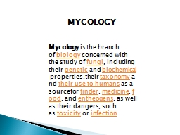
ALHEDAITHY Basic mycology 2 Superficial Mycoses 9 Pityriasis versicolor 10 Tinea Nigra 12 Piedra 14 Dermatophytoses 17 Mycetoma 25 Rhinosporidiosis 35 Lobomycosis ID: 750032

yeast hedaithy continued culture hedaithy yeast culture continued infection amp fungi cells tissue mold specimen skin candidiasis sda hyphae candida fungus etiology

Share:

Link:

Embed:

Download Presentation from below link

Download Presentation The PPT/PDF document "Medical Mycology Lecture Slides" is the property of its rightful owner. Permission is granted to download and print the materials on this web site for personal, non-commercial use only, and to display it on your personal computer provided you do not modify the materials and that you retain all copyright notices contained in the materials. By downloading content from our website, you accept the terms of this agreement.


Presentation Transcript

Slide1

Medical Mycology Lecture Slides

AL-HEDAITHYSlide2

Basic mycology 2

Superficial Mycoses 9

Pityriasis versicolor 10

Tinea Nigra 12

Piedra 14

Dermatophytoses 17Mycetoma 25Rhinosporidiosis 35 LobomycosisPhaeohyphomycosis 37Chromoblastomycosis 38Sporotrichosis 41Zygomycosis 43Aspergillosis 48Pneumocystosis (PCP) 51Candidiasis 53Cryptococcosis, 60 Trichosporonosis, Geotrichosis

List of Contents

Primary Systemic Mycoses 63Blastomycosis 65Histoplasmosis 67Coccidioidomycosis 70Paracoccidioidomycosis 73Candidosis and Oropharyngeal 75 mycosis (For Dental Students)Fungal Eye Infections 83Selected references 89

1

AL-HEDAITHYSlide3

Mycology:

Mykes = Mushroom = Fungi

Logos = Study of

= Study of fungi

Kingdom myceteae (= K. fungi)

Characteristics (distinguishing features)1) All Eukaryotic organisms2) Heterotrophic – do not have chlorophyll (Achlorophyllous)

3) The cell is surrounded by rigid cell wall

made up of chitin & complex carbohydates (Chitosan,Mannan,glucan,galactomannan)4) Have simple structure – most of them microscopic.5) Reproduce by spore formation sexually or asexually.Basic Mycology

Parasitic

Symbiotic

Saprobic

2

AL-HEDAITHYSlide4

1) Unicellular – Yeasts

Examples:

Saccharomyces cerevisiae

True yeasts

Candida albicans

cells retain individuality Cells attach to each others side - by – side forming Pseudohypha 2) Filamentous – molds Examples: Aspergillus, Penicilium Rhizopus

Hypha - Hyphae - Septum

Structure of Fungi

Septate hypha

Interwoven hyphae = Mycelium

Dimorphic: Have two forms depending on change in the environmental factor like

Temp., Medium and culture Vs Host

Yeast-like

Monomorphic: Only one form regardless of environment.

Nonseptate hypha (coenocytic hypha)

3

AL-HEDAITHYSlide5

I)

Asexual

: Only mitotic cell division

1) Somatic

Yeasts by budding Molds by hyphal fragmentation 2) Spore formation: a) Sporangiospores in sporangia b) Chlamydospores in or on hyphae

c) Conidia (conidium) on hypha or on conidiophores

PycnidiumSynnemaSporodochium

Acervulus

Reproduction in Fungi

Many types of conidia like:

Blastospore, arthrospore, aleuriospore, sympodulospore…etc

Asexual reproductive structures:

Conidiophore

Conidium

Imperfect fungi = Deuteromycetes: Do not reproduce sexually or their sexual reproduction not known

e.g.

Aspergillus, Fusarium, Candida

4

AL-HEDAITHYSlide6

II)

Sexual: Fusion, mitosis, meiosis

Sexual spores:

Oospore, Zygospore, Ascospore, Basidiospore

Zygospore Ascocarp

Ascus

AscosporeZygomycetesBasidiocarpBasidiospore

Basidium

Basidiomycetes

Gills

e.g. Truffles

Terfezia & Termania

Ascomycetes

e.g.

Rhizopus, Mucor

e.g. Mushrooms &

Podaxis pistillaris

(Agaricus campestris)

5

AL-HEDAITHYSlide7

Groups of Fungal Infections:

Superficial Mycoses

Cutaneous Mycoses

Subcutaneous Mycoses

Systemic Mycoses

Opportunistic MycosesActinomycetous Infections 6AL-HEDAITHYSlide8

Classification of Fungi

Taxonomy:

Based on morphological, Physiological, Genetic & Molecular characteristics.

Taxon

= Classification rank. Kingdom: MyceteaeDivision: AmastigomycotaSubdiv: DeuteromycotinaClass: Deuteromycetes Sub. class: Hyphomycetidae

Order: Moniliales

Family: MoniliaceaeGenus: MicrosporumSpecies: CanisVarietyStrain

Binomial system

Author

M.canis

Bodin

Primary Pathogen

Opportunistic Pathogen

Latin

7

AL-HEDAITHYSlide9

Classification of Fungi

1-

Div. Gymnomycota:

naked (No cell wall), phagotrophicMyxamoeba, slime moldsClass Acrasiomycetes, Cl. ProtosteliomycetesKingdom Myceteae

Cl. Myxomycetes –

e.g. Dictyostelium

2- Div. Mastigomycota:

flagillated, motile

Cell wall, absorptive nutrition, if mold nonseptate hyphae

Chytridiomycetes, Hyphochytridiomycetes

Plasmodiophoromycetes, Oomycetes –

e.g.

Phythium, Phytophthora

3- Div. Amastigomycota:

Non flagellated, Non-motile

Yeasts & molds: Septate hyphae & non-septate

Cl. Zygomycetes, Trichomycetes, Ascomycetes, Basidiomycetes

Deuteromycetes (Fungi Imperfecti = Imperfect fungi)

e.g.

Aspergillus, Penicillium, Fusarium, Candida

Old terms:

Phycomycetes, Aquatic, Lower fungi, Higher fungi

8

AL-HEDAITHYSlide10

Superficial Mycoses

These affect the uppermost dead layers of skin or hair shaft.

They are painless and usually do not provoke the immune system

They primarily include:

1- Tinea versicolor (= Pityriasis versicolor) Brown or discolored or white patches on the skin. 2- Tinea nigra (T.n. palmaris) Dark brown or grey macular lesions – usually on palm of hand but can be on sole of foot or others. 3- Piedra Nodules of the etiologic fungus on hair shaft; a. Black piedra b. White piedra

9

AL-HEDAITHYSlide11

Superficial Mycoses

(Continued)

Pityriasis Versicolor (= Tinea versicolor)

Brown or discolored, or white patches on skin

Affect the stratum corneum

The white lesions do not tan in the sun Endogenous source of infectionEtiology: Malassezia furfur It is a Yeast (= Pityrosporum orbiculare) Blastomycetidae, bipolar budding, Skin flora Lipophilic: oleic acid Or Mineral oilLaboratory Diagnosis: Specimen is skin scrapings

10% Or 20% KOH will show short hyphal segments and round yeast cells (spaghetti & meat ball appearance)

Culture on SDA and Mycobiotic medium (=SDA+ Chloramphenicol+ Cycloheximide = Actidione) with oily substance.10AL-HEDAITHYSlide12

Superficial Mycoses

(Continued)

Pityriasis Versicolor

In culture

M

. furfurRound cellHyphal segmentScrapings

in KOH11

AL-HEDAITHYSlide13

Superficial Mycoses

(Continued)

Tinea nigra (= T.n. palmaris)

Macular brown lesions or black stripes on palm of hand or sole of foot.

Acquired by Piercing of skin with plant material in Agricultural soil.

Etiology:Dematiaceous imperfect mold fungus; Phaeoannellomyces werneckii(=Exophiala werneckii)In vegetation debris In culture it produces annellospores from annellids,unicellular or two celled.Laboratory Diagnosis:Skin scrapings: - In 10% or 20% KOH will show brown septate hyphaeCulture on SDA & Mycobiotic. Will grow the dematiaceous fungus.

Do Lactophenol teased mount (LTM) & LPCB (Lactophenol cotton blue) for identification.

12AL-HEDAITHYSlide14

Superficial Mycoses

(Continued)

Tinea nigra

(

Exophiala

werneckii)Phaeoannellomyces werneckiiConidia

In KOH

3 wk old colonyPetri plate

13

AL-HEDAITHYSlide15

Piedra

Nodules or Encrustations on hair shaft

On scalp hair / -

mustache, beard

Black piedra

Firm, hard, dark brown nodules Piedraia hortae is the etiology Ascomycete – Loculoascomycetidae Cerebriform colony

Ascostroma, Asci, ascosporesHair with nodule

10% -20% KOH – culture on Mycobiotic & SDAAssostroma with asci & ascosporesHyphal strands

Usually in Tropics & subtropics

The nodules are composed of fungal elements

Hair

Colony of

Piedraia hortae

in culture

Black piedra nodule on hair shaft

KOH

14

AL-HEDAITHYSlide16

Piedra

(Continued)

White piedra

Soft, brown, cream less firm

Trichosporon beigelii Imperfect yeast Friable nodulesPseudohyphae, blastospores, arthrosporesCulture on SDA no cycloheximide Cream – beig Yeast with wrinkled surface

Spores & cells

Etiology:White piedra nodule on hair shaft KOH

Hair

Blastospore

Arthrospore

Pseudohypha

Yeast cell

Culture

15

AL-HEDAITHYSlide17

Treatment of Superficials

2% salicylic acid, 3% sulfur ointments, whitfield’s ointment Ketoconazole

Cut or shave hair for Piedra & clean with mild fungicide

(e.g. 1:2000 bichloride of mercury)

Or apply 2% salicylic acid Or 3% sulfur ointment.Now: Nizoral shampoo for Piedra Nizoral = Ketoconazole (Benzoic acid)16AL-HEDAITHYSlide18

Dermatophytoses

These are fungal infections of the Keratinized tissues of the body

(Stratum corneum, Hair, nail)

Necrotic scaley center

Active fungus in margin T.capitis in the scalp, barber’s itch

The skin, hair folicles, and hair are infected. Infected hair fall-off Endothrix hair infection and EctothrixT.corporis : In glabrous skin

t. circinata, t.imbricataT.cruris: In groin = jock itch = Dhobie itch t.unguium

(nail) t.barbae

(beard)t.manuum

(hand)

S/S:

Skin lesion Tinea(=Ringworm)

Causes itching – present anywhere in body

Contageous

T.pedis (=Athlete’s foot) = swimming pool itch

17

AL-HEDAITHYSlide19
Slide20

Dermatophytoses

(Continued)

Other advanced lesions in scalp:

Favus

(=t.favosa)

with scutulum (yellow crusts) + unpleasant smellEpidemiology: Wood’s lamp Emits filtered U.V. light, infected hair fluoresce especially microsporum spp. lesions.

-Acquired: from infected persons and Pets

Cats Dogs -And Livestock Animals (goats, sheep, camel, cows, horsesKerion (pustular)

-

Affect children + adults + males and females. More seen in school-age children

-

Found everywhere in the world.

-Familial cross infection occurs

-Contageous – caused by primary pathogens

18

AL-HEDAITHYSlide21

Dermatophytoses

(Continued)

Etiology:

Dermatophytes Imperfect moniliaceous mold fungi,Primary pathogens

Characteristics of dermatos:

1) Produce alkaline substance 2) Sensitive to: <20 μg/ml griseofulvin

3) Resistant to: 500 μ

g/ml cycloheximide4) Have peculiar Hyphal structures likeRaquet hyphae, nodular bodies, pectinate hyphae, hyphal coils (=spirals), favic chandeliersNatural Habitats: All are Keratinophilic fungi

- Anthropophilic, Zoophilic, Geophilic

19

AL-HEDAITHYSlide22

Dermatophytoses

(Continued)

The dermatophytes are in 3 genera:

Trichophyton

:

T.mentagrophytes in rodents,dogs,livestocks.T.violaceum in human Infect skin + hair + nailT.verrucosum in cows. T.rubrum in humanMicrosporum

: M. canis in cats. M.audouinii in human, M.gypseum in soil

Infect skin + hair, but not nailEpidermophyton floccosum in human Infect skin + nail, but not hair Perfect stage: In gymnoascaceae,

Plectomycetidae ascomycetes

Trichophyton

in

Arthroderma

Microsporum

in

Nannizzia

They reproduce asexually forming conidia by which can be identified.

20

AL-HEDAITHYSlide23

-

Elongated

-Smooth wall

-Round tip

-

Spindle-Rough-PointedMacroaleuriospore (macroconidium)Microaleuriospore

(Microconidium)

TrichophytonEpidermophytonMicrosporum-Round tip-Club shape -Smooth wall

21

AL-HEDAITHYSlide24

Dermatophytoses

(Continued)

Identification Tests:

Endothrix & Ectothrix hair infection

Hair perforation test

Urease testPigment production in PDA & CMA mediaNutrient requirement such as – Trichophyton series Agar 1-7 Dermatophyte test medium (DTM):In Acidic pH yellow, in alkaline red It has pH 5.6, Antibacterial, Antifungal, and Phenol red (Amphoteric dye)

This is a special medium for the identification of dematophytes Positive DTM test is growth of the fungus and red color. It is 98% accurate.22AL-HEDAITHYSlide25

Dermatophytoses

(Continued)

Laboratory Diagnosis:

-

Skin scrapings

, Hair, Nail 20% or 10% KOH will show hyaline Septate hyphae, or spores or both. Culture on SDA and Mycobiotic medium. IdentifySpecimens:Treatment:Griseofulvin Topical or systemic

Azoles Topical

-Miconazole (Daktrin), Clotrimazole (Canesten), EconazoleAzoles Systemic: Itraconazole - others

Allylamines Topical, Terbinafine

(Lamisil)

Tolnaftate 1% solution

(=Tinactin = Tinaderm)

23

AL-HEDAITHYSlide26

Dermatomycoses

Skin and Onychomycosis

Ear

(Otomycosis)

- Mycotic keratitis

Eye (oculomycosis) Corneal ulcer,corneal abscess- Endophthalmitis These are caused by:

Candida albicans, Scytalidium, Scopulariopsis,Fusarium, Acremonium,Aspergillus,and others

Other non-dermatophyte skin infections24AL-HEDAITHYSlide27

Mycetoma (=Madura Foot)

Chronic localized subcutaneous infection that involve underlying bone later in the disease course.

The lesions are multiple abscesses.

Main symptoms/signes are cold swelling of the affected site (tumefaction), formation of sinuses that drain pus to the surface of the skin, and presence of grains.

Grains are granules (small colonies), about 1-2 mm diameter, of the etiologic agent with different color.

The commonly affected site is the foot, however, it can be in leg, thigh, hand, arm, shoulder, or head.25AL-HEDAITHYSlide28
Slide29

Mycetoma (=Madura Foot) (continued)

Infection is acquired following trauma to the skin by plant material from trees, shrubs, or vegetation debris. Thus more seen in rural areas (in farmers, Sheppards, walking bare-foot in agricultural land or city parks).

“Madura foot” referring to the first case seen in “Madura” region of India which was in the foot of that patient.

Infection is very chronic takes months to be fully established and years to deal with. It is not contageous. More seen in tropics and subtropics.

Etiologies are fungi which cause eumycotic mycetoma (Eumycetoma) or actinomycetes which cause actinomycotic mycetoma (actinomycetoma). Their natural habitats are plant materials.

26AL-HEDAITHYSlide30

Etiology:

Eumycetoma

: caused by several mold fungi.

The color of grains in this type of mycetoma is black or white.

Fungi include: Madurella, Pseudallescheria (Scedosporium), Pyrenochaeta (Pycnidia producer), Acremonium, and the ascomycetes Leptosphaeria and Neotestudina, others.The common etiologies in Saudi Arabia and neighboring countries are:

Madurella mycetomatis causes the majority of the cases with black grains. It is imperfect dematiaceous mold with brown colonies and diffused honey – colored pigment. Produces phialoconidia from phialides, and chlamydospores

Mycetoma (=Madura Foot) (Continued)ChlamydosporePhialoconidia

Phialide

Madurella mycetomatis

Madurella grisea

Another species of

Madurella

, similar to

M.mycet

. but with grey colonies.

27

AL-HEDAITHYSlide31

Pseudallescheria boydii

– causes white grain mycetoma. It is Ascomycete mold forming cleistothecia and ascospores. The imperfect of it is the moniliaceous mold: Scedosporium apiospermum which forms annelloconidia from annellids.

Mycetoma (=Madura Foot) (Continued)

Annelloconidia

Annellid

Scedosporium apiospermum

28

AL-HEDAITHYSlide32

Actinomycetoma: Caused by about 10 species of aerobic actinomycetes.

Color of grains yellow, white, yellowish-brown, pinkish – red.

Actinomycetes are filamentous higher bacteria. The filaments (very thin about 1.0

μ

m wide) appear as long branching, beaded, or as long rods. They are Gram-positive.

Main etiologies:Streptomyces somaliensis- causes the majority of the cases – color of grains yellow to yellow-brown.Actinomadura madurae – white or yellow grains.Actinomadura pelletieri – pinkish-red grains Nocardia brasiliensis – white grains.

N.asteroides, N. caviae,

N.coeliaca – white or yellow grains.Latter species of Nocardia usually cause Nocardiosis (which is subcutaneous, pulmonary, or brain abscess infection). Mycetoma (=Madura Foot) (Continued)29AL-HEDAITHYSlide33

Nocardia is acid-fast to partially acid fast when stained by Ziehl-Nelsen stain (ZN) while

Streptomyces

and

Actinomadura

are nonacid-fast.

These actinomycetes are differentiated by their decomposition pattern of casien, tyrosine, xanthine, hypoxanthine, urea, and gelatin. Also by few other biochemical tests and colony morphology (They have adherent dry colonies). See Table of characteristics.The anaerobic actinomycete; Actinomyces israelii causes the infection: “Actinomycosis” which is subcutaneous, cervicofacial, pulmonary, abdominal, uterine, or brain abscess infection. It also causes dental caries. The organism is filamentous and will have yellow grains (sulfer granules). Mycetoma (=Madura Foot) (Continued)

30

AL-HEDAITHYSlide34

Characteristics of the main species of the above Actinomycetes

Decomposition of Ca= Casien, Ty = Tyrosine, Xa = Xanthine, Hx = Hypoxanthine

Ca

Ty

Xa

Hx

Urea

Growth in 0.4X salt

Gelatin

Colony

S. somaliensis

+

+

-

-

-

-

+

Brown

A.madurae

+

+

-

+

-

+

+

Brown

A.pelletieri

+

+

-

+

-

+

Brown

N.Brasiliensis

+

+

-

+

+

+

Orange

N.asteroides

-

-

-

-

+

-

White

31

AL-HEDAITHYSlide35

Laboratory Diagnosis:

Specimen:

Visible grains, Biopsy tissue (not skin pinch), curettings of sinuses, pus, blood for serology.

First determine color of the grains – it helps identify etiology and initiate treatment.

Make histologic sections, or grinde tissue and crush grains and make smears – stain by: Hematoxylin-Eosin, Gram, ZN; if fungi do 20% KOH or Periodic acid schiff stain.

Extract serum for serology.Direct Microscopy:Will reveal grains in tissue; Homogenous texture actinomycetes.Heterogenous texture fungi Actinomycete grains will not reveal filaments easily.Fungal grains will contain easily seen hyphae and chlamydospores.

Grains will have different morphology and color (white, black, yellow, pink, …. etc) depending on etiology.

Mycetoma (=Madura Foot) (Continued)Microscope field (10X)

Actinomycotic grain

In tissue section

Eumycotic grain

32

AL-HEDAITHYSlide36

Culture:

On SDA, Neutral – SDA, BHI-A, Blood agar and incubate at 37

o

C and at 25 -30

o

C aerobically.For actinomycosis also culture on cooked meat medium and incubate anaerobically at 37oCThe organisms will grow slowly and may be contaminated with skin flora – purify – identify.Serology:

Test for Antibody using known antigen from each etiologic agent. Methods used immunodiffusion (I.D), and /or

counterimmunoelectrophoresis (C.I.E.)Serology is good for Dx and monitoring treatment Mycetoma (=Madura Foot) (Continued)Direct microscopy of specimen from Nocardiosis and actinomycosis will show branching thin filaments by silver stain

Actinomycete filaments

from

culture (Gram +ve)

Beaded filaments

Actinomycete filaments (Silver stain)

Nocardia (Gram +ve)

33

AL-HEDAITHYSlide37

Management:

Usually actinomycetoma respond better to treatment than eumycetoma.

Generally if bone is infected the response to treatment is poor.

Actinomycetoma:

Trimethoprim – Sulfamethoxazole (Cotrimoxazole/ Septrin) + Streptomycin sulfate Or: Dapsone + Streptomycin sulfateOr: Doxycycline + CefachlorFor Actinomycosis: Penicillin G Eumycetoma:Ketoconazole (Nizoral) tablets or Itraconazole or voriconazole.

If drugs not effective and bone is infected Amputate the limb or debride tissue and continue Rx.Treatment duration is long – up to years

Mycetoma (=Madura Foot) (Continued)34AL-HEDAITHYSlide38

Rhinosporidiosis

Mucocutaneous fungal infection

Sites: Nasal, Oral

(Palate, epiglottis)

, Conjunctiva.

Lesion: Polyps, Papillomas, wart-like lesionsEtiology:Rhinosporidium seeberi

Obligately parasitic fungus

Believed to be hyphochytridiomycetes, does not grow on artificial media (e.g. SDA) But has been grown in tissue culture Cryosurgical excision of lesion-relapse common. Direct Microscopy: Stained sections or smears or KOH, will show spherules with endospores

Clinical:

More seen in communities near swamps

Laboratory Diagnosis:

Specimen: Biopsy tissue

Management:

endospores

spherule

Culture on SDA negative

35

AL-HEDAITHYSlide39

Lobomycosis

Cutaneous – subcutaneous fungal infection

Lesion: Keloidal – verrucoid - nodular

Site: face, ear, arms, legs

Etiology:

Chronic – localized

Lacazia loboi

(=Loboa loboi)Obligately parasitic fungus Does not grow in culture like SDA media or tissue cultureManagement: Surgical excision of lesion

Clinical:

Laboratory Diagnosis:

The specimen is Biopsy tissue – Direct Microscopy will show chains of cells

Culture of specimen will be negative

36

AL-HEDAITHYSlide40

Phaeohyphomycosis

Clinical:

Dematiaceous imperfect mold fungi. Mainly:

Cladosporium, Exophiala, Wangiella, Cladophialophora bantiana

(Cladosporium bantianum), Ramichloridium (Rhinocladiella) mackenziei, Bipolaris, Drechslera, RhinocladiellaBrown

C.cladosporoides, E.jeanselmei, W.dermatitidis

Subcutaneous or brain Abscess caused by dematiaceous fungiAffected site: Thigh, legs, feet, arms, ….etc, brain (cerebral)Etiology Neurotropic fungi cerebral PHM as R.mack, C.bant.

Naturally in woody plants, woods, agricultural soils

Laboratory Diagnosis:

Specimens: Pus, biopsy tissue

Direct Microscopy: KOH & smears brown septate hyphae

Culture: On SDA & Mycobiotic – very slow growing black or grey colonies.

Pus specimen in KOH

37

AL-HEDAITHYSlide41

Chromoblastomycosis (=Chromomycosis)

The lesions are Hyperkeratotic, Verrucous, Pedenculus, Violaceous,Cauliflower, Initially Ulcerative, Autochthonous spread

Phialophora verrucosa, Fonsecaea pedrosoi, Exophiala, Cladosporium

Clinical:

Affected sites: extremitees, mainly feet & legsEtiology:

Dematiaceous imperfect mold fungi in woods and woody plants.

Laboratory Diagnosis: Specimen, Biopsy tissue Direct Microscopy: KOH & smears brown cells with septa, Brown Muriform cells (=sclerotic bodies)

Culture: On SDA and Mycobiotic

Very slow growing

Dematiaceous fungi

In KOH

38

AL-HEDAITHYSlide42

Blastospore

Drechslera

E.J.

Ramichloridum mackenziei

Cladosporium

PhialophoraBipolarisFonsecaea pedrosoiExophiala

39

AL-HEDAITHYSlide43

Management:

Phaeophypho. & Chromoblasto.

Clean surgical excision of the lesion + Antifungal

Azoles

(e.g. Voriconazole, Posaconazole)

Caspofungin Subcutaneous:Cerebral phaeohypho: Aspiration of Pus - Antifungals

Amphotericin B, 5-Fluorocytosine (5-FC)

40AL-HEDAITHYSlide44

Sporotrichosis

Lymphocutaneous and subcutaneous granulomatous lesions - suppurate, ulcerate. The lesions are nodules or ulcers in local lymphatics

Affected sites: extremities, joints.

Dimorphic, imperfect fungus in trees, shrubs, plant debris

Laboratory Diagnosis: Specimen:Biopsy tissue,ulcerative materialClinical:

In agricultural communitiesEtiology:

Sporothrix schenckii. Yeast in human tissue & at 37oC in culture. Mold in culture at room temperature with flowerettes of conidia.Direct Microscopy: smear Finger-like yeast cells or Cigar Some are oval. Also asteroid bodies may be seen.

Culture: On SDA at room temperature to grow mold, and on blood agar at 37o

C to grow yeast.

Treatment:

Septrin, KI

41

AL-HEDAITHYSlide45

Yeast cells

Asteroid body

Conidium

In clinical specimen and in culture at 37

o

CMold in culture at room temperatureSporothrix schenckii

42

AL-HEDAITHYSlide46

Zygomycosis

Fast growing, moniliaceous molds, nonseptate hyphae, perfect

Infections caused by Zygomycete fungi of the orders Mucorales & Entomophthorales. Zygomycetes are:

(= Phycomycosis)

I- Subcutaneous zygomycosis (= Entomophthoromycosis)Chronic localized. Subcutaneous masses, cellulitis

Rhinofacial or other like Hand,Arm,Leg, thigh. Firm swelling of site with intact skin-Distortion.

Acquired via nasal mucosa or insect bite, Cont. debris Conidiobolus coronatus, Basidiobolus ranarum, and few mucorales. These are perfect fungi that form Sporangia (conidia) and Zygospores.Specimen: Biopsy tissue

Clinical:

In specimen

Etiology:

Laboratory Diagnosis:

Direct microscopy: stained sections or smears will show broad non-septate hyphae with eosinophils.

Culture on SDA

(no antifungals), fungi will grow fast.

Zygomycetes are inhibited by Cycloheximide

Treatment:

KI orally or KI + Ampho B or Septrin

43

AL-HEDAITHYSlide47
Slide48

Zygomycosis

Sporangium

Zygospore

Conidiobolus coronatus

Busidiobolus ranarum

44

AL-HEDAITHYSlide49

Zygomycosis

(Continued)

II- Rhinocerebral zygomycosis

(=Mucoromycosis)

Usually acute, affects compromised host especially Diabetics with acidosis. OpportunisticRhinofacial – orbital – cranealVERY SERIOUS – ACUTE - FATAL Fast growing Zygomycetes have Nonseptate hyphae of the Mucorales order maily;

Acquired Via nasal mucosa

Rhizopus, Mucor,Absidia,Rhizomucor, others. Rhizopus arrhizus Reproduce sexually and asexually forming Sporangia with sporangiospores & ZygosporesClinical:

Paranasal sinusitis, orbital cellulitis

Etiology:

45

AL-HEDAITHYSlide50

Zygomycosis

(Continued)

II- Rhinocerebral zygomycosis (=Mucoromycosis)

Culture on SDA

(no antifungal “Cycloheximide”)

.The fungi will grow fast within 2-3 days.Laboratory Diagnosis: Specimen: Biopsy tissue

Nonseptate hyphae In clinical specimen

Direct microscopy: Stained sections or smears will show broad nonseptate hyphaeTreatment: Aggressive surgical debridement + Amphotericin B – other antifungals.III- Gastrointestinal (GI) Zygomycosis

This is a chronic zygomycete infection affecting the GI., mainly liver and intestine. The lesions are masses or abscesses in these sites.

Caused by

Basidiobolus ranarum

primarily.

Specimen: fine needle biopsy Direct Microscopy will show nonseptate hyphae & culture will grow the fungus.

Treatment: Medical with Itraconazole prognosis is good

IV- Pulmonary Zygomycosis

This is chronic or acute. Other aspects similar to mucoromycosis

Prompt Dx & action are essential to save life

There is mucorales G.I. Zygo which acute & Fatal. It is rare

Seen in children

(6 -12 -Year old)

often

46

AL-HEDAITHYSlide51

Zygomycosis

Sporangium

Sporangiospore

(Stolon)

Rhizopus

Rhizoid

Absidia

RhizomucorZygosporeMucor47

AL-HEDAITHY

Nonseptate hyphae

SporangiophoreSlide52

This is any infection caused by

Aspergillus–Affecting compromised individuals.

The systemic forms of this infection are opportunistic infections.

In few occasions it is non opportunistic

The clinical manifestations vary from allergy to skin to systemic forms.

Aspergillosis1- Allergic Aspergillosis AsthmaAllergic Bronchopulmonary Aspergillosis (ABPA)

Clinical Types:

IgE antibodies present. In ABPA also IgG2- Colonizing aspergillosis (=Aspergilloma = Aspergillus fungus ball) Pulmonary aspergilloma signs include:Cough,hemoptysis, variable feverCXR will show coin – like mass in the lung There will be a radiolucent crescent (=Monod’s sign = Grelot) over the mass

3- Invasive Aspergillosis - pulmonary

Signs: Cough , hemoptysis, Fever, Pneumonia, Leukocytosis

Lab investigation (direct microscopy and culture) may be negative

especially if specimen is noninvasive like sputum.

48

AL-HEDAITHYSlide53

Aspergillosis

(Continued)

4-

Aspergillus sinusitis (= Nasal-orbital):

Nasal polyps – sinusitis – eye – craneum (Rhinocerebral)

The most common cause is Aspergillus flavus (also other fungi can cause sinusitis)5- Eye infection Corneal ulcer – endophthalmitis 6- Ear infection Otitis externa – otitis media

7- Nail & skin infection

8- Toxicosis due to ingestion of aflatoxin 9- Disseminated form – rare, in debilitated patients.Etiology:Any species of Aspergillus

. It is a moniliaceous imperfect mold – Ubiquitous in distribution

It has hyaline septate hyphae, conidiospores with chains of unicellular conidia.

The common species are

Aspergillus fumigatus

,

A. flavus, A.

niger, A.terreus

, and others

The perfect stage is:

Eurotium

species an Ascomycete fungus.

Conidia

Aspergillus

49

AL-HEDAITHY

ConidiophoreSlide54

Aspergillosis

(Continued)

Laboratory Diagnosis:

Specimen: Respiratory specimens

(Sputum, bronchoscopic, Lung biopsy)

, Surgically removed Aspergilloma, Mass, Scrapings, Blood, etc.Lab. Investigations: Direct miroscopy – culture - serology KOH, Giemsa, Grecott methenamine silver stain (GMS) Periodic Acid Schiff (P.A.S); will show Septate hyphae with Dichotomous branching

Culture on SDA (no cycloheximide) fast growing – If nonsterile specimen (e.g. sputum) rule-out contaminant possibility by repeat specimen

Serology: Primarily test for Antibody using Aspergillus polyvalent Ag, Aspergillys terreus Ag, Aspergillus nidulans Ag. Using I.D (Immunodiffusion)and/or C.I.E (Counterimmunoelectrophoresis).SP-RIA (Solid phase radioimmunoassay) more sensitive.E.I.A. test for Antigen is available.There is latex agglutination test available

Management:

Surgical + Medical – Or Medical only

Drugs Used:

Amphotericin B, Liposomal Ampho. B, Itraconazole, Voriconazole, Caspofungin

Septate hyphae with Dichotomous branching

I.D. Plate

Multiband identity lines

Direct Microscopy:

Multiband identity lines will be seen in aspergilloma

50

AL-HEDAITHYSlide55

Pneumocystosis

It is interstitial pneumonia of the alveolar area.

Signs include; Dyspnea. Cyanosis

Affect compromised host

Especially common in AIDS patients.

Infection commonly known as PCP (should be PJP)Opportunistic fungal pneumonia 51AL-HEDAITHYSlide56

Etiology:

Previously thought to be a protozoan parasite.

It has been proven to be a fungus based on:

1- RNA studies – similar to fungi

Pneumocystosis

(Continued)Pneumocystis jirovecii 2- Chitinase enzyme attacks the cell wall of the cyst so it has chitin like fungiDoes not grow in media like SDA, others

Other species naturally found in rodents and other mammals.

P. cariniii in rats. Humans contract it during childhood.Laboratory Diagnosis: Patient specimen: Bronchoscopic specimens (B.A.L.), Sputum, Lung biopsy tissue. Histologic sections or smears stained by Silver stain (GMS). If (+) there will be cysts of hat-shape, cup shape, crescent, parentheses, comma

Can be detected by specific antibodies

Treatment: Trimethoprim – sulfamethoxazole

(septrin)

Cysts (4-5

μ

m)

52

AL-HEDAITHYSlide57

Candidiasis (=Candidosis)

This is any infection caused by any species of the yeast fungus

Candida

or few other yeasts.

General :

It is considered opportunistic infection affecting compromised individuals.Young age and elderly, Cancer patients (Malignancies, Lymphoma, Leukemia), Broadspectrum antibacterial antibiotics, Altered immunity (AIDS, Inhereted).Predisposing factors:(Old name Moniliasis)

Certain drugs (steroids, immunosupressives, cytotoxic)

For Vaginitis, Pregnancy – B.C.P. – IUCD – Adult femaleMalnutrition & Iron deficiencies, Diabetes, Dentures, Xerostomia, Intensive care patients & other hospitalized patientsThe source of infection is Endogenous because Candida species are normal flora of the body.53

AL-HEDAITHYSlide58

Candidiasis

(Continued)

A- Mucocutaneous & Cutaneous:

Clinical types:

Oral thrush: White or grey Pseudomembranous patches on oral surfaces especially tongue with underlying erythema. Common in neonates, infants, children, elderly, compromised host, AIDS.

Diaper (Napkin) rash: Rash on groin of diapeer wearers.Mycotic vaginitis (vulvovaginitis) = vaginal thrush

Whitish or erythematous patches on vaginal mucosa.

Intertrigneous candidiasis: erythematous lesions on body skin folds. More seen in over weight individuals.Paronychea – Infection of tissue distal to nailOnychomycosis: Nail infection Signes: vaginal discharge (whitish-yellow) & pruritus.Adult ladies especially pregnant, contraceptive users (B.C.P., IUCD), even virgin girls, Husbands may develop balanitis.

54

AL-HEDAITHYSlide59

Candidiasis

(Continued)

B- Bronchopulmonary candidiasis: Bronchitis/Pneumonia in compromised host – Diagnosis difficult

Urinary Tract Infection – 10

5

c.f.u./ml msuTransient candidemia (Yeast fungemia)-Only one (+) cultureSepticaemia: - Blood infection – more than one (+) cultureMeningitis in compromised host.

Endocarditis in compromised host.

C- Other opportunistic systemic candidiasis:More seen in catheterized patients55AL-HEDAITHYSlide60

Candidiasis

(Continued)

Etiology:

Any species of the genus

Candida. Candida

is a yeast fungus. It is imperfect, reproducing asexually by budding.It has cream moist colony, Fast growing on Sabouraud Dextrose agar (SDA) and Blood agar.Structure on Cornmeal agar (CMA): Budding yeast cells Pseudohyphae, and blastospores.Blastospores

PseudohyphaBudding yeast cell

In CMA mediumIt is part of the body flora & other habitatsThere are many species of

Candida. The common ones to cause infection are

Candida albicans, C.glabrata, C.tropicalis, C.Krusei, C.parapsilosis.

56

AL-HEDAITHYSlide61

Candidiasis

(C

ontinued)

C.albicans

is the most common species to cause infection among all yeasts (causes about 50% to 60% of the cases), therefore there are short-cut tests to identify it which are:

Germ tube test in serum2. Chlamydospore production in CMA3. Resistance to 500 μg/ml Cycloheximide (will grow on Mycobiotic Medium)

If these 3 are (+), yeast is C.albicans, if not other yeasts.

The test to identify any yeast in the clinical lab. is Carbohydrate assimilations. There are commercial kits available for this like: API 20C, ID32.Yeasts other than Candida that may cause Candidiasis include:

Saccharomyces cerevisiae, Trichosporon beigelii, Rhodotorula

species

Chlamydospore

C.albicans

in CMA

G.T.

Germ tube test

57

AL-HEDAITHYSlide62

Candidiasis

(Continued)

Laboratory Diagnosis:

Specimen obtained depend on site of infection.

-

Swabs, Urine, Blood, Respiratory specimens, CSF, Blood for serology If available In the Lab. Direct microscopy (D.M.), culture & SerologyDirect M. If (+) Pseudohyphae and budding yeast cells will be seen in stained smear or KOH.

Culture: on SDA & Blood agar at 37

oC, If (+) creamy moist colonies in 24 - 48 hours. Identify: GTT, Chlamydospore, Cycloheximide, Carbohydrate assimilation. Serology: patient serum: test for Ag (Mannan, enolase, proteinase) or Ab.Use Immunodiffusion (I.D) or Counterimmunoelectrophoresis (C.I.E.). Also latex agglutination test.Pseudohyphae

Budding yeast cell

In clinical specimen (Direct Microscopy)

58

AL-HEDAITHYSlide63

Candidiasis

(Continued)

Treatment:

Oropharyngeal: Topical Nystatin suspension,

Clotrimazole troches (Lozenges),Miconazole, Fluconazole suspension.

Vaginitis: Topical; Miconazole, Clotrimazole, NystatinSystemic Rx: Amphotericin B, Liposomal amphotericin B, Fluconazole, Caspofungin, VoriconazoleIf resistant to Rx request antifungal sensitivity.

59

AL-HEDAITHYSlide64

Cryptococcosis

Opportunistic Yeast infection, caused by

Cryptococcus neoformans

Meningeal (cryptococcal meningitis) and / or Pulmonary

Cryptococcus neoformans. True Yeast.Clinical: Etiology: It affects compromised host.

Skin infection by C.albidus

Perfect stage is Filobasidiella neoformans which is a BasidiomyceteTrue yeast with encapsulated yeast cells.Mucoid yellowish colonies on SDANaturally in Pigeon habitats-it is urease (+) has melanin pigment

It produces phenol oxidase / hence forms chocolate colonies on Caffeic acid / Bird seed agar media

60

AL-HEDAITHYSlide65

Direct Microscopy: - India ink (Or Nigrosin negative stain) will show encapsulated budding yeast cells

- Or stained smears

- Lunar cells may be seen in tissue (=Crescent cells)

Laboratory Diagnosis:

Specimens: C.S.F. / Body fluids / tissueCryptococcosis (Continued)Culture on SDA – BHI-A at 37oC will grow fast 1-2 days.Serology: - Latex agglutination – Rapid

I.D. /C.I.E. tests for antibodies; Cross reactivity with Rhumatoid factor (RF)

Treatment: Amphotericin B – and 5–FC. Others. 61AL-HEDAITHYSlide66

Opportunistic Yeast infection usually pulmonary.

Trichosporonosis

Etiology is

Trichosporon beigelii

In culture: Yeast with Pseudohyphae, arthrospores and blastspores. It is urease (+)Opportunistic yeast infection, usually pulmonary Etiology is Geotrichum candidum

In culture: Pseudohyphae, hyphae & arthrospores.

Treatment: Amphotericin B – others In clinical specimen: budding yeast cells and Pseudohyphae.In clinical specimen: Pseudohyphae and budding yeast cells. Geotrichosis

62

AL-HEDAITHYSlide67

Primary Systemic Mycoses

General:

These are primary fungal infections. They do not require risk factors to occur. They affect normal and compromised subjects.

They start as respiratory diseases, and if not cured, they disseminate to other body sites like: bone, skin and subcutaneous tissues, central nervous system, bone marrow, …etc.

Contracted by inhalation of fungal elements in dust.

Symptoms include flue signs initially, then fever, cough, chest pain, loss of weight, night sweats –other signs of sites they disseminate to.63AL-HEDAITHYSlide68

Primary Systemic Mycoses

(Continued)

Common in North America and to a lesser extent South America. Not common in other parts of the World.

Etiologies are dimorphic fungi. In nature found in soil of restricted habitats.

Drugs of choice for treatment: Amphotericin B, Liposomal amphotericin B,Voriconazole, Caspofungin, other new antifungals.

They include: Blastomycosis, Histoplasmosis, Coccidioidomycosis, and Paracoccidioidomycosis. 64AL-HEDAITHYSlide69

Blastomycosis

The general statements about the primary systemics apply to this infection.

The pulmonary form is progressive, and if not treated it disseminates to skin, subcutaneous tissue, bone, central nervous system (CNS).

Etiology:

Blastomyces dermatitidisDimorphic, imperfect, moniliaceous fungus.It is mold in nature and in-culture at ≤30oC. But yeast in human body and in-culture at 37oC. The mold is white with

septate hyphae and lateral unicellular conidia.The yeast cells are large 8-15μ

m with broad-base attachment of bud to mother cell.In nature, it is present in soil rich in organic matterYeast phaseConidium

Mold phase

65

AL-HEDAITHYSlide70

Blastomycosis

(Continued)

The perfect stage of the fungus has been discovered and it is an ascomycete reproduce sexually forming ascospores. Named,

Ajellomyces dermatitidis

Laboratory Diagnosis:

Specimens: respiratory (sputum, bronchoscopic) or biopsy tissue from site, blood for serologyDirect microscopy: Yeast cells with broad-base buddingCulture: On SDA, blood agar, BHI-A At ≤30oC Mold; at 37

oC Yeast

Serology: Test for Ab using known Ag (Blastomycin) Methods: I.D, C.I.E, complement fixation (CF). There is cross-reactivity with others.Treatment: As mentioned in the general statements.

66

AL-HEDAITHYSlide71

Histoplasmosis

(Cave Disease)

This is an intracellular infection of the reticuloendothelial system (RES).

Starts as respiratory- could be self-limiting.

The pulmonary form similar to tuberculosis – there is caseation and fibrosis. Disseminates to RES (liver, spleen, bone marrow …. Macrophages).

Seen more in U.S.A., reported from other parts of the World.Etiology: Histoplasma capsulatumDimorphic, imperfect, moniliaceous fungus.Mold in nature and in culture at ≤30oCYeast in human body and in culture at 37oC

There are two varieties of the species, differing mainly in the yeast phase and having same mold phase. These varieties are:H.cap

. var. capsulatum; Has small (2-3 x 3-4 μm) oval yeast cells. Causes the usual histoplasmosis.H.cap. var. duboisii; Has large yeast cells (7-15μm), causes African histoplasmosis.

67

AL-HEDAITHYSlide72

Histoplasmosis

(Cave Disease) (Continued)

The mold phase has white colonies, septate hyphae, produces two types of conidia; Tuberculated macroconidia (8-14

μ

m), and smooth microconidia (2-5

μm). The natural habitat of the fungus is specific soils rich in animal excreta especially bat guano and droppings of certain birds. Because caves harbor bats – Thus called “Cave Disease”.The perfect stage of the fungus has been known; it is ascomycete producing ascospores sexually called Ajellomyces capsulatus.

Tuberculated macroconidium

MicroconidiumH.cap.var.dub. Yeast phaseH.cap.var.cap

. Yeast phase

Histoplasma capsulatum

Mold phase

68

AL-HEDAITHYSlide73

Histoplasmosis

(Cave Disease) (Continued)

Laboratory Diagnosis:

Specimen: Respiratory, biopsy tissue of affected site, blood, bone marrow.

Direct microscopy: Intracellular yeast cells in macrophages – small for var. capsul. and large for var. douboisii.Culture: On SDA, BHI-A, BHI-A-blood (biphasic medium) Incubate at 30oC and 37oC. Slow growth for primary isolation- may take weeks.

Serology: Test for Ab in patient serum using known Ag (Histoplasmin Ag, H and M Ags.)

using I.D., C.I.E, C.F. There is cross reactivity.Treatment: As mentioned in the general statements. 69AL-HEDAITHYSlide74

Coccidioidomycosis

(Valley Fever)

Starts as respiratory – could be self limiting

If pulmonary not cured – It may disseminate

Endemic in Southwestern U.S.A. (Southern California, and Arizona), where it is known as Valley fever, children summer sickness and adults flue.

Rarely seen out of AmericaEtiology: Coccidioides immitisDimorphic, imperfect, moniliaceous fungus.

Mold in nature and in culture at ≤30o

C The other phase is spherules with endospores in human body and in modified converse medium at 37oC. 70AL-HEDAITHYSlide75

Spherules

Endospores

Mold phase

Coccidioides immitis

Arthrospore

Disjunctor cell

The natural habitat of the fungus is soil in rodent-burrows or around them in hot dry deserts.

The mold phase has white colonies with septate hyphae. It produces berrel-shape arthrospores (2.5 - 4 x 3 - 6 μm) that alternate with disjunctor cells.The spherule phase will have large spherules (30-60 μm) upon maturity with endospores.Coccidioidomycosis (Valley Fever) (Continued)

71

AL-HEDAITHYSlide76

Coccidioidomycosis

(Valley Fever) (Continued)

Laboratory Diagnosis:

Specimens: Respiratory (Sputum, bronchoscopic), biopsy tissue from site of infection, blood for serology)

Direct Microscopy:

Presence of spherules, mature spherules with endospores.Culture: Grows readily on SDA at room-temperature or 37oC. On modified converse medium at 37oC and reduced oxygen the spherules will be produced readily.Serology:Test for Ab in patient serum using known Ag (Coccidioidin Ag). Methods include C.F., Tube precipitin test, I.D, and C.I.E. Serology is good-rising titers Infection. Declining titers remission.

Treatment: As in general statement

72AL-HEDAITHYSlide77

Paracoccidioidomycosis (South American Blastomycosis)

General Statements about primary systemics

Additional symptoms: Ulcers in buccal mucosa and lymphadenopathy

The infection is more seen in South American countries (Brazil, Venezuella, Argentina, Chile, …etc.) It is rarely seen elsewhere.

Etiology:

Paracoccidioides brasiliensisDimorphic, imperfect, moniliaceous fungus. It is mold in nature and in culture at ≤30

oC, And large yeast in human body and in culture at 37oC on blood agar.

The mold grows as white colonies with septate hyphae having Chamydospores and lateral unicellular conidia.The yeast phase has large yeast cells (some up to 30 μm diam) with multiple nuclei and multiple buds; known as mariner’s wheele cell or micky mouse cell.Mickey mouse cellMariner’s wheele cell

Yeast phase

Conidium

Chlamydospore

Mold phase

Paracoccidioides brasiliensis

73

AL-HEDAITHYSlide78

Laboratory Diagnosis:

Specimens: Respiratory, Aspirates, ulcerative material, biopsy tissue from site of infection, blood for serology.

Direct Microscopy:

Presence of budding yeast cells some large with multiple nuclei and buds; Marriner’s wheele cells/ Mickey mouse cells

Culture:

On SDA incubate at room temperature to grow mold phase, and on Blood agar at 37oC to grow the yeast phaseSerology: Test for Ab Treatment: As in general statement. Mild cases can be treated with sulfonamides.Paracoccidioidomycosis (South American Blastomycosis) (Continued)

74

AL-HEDAITHYSlide79

Definition:

Any infection caused by any species of the yeast fungus Candida or similar other yeasts..

It is considered opportunistic infection affecting compromised individuals.

Predisposing factors:

Young age and elderly, Cancer patients (Malignancies, Lymphoma, Leukemia), Broad spectrum antibacterial antibiotics, Altered immunity (AIDS, genetic), Certain drugs (Steroids, immunosupressive, cytotoxic), Diabetes, Pregnancy – B.C.P. – IUCD, Malnutrition & Iron deficiencies, Dentures, Xerostomia, Intensive care patients and other hospitalized patients.

Candidiasis (=Candidosis ) (Old name Moniliasis)(For Dental Students)75

AL-HEDAITHYSlide80

Clinical Types (Features)

Mucocutaneous

and

cutaneous

Systemic InfectionsThe first group include a common mucocutaneous infection: Oropharyngeal Candidiasis These are infections of mouth; tongue, throat, palate, pharynx, buccal

mucosa, gingiva by Candida species.

More seen in malnorished children, Infants, AIDS patients, Hospitalized patients ….etc.Candidiasis (=Candidosis ) ContinuedThere are many clinical types of Candidiasis

The source of infection is Endogenous because Candida species

are normal flora of the body.

76

AL-HEDAITHYSlide81

Oropharyngeal

Candidiasis (Continued)

There are 3 Clinical types of Oropharyngeal Candidiasis:

Oral thrush (=Pseudomembraneous)

Appears as white-creamy plaques on the mucosa with underlying erythema when white patches wiped the erythematous (red) surface is exposed and may bleed.Atrophic (erythematous) candidiasis: Appears as red patches on palate or tongue more seen in denture wearers and xerostomia.Leukoplakia (chronic hyperplastic candidiasis)Lesions are whitish plaques but cannot be wiped off as in thrush. Often involve tongue and inner commissures of lips.More seen in smokers and patients with secretor blood group.In AIDS patients differentiate from Hairy leukoplakia which is caused by Epstein Barr virus.

77

AL-HEDAITHYSlide82

Oropharyngeal Candidiasis (Continued)

Candida

also can infect the commissures of lips producing fissured red lesions that cause pain and burning It is called:

Perleche or angular cheilitis (=stomatitis)

Complications of Oropharyngeal candidiasis:

(a) esophagitis (b) SepticaemiaOther Infections by Candida Diaper rash, Mycotic vaginitis (vaginal thrush), Intertrigeounus Candidiasis,Paronychia and onychomycosis, Systemic Infections (In hospitalized patients), Bronchopulmonary, UTI, Septicaemia, Meningitis, Endocarditis.

78

AL-HEDAITHYSlide83

Oropharyngeal Candidiasis (Continued)

Etiology:

Any species of the genus

Candida. Candida

is a unicellular yeast fungus. It is imperfect reproducing by Asexual means budding

Structure: Budding yeast cells, blastospores, and Pseudohyphae. Creamy colony,fast growing on Sabouraud Dextrose agar (SDA), Blood agar, and on CMA (Cornmeal agar)It is part of the body flora in mucocutaneous tissue (Mouth, Vagina), RT, GI, UT., Skin, also other habitats .

There are many species of Candida The common species are:

Candida albicans, C.glabrata, C.tropicalis, C.Krusei, C.parapsilosis79AL-HEDAITHYSlide84

Oropharyngeal Candidiasis (Continued)

C.albicans

is the most common species to cause infection among all yeasts (causes

~

50% - 60% of the cases)

Therefore there are short-cut tests to identify it: 1) Germ tube 2) Chlamydospore production in CMA. 3) Resistance to 500 μg/ml Cycloheximide (growth on Mycobiotic)If these 3 (+), yeast is C.alb. If not other yeasts.The test to identify any yeast in the clinical lab. is by Carbohydrate assimilation tests. There are commercial kits for this; API 20C, ID 32.There are yeasts other than Candida that may cause Candidiasis, of these

Saccharomyces cerevisiae, Trichosporon beigelii, Rhodotorula spp.

Chlamydospore Yeast cellPseudohypha

C.albicans

Yeast cell

Germ tube

80

AL-HEDAITHYSlide85

Laboratory Diagnosis:

Specimen:

Depend on site of Infection.

Oropharyngeal: Swab from lesion

Blood for serology If available

Syst. Inf.: Other systemic specimens (e.g. C.S.F. respiratory specimens, MSU)In the Lab. Direct microscopy (D.M.), culture, and serology Direct M. If (+) Pseudohyphae and budding yeast cells in stained smear or KOH

Culture: On SDA and Blood agar at 37o

C and at 25-30oC. If (+), creamy moist colonies will develop within 24 – 48 hrs.Identify: GTT, Chlamydo, Cycloh., Carboh. assim.Serology: Patient serum test for Mannan antigen (Ag) or antibody(Ab). Use Immunodiffusion (I.D.) or (C.I.E.) counterimmunoelectrophoresis. Ag Serol. Good for Dx.Candidiasis (Continued)

Pseudohypha

Budding Yeast cell

Candida

in patient specimen

81

AL-HEDAITHYSlide86

Treatment:

Oropharyngeal:

Topcial Nystatin suspension, Clotrimazole troches (Lozenges),Miconazole, or Fluconazole suspension

Systemic:

Amphotericin B, Fluconazole, Caspofungin, Voriconazole

If resistant to Rx request antifungal sensitivity Other Oral Fungal InfectionsYeast: Cryptococcus neoformans Mold infections: Aspergillosis, Zygomycosis Histoplasmosis, Blastomycosis, Rhinosporidiosis.Candidiasis)

(Continued)

82AL-HEDAITHYSlide87

Fungal Eye Infections

THREE TYPES:

I-

Mycotic Keratitis

(= keratomycosis) Fungal infection of cornea Corneal ulcer, corneal abscess Risk Factors: Canaliculitis. Trauma, Surgery, Corticosteroids, none Lesion with hyphated margin Hypopyon usually present fungi usually (Shaggy margin) deep in C.tissue not in surface Oculomycoses83

AL-HEDAITHYSlide88

Fungal Eye Infections

(Continued)

Etiology:

Environmental fungi

Commonly, fast growing moniliaceous fungi viz,

Fusarium solanii, F.oxysporum, Aspergillus fumigatus, A. flavus, A.niger Candida albicans, Candida sp., Acremonium, Pseudallescheria (Scedosporium)Dematiaceous fungi: Scytalidium, Curvularia, Exophiala.Rhinosporidiosis – Rhinosporidium seeberi84

AL-HEDAITHYSlide89

Fungal Eye Infections

(Continued)

Laboratory Diagnosis:

Specimens: Corneal scrapings – swabs no good, conjunctival scrapings

Smears, 10% KOH, Calcofluor

Gram stain, Giemsa, Silver stain (GMS)If positive : Septate hyphae, Hyphal fragments, Yeast cells, PseudohyphaeCulture on Sabouraud dextrose agar (SDA) with no Cycloheximide, BHI, BA. C-cut inoculation Diagnostic:Direct microscopy (+) & culture (+) or (+) culture from repeated specimens, or (+) culture from multiple sites of inoculation.Direct microscopy Exam

85

AL-HEDAITHYSlide90

Fungal Eye Infections

(Continued)

II-

Endogenous Oculomycosis

(Endophthalmitis):

This is ocular infection by dissemination of systemic disease. Dissemination usually hematogenous Affect any eye tissue; eye orbit, conjunctiva, Sclera, Iris,Lenz,retina, optic nerve …Endophthalmitis infection. May occur with no systemic disease. Common etiology; Candida but also disseminated Histoplasmosis, Blastomycosis, Cryptococcosis & others. Fusariosis, Scytalidium has caused endophthalmitis.Specimen for Lab.: Aquous & vitreous fluids Biopsy tissue.86

AL-HEDAITHYSlide91

Fungal Eye Infections

(Continued)

III-

Extension Oculomycosis

:

Extension of a fungal infecion in tissue adjacent to eye. It extends to involve eye orbit The common type is rhinocerebral zygomycosis (rhinocerebral mucoromycosis) caused by Zygomycete fungi very serious. Have broad nonseptate hyphae in tissue Rhizopus, Mucor Subcutaneous zygomycosis: Basidiobolus, Conidiobolus Nasal-orbital aspergillosis, Sinusitis Brain involvement serious Act promptly – aggressive measures

Also:87AL-HEDAITHYSlide92

Fungal Eye Infections

(Continued)

Treatment:

Pimaricin (natamycin) (5% solution)

Q

1o – 3o Nystatin Miconazole (subconjunctival injects & drops)Amphotericin B (fungizone), Flucytosine or AmbisomeOr Medical + surgical Other systemic antifungals: Itraconazole, FluconazoleVoriconazole

Caspofungin

88AL-HEDAITHYSlide93

Kwon Chung, J.J. and Bennett, J.E. 1992.

Medical Mycology

. Lea & Febiger, Philadelphia.

Rippon, J.W. 1988.

Medical Mycology

, The pathogenic fungi and the pathogenic actinomycetes. 3rd ed. W.B. Saunders Co., PhiladelphiaAnaissie, E., McGinnis, M., Pfaller, M. 2003. Clinical Mycology. Churchill Livingstone. Philadelphia, U.S.A.Evans and Richardson. 1989. Medical Mycology, a practical approach. IRL Press, Oxford.Larone, D. 2002. Medically Important fungi, A guide to identification. American Society for Microbiology. Washington DC.Campbell MC, Stewart JL. 1980. The Medical Mycology Handbook. John Wiley and Sons. New York, U.S.A.SELECTED REFERENCES

Koneman et al

. 3rd ed. Practical Laboratory Mycology. Williams and Wilkins. Baltimore, London.Barnett, H.L. and Hunter, B.B. 1972. illustrated genera of imperfect fungi. 3rd ed. Burgess Publishing Comp., Minneapolis.McGinnis, M.R. 1980. Laboratory Handbook of Medical Mycology. Academic Press, London, N.Y. 89AL-HEDAITHYSlide94

Beneke, E.S. and Rogers, A.L. 1980.

Medical Mycology Manual.

4th ed. Burgess Publishing Comp., Minneapolis.

Chandler, F., Kaplan,W., Ajello, L. 1980.

Color Atlas and Text of the Histopathology of Mycotic Diseases

. Year Book Medical Publishers, Inc. Chicago, U.S.A.Moore, G.S. and Jaciow, D.M. 1979. Mycology for the Clinical Laboratory. Reston Publishing Comp., Inc. Reston, Virginia, U.S.A.Al-doory, Y. 1980. Laboratory Medical Mycology. Lea & Febiger. Philadelphia, U.S.A.Murray, P. et al. 2003. Manual of Clinical Microbiology. 8th ed. American Society for Microbiology. Washington D.C.Isenberg, H.D. 2004. Clinical Microbiology Procedures Handbook, Vol. I and II. 2nd ed. American Society for Microbiology. Washington, D.C.Alexopoulos, C.W. and Mims, C.W. 1979.

Introductory Mycology. 3rd ed. John Wiley & Sons, N.Y.Ainsworth, G.C. 1983. Ainsworth & Bisby’s Dictionary of the fungi

. 7th ed. Commonwealth Mycological Institute, Kew, Surrey.Some Periodicals In Medical MycologyMedical Mycology. Taylor & Francis, Abingdon, Oxon. U.K.Mycoses. Blackwell Verlag, Berlin. Vienna.Journal de Mycologie Medicale. Paris.

90

AL-HEDAITHY