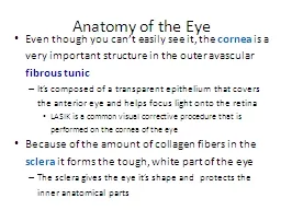
cornea is a very important structure in the outer avascular fibrous tunic Its composed of a transparent epithelium that covers the anterior eye and helps focus light onto the retina LASIK is a common visual corrective procedure that is performed on the cornea of the eye ID: 775211
Download Presentation The PPT/PDF document " Anatomy of the Eye Even though you can..." is the property of its rightful owner. Permission is granted to download and print the materials on this web site for personal, non-commercial use only, and to display it on your personal computer provided you do not modify the materials and that you retain all copyright notices contained in the materials. By downloading content from our website, you accept the terms of this agreement.
Slide1
Anatomy of the Eye
Even though you can’t easily see it, the
cornea
is a very important structure in the outer avascular
fibrous tunic
It’s composed of a transparent epithelium that covers the anterior eye and helps focus light onto the retina
LASIK is a common visual corrective procedure that is performed on the cornea of the eye
Because of the amount of collagen fibers in the
sclera
it forms the tough, white part of the eye
The sclera gives the eye it’s shape and protects the inner anatomical parts
Slide2Anatomy of the Eye
Of the 3 parts of the
middle tunic
the
choroid
forms the major
vascular
portion that lines the internal surface of the sclera
The
ciliary body
consists of two parts:
The ciliary processes that secrete aqueous humor
The ciliary muscle that changes the shape of the lens to adapt to near and far vision
The
iris
is the colored portion of the eyeball consisting of circular and radial smooth muscle fibers
Slide3The inner nervous tunic (retina) lines the posterior 2/3 of the eyeThe retina consist of a layer of melanin pigmented epithelium that allows light to be absorbed rather than scattered. Without the melanin, scattered light in our eye would cause us to always be squinting, even in a moderately lit room
Anatomy of the Eye
Slide4The exact center of the
retina
is called the macula lutea, and in its center is a small depression called the central fovea (or fovea centralis)There are no rods or nerve cells in the fovea, only a high concentration of cones - this gives us the sharp central vision necessary in any activity wheredetail is of primary importance
Anatomy of the Eye
Slide5The retina can be viewed through the pupil using an ophthalmoscope, allowing direct inspection of the retinal vessels for any pathological changes. This is the only place in the body where arterial vessels can be so viewed (without opening the body)
Anatomy of the Eye
Slide6Anatomy of the Eye
The
optic disc
is where the optic nerve and retinal vessels enter and exit the eyeball. Its existence creates a necessary defect on the retina – an area where there are no cones or rods. Bilateral vision, and
saccade (involuntary,
quick) muscle movements
allow our brain to correct
for this “blind spot”, and
most are not even aware
they have one
(try the test on the next page)
Slide7Slide8Anatomy of the Eye
The retina consists of two types of photoreceptor cells, rods and conesRods are abundant in the periphery of the retina whereas cones are found more frequently in the central areas
Slide9Anatomy of the Eye
Each eye contains ≈ 120 million rod-shaped photoreceptors that are adapted for a low light threshold (high sensitivity) - they produce low resolution, black and white imagesa loss of rods with age makes it difficult to drive at night
Slide10Anatomy of the Eye
Cone-shaped photoreceptors
function in bright light to produce high resolution color images
They exists in three varieties,
corresponding to the type of
pigment they contain:
red, green
or
blue
The photopigments are concentrated in
the outer segment of the receptor, while the
inner segment contains the
nucleus and organelles
Slide11Eye Cavities and Chambers
The
lens
is an avascular
refractory
structure situated posterior to the pupil and iris. It consists of a capsule with crystallin proteins arranged in layers, and like the cornea, the lens is
transparent
It attaches to the ciliary muscle
of the ciliary body
by suspensory
ligaments that fine
tune the focusing of
light on the retina
Slide12Eye Cavities and Chambers
The lens
divides the eyeball into two cavities:
An anterior cavity anterior to the lens, and a
posterior cavity
(vitreous chamber) behind the lens
The
anterior cavity
is further
divided at the level of the
iris into anterior and
posterior chambers
(both filled with
aqueous humor)
Slide13Eye Cavities and Chambers
The much larger posterior cavity of the eyeball
(vitreous chamber)
lies between the lens and the retina
Within the vitreous chamber is the
vitreous body,
a transparent jellylike substance that holds the retina flush against the choroid, giving the retina an even surface for the reception of clear images
occasionally, collections of debris called vitreal floaters cast shadows on the retina and create a spot in our field of vision (they are usually harmless and do not require treatment)
Slide14Eye Cavities and Chambers
This cow eye dissection shows an eye bisected into anterior and posterior sections along its coronal axis. The anterior structures of the
iris and pupil are seen in
the bottom half; the
posterior retina,
choroid, and optic
disc are seen in the
top half
Slide15Aqueous Humor
The eye requires a constant bath in a nourishing fluid to deliver enough
O
2
to support the avascular lens and cornea
It also needs fluid to help “inflate” the walls of the eyeball (maintain a constant intraocular pressure –
IOP
) and support the vitreous body
this need is accomplished through the production of
aqueous humor,
which flows through the anterior cavity of the eye and is replaced every 90 minutes
Slide16Aqueous Humor
Aqueous humor
is produced at the
ciliary body
and flows first through the posterior chamber (of the anterior cavity of the eye)
Traveling along the posterior surface of the iris it passes through the pupil to enter the anterior chamber
It proceeds along the anterior surface of the iris until it is reabsorbed into the
scleral venous sinus
(canal of Schlemm) and returned to the venous system
Slide17Aqueous Humor
Any sort of blockage to aqueous humor flow, or overproduction at the ciliary body may result in an
increase of pressure
inside the eye –
a condition called
glaucoma
If not treated,
glaucoma can lead
to a degeneration
of eye function
Slide18Retinal Detachment
The vitreous body (humor) also contributes to maintain proper intraocular pressure
as it holds the retina against the choroid. The vitreous humor, however, is only formed during embryological development and is not replaced. As we age, shrinkage of the vitreous body may lead to a detachment of the retina from the choroid
A
retinal detachment
is considered a medical emergency and needs immediate repair before vision loss becomes permanent
Slide19The pupil is an opening in the center of the iris. It is composed of a radial muscle that “radiates” away from the center, and a circular muscle that is in the center
Contraction of the inner circular muscle fibers cause the pupil to constrict while contraction of the radial fibers cause it to dilate
The Pupillary Response
Slide20Refraction and Image
Normal image formation depends on refraction of light waves, accommodation of the lens, constriction of the pupil, and convergence of the two eyesRefraction is the process of bending light rays. Both the cornea and the lens refract light rays, and both must be functioning in order to properly focus light onto the right spot on the retina to produce clear vision
Slide21Refraction and Image
Since the cornea has a fixed shape, its “focal length” is also fixed; and its ability to refract light is likewise fixedIn order to focus light that has already been bent by the cornea the lens must change shape – the amount depending on the type of light rays we are trying to “see”
Slide22Refraction and Image
An increase in the curvature of the lens for near vision is called
accommodation
The near point of vision is the minimum distance from the eye that an object
can be clearly
focused - about 4 in
(a distance that
increases with age
due to a loss of
elasticity in the lens)
Slide23Refraction and Image
Convergence
is the inward movement of the eyes so that both are directed at the object being viewed - becoming a little cross-eyed when viewing things close up
The nearer the object, the greater the degree of convergence needed to maintain binocular vision
the coordinated action of the extrinsic eye muscles brings about convergence.
Convergence helps us maintain our binocular vision and see in three dimensions
Slide24Refraction and Image
With nearsightedness
(myopia),
only close objects can be seen clearly: Light rays coming in from distant objects are naturally focused in front of the retina and appear blurry
Correction involves the use of a concave
(negative) lens
With farsightedness
(hyperopia),
only distant objects can be seen clearly: Light rays coming in from nearer objects are naturally focused behind the retina
Correction involves the use of a convex (positive) lens
Slide25Abnormal refractive capabilities of the eye are the result of a
misshapen eyeball (usually too long or too short), or because the lens becomes stiff (usually with age). Corrections areaccomplished using either a positive (convex) or negative (concave) lens (eyeglasses, contacts, or lens replacements)
Refraction and Image