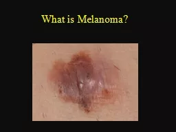
Melanoma Patient Symposium YNHH Smilow Cancer Hospital Sept 11 2014 What is Melanoma Cancer of cells which are responsible for all types of body pigmentation melanocytes Melanocytes are primarily present in skin but are also present in the eye and mucous membranes head sinuses o ID: 776699
Download Presentation The PPT/PDF document " Melanoma – Natural History and Princi..." is the property of its rightful owner. Permission is granted to download and print the materials on this web site for personal, non-commercial use only, and to display it on your personal computer provided you do not modify the materials and that you retain all copyright notices contained in the materials. By downloading content from our website, you accept the terms of this agreement.
Slide1
Melanoma – Natural History and Principles of Treatment
Melanoma Patient Symposium
YNHH –
Smilow
Cancer Hospital
Sept 11, 2014
Slide2What is Melanoma?
Cancer of cells which are responsible for all types of body pigmentation (melanocytes)
Melanocytes are primarily present in skin but are also present in the eye and mucous membranes (head sinuses, oral cavity, rectum/anus, vulva/vagina)
Some types of melanoma are related to sun exposure and sunburns
Malignant cells gain special properties through genetic (mutations) and other cell changes
Uncontrolled growth
Ability to travel in blood and
lymphatics
to other organs
Can implant in other organs and divide and grow (metastases)
Can remain dormant for years before growth is triggered
Dormant state cannot be detected by scans or other tests
Slide3A - asymmetry
B – borders irregular
C – color variation
D - diameter
E- evolution
F – funny looking
Slide4Skin
Ocular
Nasal
Vulvar
Anorectal
Acral-lentiginous
Slide5Primary Tumor SkinMucosaOcular
Local (Lymphatic) DisseminationLocal recurrenceIn-transit metastasesRegional node involvement
Hematogenous Dissemination
SkinLungLymph nodeLiverBoneGI/mesenteryCNS (+ leptomeninges)
?
years
Biology of Dormancy Not Understood
Slide6Slide7Slide8Slide925-30% at presentation of systemic
mets
, 60-70% of all patients subsequently
Slide10Treatment of Primary Melanoma
Excisional
biopsy by dermatologist
Greater than 90% present without distant metastases
Referral to
surgeon
Wide
local excision – margins ≥ 2cm for lesions > 1mm thick
Sentinel Node Biopsy in regional basin
For
lesions > 0.75 thick
For prognostic information – does not affect outcome
In very high risk patients, CT scans or PET scans to rule out distant
metastases
Completion
lymph node dissection (for positive SLNB)
Slide11The Three Important Questions After Complete Resection of Primary Melanoma (and Regional Nodes)
What is my risk that distant metastases will be found in the future?
What can be done to lower the risk that my cancer might recur?
How will I be monitored to detect the cancer if it recurs?
Slide12Staging is Used to Provide Risk for Distant Recurrence (Distant Metastases)
Risk FactorsDepth of primary Ulceration under the microscope Presence of cancer cells in the regional nodes (from the sentinel node biopsy and complete lymph node dissection)
Slide13Prognostic Factors for Patients with Metastatic Disease
Slide14Principles of Monitoring for Recurrence
No (good) data to understand the impact of frequency of type of monitoring on outcome
Both determined by risk
Views on monitoring may change as more effective therapies are introduced for advanced disease
Evaluation by oncologist every 3 months to 1 year
History, exam, blood work (CBC, liver function, LDH)
CT scans and/or PET-CT scans in high risk individuals every 6 to 12 months
Usually stop monitoring at 5-7 years
Dermatology evaluation 2-4x yearly for detection of second primaries (10% risk)
Slide15Options to Reduce Recurrence Risk
Observation
Interferon-
alfa
Different dose and schedules
Administration for up to one year
Increases time to recurrence
Reduces overall risk of recurrence by about 10%
Can induce moderate to severe toxicity in some (fever, chills, fatigue, loss of appetite, depression, difficulty in concentration)
Possible new options
Ipilimumab
(
Yervoy
) – not yet approved, data so far similar to interferon, potential for severe toxicity
Clinical Trials – compare potential better agents to standard of care
Slide16Management of Advanced Disease
Treat both the lesions seen on scans
and areas of disease that have not yet appeared on scans
Surgery, local injection, or radiation not sufficient to eliminate the disease
Requires systemic (intravenous or oral) medications
Use systemic therapies first that can induce long term remissions
Control pain and manage lesions early that may cause early morbidity (pain, bleeding, limitation of function, unacceptable cosmetic appearance)
Screen the brain at baseline and every 8-12 weeks
CT scans of chest/
abd
/pelvis or CT chest + MRI abdomen/pelvis to ‘stage’ disease
Repeat scans every 6-12 weeks (depends of treatment)
Slide17Options for systemic therapies
Clinical Trials
Immune therapies (can give long term remissions)
High dose interleukin-2 (
Proleukin
)
Ipilimumab
(anti-CTLA-4) (
Yervoy
)
Pembrolizumab
(anti-PD-1) (
Keytruda
)
Nivolumab
(anti-PD-1)
(
Optiva
) – approval pending
Targeted therapies (rapid response in most but few have long term control)
BRAF mutation–
dabrafenib
(
dafinlar
)/
trametinib
(
mekinist
),
vemurafenib
(
zelboraf
)
NRAS mutation – investigation MEK + CDK4 inhibitors
C-kit (mucosal and
acral-lentiginous
melanomas) –
imatinib
,
dasatinib
,
sorafenib
, others
Cytotoxic Chemotherapy (can work rapidly but only in a few and rarely achieve long term control)
Temozolomide
(
temodar
) or
dacarbazine
Carboplatin and paclitaxel
Biochemotherapy
Targeted therapies
Immune therapies
Chemotherapy
Biochemotherapy
mBRAF
m
NRAS
mCKIT
Vemurafenib
Dabrafenib
Trametinib
(
MEKi
)
Dabrafenib
+
Trametinib
Vemurafenib
+
cometinib
CDK4i +
MEKi
cKITi
Interleukin-2
Ipilimumab
(anti-CTLA4)
Anti-PD1
(nivolumab)(pembrolizumab)
Nivolumab
+
ipilimumab
2014 – Treatment Options for Metastatic Melanoma
Slide19Eligible for Immunotherapy
Yes
No
Anti-PD1
Anti-CTLA-4
High dose IL-2
Adoptive ImmunotherapyOther Investigational Immunotherapy Trials
BRAF mutation
Yes
BRAFi
+
MEKi
BRAFi + otherERKi
NO mutation
Phase 1
Other targeted Rx
Anti-angiogenesisSupportive careChemotherapy
PD
Brain mets
GKS/SRS
yes
no
C-kit mutation
Ckit
inhibitor
Yale Cancer Center Melanoma
Treatment Algorithm
Mutation
analyses
NRAS
mutation
CDK4i +
M
EKi
Slide206-24-05
11-23-05
Il-2 Induced Regression of Melanoma Liver Metastases
Persistent/progressing disease in spleen, SQ buttock, and lung removed; NED x 7 years
Slide21Response to Ipilimumab 10 mg/kg x 2 doses
2 baseline brain
mets
regressed also:
No disease progression 5+ years
Slide22Metastatic Melanoma,
Anti-PD1 1 mg/kg every other week
Slide23Slide24Slide25Slide26Response to
ipi
/anti-PD1, 3/1 dose level
Slide27Response to
ipi
/anti-PD1, 3/1 dose level
Slide28Response to
ipi
/anti-PD1, 3/1 dose level
Slide29Cohort 8 response at 12 weeks
Slide30Presented by:
Overall Survival for Concurrent Therapy by Dose Cohort
Censored
14
17
16
653
131716652
111615648
101515646
81515644
71413640
7144631
7132628
790319
740011
53008
23005
23005
22004
10001
10001
00000
Pts at RiskNivo 0.3_IPI 3Nivo 1 _IPI 3Nivo 3_IPI 1Nivo 3_IPI 3Concurrent
100
90
80
70
60
50
40
30
20
10
0
Survival (%)
Months
2 Yr OS 88%
0
3
6
9
12
15
18
21
24
27
30
33
36
39
42
45
48
2 Yr OS 50%
2 Yr OS 79%
Concurrent Cohorts 1-3 (n=53)
Nivo 0.3 mg/kg + IPI 3 mg/kg (n=14)
Nivo 1 mg/kg + IPI 3 mg/kg (n=17)
Nivo 3 mg/kg + IPI 1 mg/kg (n=16)
Nivo 3 mg/kg + IPI 3 mg/kg (n=6)
1 Yr OS 94%
1 Yr OS 85%
1 Yr OS 57%