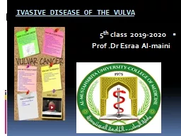
/
British Gynaecological Cancer Society BGCS Vulval Cancer British Gynaecological Cancer Society BGCS Vulval Cancer

British Gynaecological Cancer Society BGCS Vulval Cancer - PDF document

patricia
patricia . @patricia
Follow
342 views
Uploaded On 2022-08-23

British Gynaecological Cancer Society BGCS Vulval Cancer - PPT Presentation

Guidelines Recommendations for Practice 15 May 2020 Jo Morrison Peter Baldwin Lynn Buckley Lucy Cogswell Katharine Edey Asma Faruqi Raji Ganesan Marcia Hall Kathryn Hillaby Nick Reed Phil ID: 940573

disease cancer treatment vulval cancer disease vulval treatment node patients carcinoma vulvar grade cell surgery lymph squamous risk vulva

Share:

Link:

Embed:

Download Presentation from below link

Download Pdf The PPT/PDF document "British Gynaecological Cancer Society BG..." is the property of its rightful owner. Permission is granted to download and print the materials on this web site for personal, non-commercial use only, and to display it on your personal computer provided you do not modify the materials and that you retain all copyright notices contained in the materials. By downloading content from our website, you accept the terms of this agreement.


Presentation Transcript

British Gynaecological Cancer Society (BGCS) Vulval Cancer Guidelines: Recommendations for Practice 15 May 2020 Jo Morrison, Peter Baldwin, Lynn Buckley, Lucy Cogswell , Katharine Edey, Asma Faruqi, Raji Ganesan, Marcia Hall, Kathryn Hillaby, Nick Reed, Phil Rolland , Christina Fotopoulou. The purpose of this guideline is to collate evidence and propose evidence - based guidelines for the diagnosis and management of adult patients with vulva carcinoma treated in the UK. Malignant melanoma may present via similar routes and will be discussed. The reader is referred to the Ano - uro - genital Mucosal Melanoma Full Guideline (1) for more detailed recommendations. The management of vulval sa rc oma is outside of the scope of this guideline. Reviewers: Shahram Abdi, Jonathan Frost, Cathrine Holland, Sean Kehoe, Tracie Miles, Andy Nordin, Nithya Ratnavelu, Partha Sengupta, John Shepherd, Sudha Sundar. International Reviewers : Amanda Tristram; Linn Woelber . Table of Contents 1 INTRODUCTION ................................ ................................ ................................ .......................... 4 1.1 G RADES OF RECOMMENDATIONS ................................ ................................ ............................. 4 1.2 G UIDELINES DEVELOPMENT PROCESS ................................ ................................ ...................... 4 2 BACKGROUND AND EPIDEMIOLOGY ................................ ................................ ...................... 6 3 PREVENTION, SCREENING, PRESENTATION AND DIAGNOSIS ................................ ........... 6 3.1 P REVENTION AND TREATMENT OF PRE - DISPOSING CONDITIONS ................................ ................. 6 3.1.1 HPV - related squamous cell carcinoma ................................ ................................ ............ 7 3.1.2 Squamous cell carcinoma on a background of lichen sclerosus (LS)/lichen planus (LP) 8 3.1.3 Mucosal malignant melanoma ................................ ................................ ......................... 8 3.2 S CREENING ................................ ................................ ................................ ........................... 8 3.3 P RESENTATION ................................ ................................ ................................ .................... 10 3.4 D IAGNOSIS ................................ ................................ ................................ .......................... 11 3.5 P RE - OPERATIVE INVESTIGATIONS ................................ ................................ .......................... 14 3.5.1 Squamous Cell Carcinoma (SCC) ................................ ................................ ................. 14 3.5.2 Melanoma ................................ ................................ ................................ ...................... 14 3.5.3 Basal Cell Carcinoma ................................ ........................

........ ................................ .... 14 3.5.4 BarPholin’s Gland Carcinoma ................................ ................................ ......................... 14 3.5.5 PageP’s disease of Phe vulva ................................ ................................ .......................... 14 4 PATHOLOGY ................................ ................................ ................................ ............................. 15 4.1 P RECURSOR LESIONS ................................ ................................ ................................ ........... 15 4.1.1 Usual/classical VIN (HPV - associated neoplasia) ................................ ........................... 15 4.1.2 Differentiated VIN ................................ ................................ ................................ .......... 15 4.1.3 Other putative precursors ................................ ................................ .............................. 16 4. 2 P ATHOLOGY OF SQUAMOUS CELL CARCINOMA ................................ ................................ ........ 16 4.2.1 Types of squamous cell carcinomas ................................ ................................ .............. 16 4.2.2 Macroscopic features of importance ................................ ................................ .............. 16 4.2.3 Microscopic features of importance ................................ ................................ ............... 16 4.3 S PREAD ................................ ................................ ................................ .............................. 17 4.3.1 Lymph node metas tasis ................................ ................................ ................................ . 17 4.3.2 Sentinel lymph nodes (SLN) ................................ ................................ .......................... 17 4.3.3 Extranodal extension ................................ ................................ ................................ ..... 18 4.4 A N CILLARY STUDIES ................................ ................................ ................................ ............. 18 4.4.1 Ancillary studies in uVIN ................................ ................................ ................................ 18 4.4.2 Ancillary studies in dVIN ................................ ................................ ................................ 18 4. 5 P ATHOLOGY OF V ULVAL P AGET ’ S DISEASE AND I NVASIVE ADENOCARCINOMA OF THE VULVA ...... 18 4.6 P ATHOLOGY OF VULVAL MELANOMA ................................ ................................ ....................... 19 5 TREATMENT OF PRIMARY DISEASE ................................ ................................ ..................... 20 5.1 S URGERY ................................ ................................ ................................ ............................ 20 5.1.1 Management of primary site ...........................

..... ................................ .......................... 20 5.1.2 Surgical management of other vulval cancers ................................ ............................... 24 5.1.3 Management of inguinal lymph nodes ................................ ................................ ........... 28 5.1.4 Reconstructive surgery ................................ ................................ ................................ .. 32 5.1.5 Vacuum - assisted Closure (VAC) ................................ ................................ ................... 34 5.2 R ADIOTHERAPY ................................ ................................ ................................ .................... 35 5.2.1 Adjuvant radiation / chemoradiation therapy ................................ ................................ .. 35 5.2.2 Primary site irradiation - primary treatment ................................ ................................ ...... 36 5.2.3 Palliative radiotherapy ................................ ................................ ................................ ... 37 5.3 C HEMOTHERAPY ................................ ................................ ................................ .................. 37 5.3.1 Squamous Cell Carcinoma ................................ ................................ ............................ 37 5.3.2 Neoadjuvant chemotherapy for invasive squamous cell carcinoma .............................. 37 5.3.3 Adjuvant chemotherapy ................................ ................................ ................................ . 38 6 TREATMENT OF RECURRENT DISEASE ................................ ................................ ............... 40 6.1 R ECURRENCE RATES AND SURVIVAL ................................ ................................ ...................... 40 6.2 L OCALISED RECURRENCE ................................ ................................ ................................ ..... 40 6.2.1 Surgery ................................ ................................ ................................ .......................... 40 6.2.2 Radiotherapy ................................ ................................ ................................ ................. 41 6.3 P ALLIATIVE CHEMOTHERAPY ................................ ................................ ................................ . 41 6.4 L OCAL NODAL RECURRENCE ................................ ................................ ................................ . 42 7 PSYCHOLOGICAL/PSYCHOSEXUAL SUPPORT ................................ ................................ ... 43 7.1 P SYCHOSOCIAL ................................ ................................ ................................ .................... 43 7.2 L YMPHOEDEMA ................................ ................................ ................................ .................... 43 7.3 S EXUALITY /S EXUAL M ORBIDITY ................................ ...

............................. ............................ 44 7.4 B OWEL / BLADDER FUNCTION ................................ ................................ ................................ .. 44 8 FOLLOW UP ................................ ................................ ................................ .............................. 45 8.1 F OLLOW UP OF VSCC ................................ ................................ ................................ .......... 45 8.2 F OLLOW UP OF B ASAL CELL CARCINOMA OF THE VULVA ................................ ........................... 45 8.3 F OLLOW UP OF V ULVAL P AGET ’ S D ISEASE ................................ ................................ ............. 45 8.4 F OLLOW UP OF V ULVAL M ALIGNANT M ELANOMA . ................................ ................................ .... 46 9 REFERENCES ................................ ................................ ................................ ........................... 46 1 Introduction 1.1 Grades of recommendations Recommendations are graded as per the Royal College of Obstetricians and Gynaecologists document. Clinical Governance Advice No. 1: Guidance for the Development of RCOG Green - top Guidelines, available on the RCOG website at: https://www.rcog.org.uk/en/guidelines - research - services/guidelines/clinical - governance - advice - 1a/ . Evidence was searched in the Cochrane Central Register of Controlled Trials (CENTRAL, The Cochrane Library 2014, Issue 12), MEDLINE and EMBASE up to April 2018, registers of clinical trials, abstracts of scientific meetings, reference lists of included studies and contacted experts in the field. 1.2 Guidelines development process The guideline development process is detailed below: • Chair, officers, council and guidelines committee (GC) nominated a lead for each guideline topic; • Lead then identified a team called the guideline team (GT) to develop the 1 st draft; • 1 st draft was submitted to the GC; • GC approved draft and recommended changes; • Changes were accepted by the GT who produced the guidelines; • 2 nd draft was then submitted to council members and officers; • Council and officers approved 2 nd draft and recommended changes; • Changes were then accepted by GC and GT; • 3 rd draft was sent to national and international peer review; • GC and GT then made changes based o n peer review comments; • 4 th draft was sent back to council for approval; • 4 th draft was sent to BGCS members for feedback; • GC and GT Phen made changes Nased on memNers’ feedNack; • 5 th draft was sent to public consultation including patient support groups; • GC and GT then made changes based on non - memNers’ feedNack; • Final draft approved by council and officers. 2 Background and epidemiology V ulval cancer is a rare disease with 1,300 new cases registered in the UK during 201 5 , representing less than 1% of all n

ew cancer cases registered that year. In the UK it is the 20 th most common female cancer and 4 th most common gynaecological cancer , with a crude incidence rate of 3.9/100 000 . (2) Incidence in the UK is highest in females over 90 years of age . The Incidence of vulval cancer has increased by 18% since the early 1990s, mainly due to an increase in incidence of over 50% in those under 60 years. (3) While most vulval cancer is still diagnosed in women aged over 70 years, age standardized rates have increased by 92% within the 50 - 59 cohort between 1993 - 1995 and 2012 - 2014. This increase in incidence in younger cohorts is most lik e l y due to an increase in human papilloma virus ( HPV ) - related VIN within those groups . (3) However, Dutch Registry data demonstrate an almost two - fold rise in incidence of lichen scleros u s between 1991 and 2011 , so the rise may not be solely HPV - related. (4) Approximately 90% of vulval cancers are squamous cell carcinomas, with the main risk factors for disease being infection with high risk HPV and inflammation due to vulval dermatoses, such as lichen sclerosus and lichen planus. The remaining 10% ar e made up of primary vulval melanoma, basal cell carcinoma, BarPholin’s gland carcinoma, adenocarcinoma, an d rarely, sarcoma. In 2014 there were 450 vulval cancer - related deaths in the UK, representing less than 1% of all cancer - related deaths in females that year. The mortality rate increases with age with the majority of deaths occurring in women over 70 years of age. However, m ortality rates in this group have reduced by 30% since the early 1990s . (5) The increased incidence of squamous cell vulval cancer is mirrored by data from Germany and Australia, where rates have nearly doubled in the past decade . (6, 7) It is likely to be decades before the effects of HPV vaccination on reducing vulval cancer are known ; nevertheless, it is antici pated that a decrease will occur, as HPV16 is the most common viral subtype associated with vulval cancer. Unfortunately, this is likely to be less dramatic than for other HPV - related malignancies, as vulval dermatoses account for a large proportion of vul val cancers. 3 Prevention, s creening, presentation and diagnosis 3.1 Prevention and treatment of pre - disposing conditions For a summary of recommendations on prevention, screening, please see Table 1 . The most common type of vulval cancer is squamous cell carcinoma (VSCC). This may be HPV - independent, developing on a background of vulval dermatoses (lichen sclerosus and lichen planus) and differentiated vulval intraepithelial neoplasia (dVIN) or it may HPV - depend e nt with a background of usual type vulval intraepithelial neoplasia (uVIN). The combination of the two may increase the risk as the risk of developing VSCC in women with VIN and L S was 19% in one Dutch cohort study over 10 years. (4) 3.1.1 HPV - related squamous cell carcinoma HPV Vaccination The majority of HPV - related V SCC is caused by HPV16 .

(7) HPV vaccination will likely provide significant protection to those vaccinated prior to HPV exposure. However, since the natural history of developing VIN and vulval carcinoma is often decades from original exposure, the effect s of HPV vaccination progra mmes are likely to take many years to become apparent. HPV vaccination has already had a significant effect on rates of genital warts and cervical intraepithelial neoplasia in vaccinated populations . (8) The time to development from exposure is much lower for benign warts than for u sual type vu lval intraepithelial neoplasia (u VIN ), the pre - malignant lesion for HPV - related V SCC , so these benefits will take longer to realise . Prophylactic vaccination against HPV6, 11, 16 and 18 has been shown to result in a substantial decrease in the development of preinvasive vulval lesions and it is anticipated that the relative proportions of HPV - and non - HPV - associated malignancy may alter as vaccine coverage increases and with the use of vaccines protecting against additional HPV types. (9) Studies are ongoing to determine whether HPV - vaccination following diagnosis of uVIN can reduce the risk of recurrence or development of SCC . ( 7) S tudies looking at the effect of HPV vaccination on developme nt of CIN in those already exposed to HPV did not suggest a significant benefit overall in the incidence of CIN2+ . (10) In contrast , retrospective subgroup analysis of a randomised control trial (RCT) of HPV vaccination demonstrated a 46.2% reduction in incidence of further HPV - related disease (95% confidence i nterval 22.5% to 63.2% ) in those vaccinated prior to initial treatment for HPV - related disease, compared to the unvaccinated cohort. (11) Other studies suggest that HPV vaccination after treatment for CIN may reduce the risk of recurrence and other HPV - related disease (12) and RCTs to look at this specifically are on - going. Treatment of uVIN A systematic review of the natural history of VIN3 found 97 articles including a total of 3 , 322 women . T here was an occult cancer rate of 3.2% in those with suspected VIN3 and 3.3% went on to develop VSCC during follow up. (13) Of 88 women with untreated VIN3, 9% went on to develop VSCC o ver 12 to 96 months. However, they concluded that the progression rate to VSCC is likely to be over - estimated. A Cochrane review of intervention for treatment of uVIN examined effects of imiquimod, cido fo vir, indole - 3 carbinol and surger y. (14) They found that topical imiquimod, an immune modulator was more effective than placebo in achieving a response (complete or partial) to treatment 5 - 6 months after randomisation (risk ratio (RR) 11.95, 95% confidence interval (CI) 3.21 to 44.51; high - certain ty evidence). A complete response occurred in 58% of women in the imiquimod groups and none in the placebo groups (RR 1 4.40, 95% CI 2.97 to 69.80). Persistent responses after 12 months were present in just over a third of women. Only one study reported vulval cancer rates at

12 months follow up (1/24 and 2/23 in imiquimod and placebo groups, respectively). Adverse events were more common with imiquimod than placebo (RR 7.77, 95% CI 1.61 to 37.36; high - certain ty evidence). One very small, long - term follow - up study of those with complete response to imiquimod demonstrated very low recurrence rates of uVIN. (15) Complete response rates after 6 months were similar for a 16 - week course of imiquimod and cidofovir (imiquimod 45% and cido fo vir 46%; RR 1.00, 95% CI 0.73 to 1.37; moderate - certain ty evidence). A follow up study found that responses for complete responders were maintained after 18 months, especially in the cido fo vir group (94% for cido fo vir (95% CI 78.2 - 98.5) versus 71.6% for imiquimod (95% CI 52.0 - 84.3)). (16) Side effects, mainly h eadache, fatigue and discontinuation due to pain, were slightly more common with imiquimod than cidofovir . Cidofovir cream is curr ently not licenced for use in uVIN. The same Cochrane review look ed at evidence for surgical treatment of uVIN and found low - quality evidence from the better studies where data were adjusted for confounders. (14) There was little or no difference in the risk of VIN recurrence between surgical excision and laser vaporisation ( 51% (37/70) of women overall, at a median of 14 months ) . Recur r ence was , unsurprisingly , more common in those with multifocal uVIN (66% versus 34%) . There was only very low - certain ty evidence for other treatments such as photodynamic th er apy, Cavitron ultrasonic surgical aspiration and loop electrosurgical excision. In the small surgical studies included in the Cochr ane review, v ulval cancer occurred in 11 women (15.1%) overall at a median of 71.5 months (9 to 259 months) . They concluded that , if cancer is suspected, despite a biopsy showing uVIN only , ‘surgical excision remains Phe PreaPmenP of choice’ . However, i f a n occult cancer was not suspected , treatment of uVIN can be individualised , tak ing into account wom e n's preferences and the site and extent of disease, using a combination approach to optimise outcomes , which can include conservative treatment and close fo llow up with vulvoscopy in selected patients . It should be emphasised that the volume of data in this area, as with much of the vulval field, is limited . 3.1.2 Squamous cell carcinoma on a background of lichen sclerosus (LS) /lichen planus (LP) Lichen sclerosus is associated with an increased lifetime risk of developing vulval cancer, with estimates of the risk varying between 2.6 - 6.6%. (4, 17) However, these data are not based on populat ion - level data and lichen sclerosus is under diagnosed, so these data are likely to be at high risk of bias and are likely to over - estimate the risk. D ata from non - randomised studies suggest that good control of LS/LP with ultra - potent topical steroids (su ch as Clobetasol 17 - propionate 0.05%) may reduce the risk of progression to SCC. (17 - 19) There are yet no RCT level dat

a to support this, although control of active LS/LP should be recommended to improve symptoms , reduce scarring and may reduce the risk of developing malignancy B OfPen women are fearful of using ‘Poo much’ sPeroid cream and Phey should be reassured that appropriate usage (less than 30 g tube of ultra - potent steroid ointment/cream, such as Dermovate ( clobetasol propionate 0.05% ) , over a 3 - month period) is unlikel y to be harmful and may be of benefit , both to scarring/vulval appearance as well as longer term risk of cancer . For the same reasons, women should be advised to avoid irritants that can exacerbate LS/LP, particularly detergents, such as soap. 3.1.3 Mucosal mal ignant melanoma Unlike cutaneous melanomas, vulval mucosal melanomas are not related to ultraviolet light exposure. A small minority may be related to c - kit mutations , which are more common than in cutaneous melanoma . (20) 3.2 Screening There are currently no proven screening test s for vulval carcinoma. Thos e with known VIN and lichen scle rosus/lichen planus are at higher risk. R ates of progression to vulval carcinoma were 5.7 % in a 14 - year series for u VIN (21) and a 2.6 - 6.6% overal l risk for those with lichen sclerosus/lichen planus (17, 22) , increased significantly when associated u VIN. (4) However, differentiated VIN (dVIN) , which arises on a background of lichen sclerosus, has a very high risk of progression to cancer compared to uVIN and should ideally be surgically excised (relative risk (RR) of progression: dVIN RR= 38. 5 (9. 8 - 150.8) ; uVIN = 0.06 5 (0.0 3 - 0.15) . (23, 24) Current guidelines from the British Association of Dermatologists recommend annual review in primary care in those with lichen sclerosus , following review at 3 - months to check response to initial treatment and a 6 - month follow up to check compliance and understanding of self - management . (25) I mportantly, patients should be aware of the small risk of developing vulval cancer and report new lesions to their GP, especially if these symptomatic . Re cent data suggest that the risk of vulval cancer in the presence of a lesion is around 13 % and presence of a suspicious vulval lesion should prompP rapid ‘Cancer W ait T ime’ referral to secondary care . (26, 27) Women with u VIN should receive follow up with formal vulvoscopy. These women are at increased risk of multi - focal disease , so it is important to ensure that they have appropriate follow up with time ly cervical screening. Patients are also at risk of anal/ peri - anal intraepithelial neoplasia and those affected may need a multi - disciplinary approach to follow up and management. Those with multi - focal disease should be followed up in colposcopy by some one experienced in treatment for CIN and u VIN , although the NHS Cervical Screening Programme guidelines do not specify a frequency for follow up . (28) The risk of recurrent disease is high, particularly in the first two years. (29) Follow - up reg

imens should reflect this fact, with increased surveillance in the first two years, particularly for those with multifocal disease. Recommended minimum follow - up intervals after successful treatment would be six - monthly in the first tw o years and annual follow - up to 5 years. Patients with unifocal, treated disease may be discharged at that time, with instructions to return if new lesions or symptoms develop. Patients with multifocal or recurrent disease may require more long - term follow - up . HIV testing should be considered as per the 2008 recommendations from the British HIV Association, British Association of Sexual Health and HIV British Infection Society ( https://www .bashhguidelines.org/media/1067/1838.pdf ). The effectiveness of anal screening in this population has not been proven and most data on anoscopy and anal cytology is limited to higher risk populations (Human Immunodeficiency Virus - positive (HIV+) men who h ave sex with men (MSM)), reviewed in (30) . An expert review group of American Society Colposcopists and Cervical Pathologists and the International Anal Neoplasia Society examined the data and made recommendations on anal H PV infection, anal intraepithelial neoplasia (AIN) and anal cancer in women. They did not find data to support routine anal cytology or anoscopy in women with uVIN or vulval cancer, although noting that they were at higher risk than the general population. (31) They recommended s creening for anal cancer with digital ano - rectal examination and assessment if anal cancer symptoms developed, such as pain or bleeding. They noted that routine screening and treatment of AIN2/3 was not proven to be effective in reducing anal cancer in this population . Recommendation Grade of recommendation HPV vaccination is likely to reduce the incidence of uVIN and HPV - related vulval SCC in the future. Grade C Imiquimod, Cidofovir and surgical excision are treatment options for high grade ✓HN all wiPh ~D0% responseCrecurrence raPes Ny 12 monPhs’ posP - tre atment. However, cidofovir is currently unlicensed for use in uVIN. Grade A Good control of lichen planus and lichen sclerosus with ultra - potent topical steroids improves symptom and may reduce the incidence of developing SCC. Grade C There is currently no proven screening test to prevent vulval cancer. Grade D Recommendation Grade of recommendation Women with multi - focal HPV related disease should be followed up colposcopy of the lower genital tract and digital ano - rectal examination with prompt referral should symptoms of anal cancer develop. Grade D Women with multi - focal HPV - related disease should be offered HIV testing. Grade D Women with high grade uVIN should be followed up with careful clinical inspection +/ - vulvoscopy. Grade D Women with uncomplicated lichen sclerosus or lichen planus can be followed up in primary care and once quiescent and confident of self - management, 12 - monthly review is suggested. Grade C Women with lichen sclerosus who develop new lesions should be referred to sec

ondary care, via a Cancer Wait Pathway, if these do not respond to nightly ultra - high potency steroids within 1 - 2 weeks. Grade C Table 1 . Recommendatio ns for prevention and screening 3.3 Presentation For recommendations on presentation and diagnosis, see Figure 1 and Table 2 . Most vulval carcinomas will present with a specific lesion. The risk of cancer , in Phe presence of a ‘suspicious vulval lesion’ , was 12.8 % in a recenP sPudy of women referred Po a secondary care ‘rapid access clinic’ with vulval symptoms . (26) This risk of invasion was higher , if the lesion was symptomatic (pain and/or bleeding) . Women with generalized vulval irritation without a visible lesion on careful examination, were extremely unlikely to have a cancer diagnosis. Women with clinical features highly suspicious of vulval cancer, for example a fungating lesion +/ - palpable groin nodes, should be referred to a cancer centre without the need to await biopsy results. Punch biopsie s may not adequately sample the lesion, especially if it is large and/or deep, and delay for diagnostic biopsy is not warranted. Vulval melanoma is rare, presents as a vulval lesion, which may or not be pigmented and may or may not develop in the backgrou nd of melanocytic dysplasia. Symptoms may include altered vulvo - vaginal pigmentation, itching or bleeding. Alternatively, an asymptomatic lesion is noted, which may occur as an irregularly outlined pigmented or non - pigmented macule, papule, patch or nodul e with or without ulceration. Some lesions will be found on clinical examination after noticing groin lymph node(s) enlargement. Basal cell carcinoma of the vulva ten d s to present with a discrete vulval lesion or classical raised, rolled - edge ulcer, wi thout a background dermatosis or evidence of uVIN. BarPholin’s gland carcinoma is rare and may presenP wiPh a mass in Phe vulvaClower vagina over Phe area of Phe BarPholin’ s gland. These lesions are often painful and may be mis - diagnosed as a BarPholin’s cyst or abscess. The diagnosis should be suspected and excluded in those aged over 40 years presenPing wiPh a ‘BarPholin’s aNscess’, since inadverPenP BarPholin ’ s gland ‘excision’ or marsupilisation can delay diagnosis and/or make further surgical treatm ent more challenging. 3.4 Diagnosis Incisional biopsy (punch or wedge biopsy) ideally including the edge of a lesion , where there is a transition from normal to abnormal tissues. Biopsies should avoid a central ulcer , since this may not be diagnostic . Biopsies should be of adequate depth to allow differentiation between superficially invasive and those with invasion � 1 mm, since this will inform subsequent management. Excision biopsy should be avo ided , where possible, since this can limit options for more conservative treatment with wide local excision and sentinel node biopsy. This is especially the case , if the lesion is small , as the vulva can heal well and the original site be hard to determine at the time of

more definitive treatment . However, there may be exceptions to this, for instance in someone who is very elderly or frail it may be acceptable to excise a small, symptomatic lesion under local anaesthetic for palliation and planning of subsequent treatment. T his should ideally b e performed by the gynaecological oncologist who will perform subsequent treatment . H istological confirmation is required prior to consideration and planning more radical treatment. At a minimum, a detailed diagram of the vulva, indicating each biopsy sit e, should be drawn. Use of a schematic diagram, which can be annotated is encouraged (e.g. https://www.nva.org/what - is - vulvodynia/vulvar - anatomy/ ). Hdeally, vulvoscopic ‘Nefore and afP er’ Niopsy phoPos should Ne Paken (with a scale indication) . This will help to localize the lesion for the treating gynaecological oncologist an d a ssist pre - planning of more definitive treatment. GMC and local guidance on the capture and storage of imaged should be followed. If more than one lesion is present, each individual biopsy should be sampled separately, sent in separate pots and carefully labelled , so that lesion site can be identified at a later date. Recommendation Grade of recommendation Women with suspicious vulval lesions should be referred to a rapid access clinic for urgent assessment, as per NICE guidelines. (169) Grade C Women highly likely to have vulval cancer on clinical grounds should be referred to a gynaecological cancer centre without waiting for biopsy results. Gra de D Clear documentation of clinical exam size of lesion, distance to the midline/clitoris/anus/vagina/urethra and palpation of lymph nodes is mandatory. Imaging, with indication of biopsy sites and/or clinical drawing is essential for further treatment plannin g. Grade D Suspicious vulval lesions should ideally be sampled with a punch or wedge biopsy and excisional biopsy avoided until a diagnosis is made. Grade D Biopsies should include the edge of a lesion to ascertain the background condition. Grade D At a minimum, a detailed diagram, indicating lesion and biopsy sites, should be drawn. Grade D Ideally, clinical photographs, before and after biopsy should be taken, with an indication of scale. Grade D Biopsies from separate lesions should be sent i n separate pots and clearly labelled. Grade D All cases vulval cancer should have diagnosis confirmed by a specialist multi - disciplinary team (MDT) prior to planning radical treatment. Grade D Table 2 . Recommendations for prese ntation and diagnosis Figure 1 . Flowchart demonstrating investigation of suspicious vulval lesion. LSA= Lichen Sclerosus Atrophicus; LP = Lichen Planus; VIN = vulval intraepithelial neoplasia; uVIN = usual - type VIN; dVIN = differentiated VIN; D/C = discharge 3.5 Pre - operative investigations For recommendation on pre - operative investigations, see Table 3 . 3.5.1 S quamous Cell Carcinoma (SCC) Squamous cell carcinoma most commonly spreads via inguinal lymph nodes and rarel

y presents at distant sites , if regional nodes are negative. Imaging is poor at excluding microscopic g r oin node metastases, hence groin node surgery is recommended for those with greater than FIGO Stage Ia SCC. Prior to sentinel lymph node (SLN) biopsy, clinical examination and imaging of the groins are required to identify metasta ti c disease, since obvious groin node involvement would be a contraindication to SLN biopsy . The groins can be assessed with ultrasound, but cross - sectional imaging (with computerised tomography of chest, abdomen and pelvis (CT CAP) , or magnetic resonance i maging (MRI) ) , will provide additional information on the presence of pelvic lymphadenopathy and distant disease and should be employed prior to lymphadenectomy. If radical surgery is proposed , there may be a role for positron emission tomography CT (PET - C T). Those with suspicious groin nodes on clinical examination and/or imaging may be further investigated with USS - guided fine needle aspiration (FNA) or core biopsy , where node positivity would change management. Evaluation of the pelvic nodes with cross sectional imaging (MRI or CT) is recommended before undertaking lymphadenectomy. S taging with full body, cross - sectional imaging (CT CAP) should be considered for all those diagnosed with more than minimally invasive disease, as the presence of distant metastatic disease will influence the extent of loco - regional treatment options. MRI (pelvis) can be used for further assessment of the loco - regional disease, if loca lly extensive . CT is also suggested for those who are not fit for radical treatment , to aid discussion and planning of treatment options. (32) 3.5.2 Melanoma Vulval melanoma commonly presents with a more locally advanced lesion than cutaneous melanoma, since the area is difficult to visualise . The risk of metastatic disease (both lymphatic and haematogenous spread) is high. Recommended imaging at diagnosis would include CT CAP and also CT or MRI head, since systemic disease and intra - cranial lesions are not uncommon. Please see the Ano - uro - genital Mucosal Melanoma Full Guideline for further details . (1) 3.5.3 Basal Cell Carcinoma Distant disease spread is rare and no specific imaging is required , unless there is clinical suspicion of nodal disease. 3.5.4 Bartholin’s Gland Carcinoma BarPholin’s gland carcinoma may presenP wiPh more advanced disease, since Phey arise deep Po Phe surface of the skin and are less clinically obvious. Pre - operative imaging with CT CAP is therefore recommended, since these lesions are not suitable for a SL N approach and there is an increased r isk of locoregional spread at di agnosis. MRI pelvis may help to delineate the local degree of involvement. 3.5.5 Paget’s disease of the vulva See section 5.1.2 for discussion of managemenP of PageP’s disease of Phe vulva, including pre - operative i nvestigation s . Recommendation Grade of recommendation Gross nodal involvement should be excluded by clinical examin

ation and appropriate imaging / radiologic staging. Grade D If sentinel lymph node biopsy is considered, imaging of the groins either by ultrasound, CT, or MRI is suggested to identify potenti al lymph node metastases. Grade D FNA or core biopsy can be used to evaluate suspicious nodes when this would alter primary treatment, although removal of involved lymph nodes should be considered standard of care. Grade D Further staging with CT/PET - CT is recommended in the presence of proven metastatic disease (i.e. positive lymph nodes) and/or in advanced disease prior to radical treatment/surgery . Grade D No additional imaging is required in the preop assessment of BCC lesions, unless there is a clinical suspicion of nodal disease . Grade D Melanoma and BarPholin’s cancers should Ne assessed wiPh comNinaPion imaging (MRI and CT) to provide information on the extent of local disease and metastatic disease. PET - CT may b e appropriate in selected cases. Grade D Table 3 . Recommendations for pre - operative investigations 4 Pathology For a summary of pathological subtypes, please see Table 4 . 4.1 Precursor lesions 4.1.1 Usual/classical VIN (HPV - associated neoplasia) The nomenclature of precursor lesions in HPV - related neoplasia is varied and includes the terms usual type VIN and classical VIN. The terminology recommended by the World Health Organisation (WHO) and ISSVD refers to LSIL (low - grade squamous intraepithelial lesion that includes HPV - related changes and VIN1) and HSIL (high - grade squamous intraepithelial lesion, which encompasses VIN2, VIN3). (33) The terms LSIL and HSIL are not widely used in the UK and the use of the alternative terms low - and high - grade VIN can be used with sub - categorisation as VIN2 or VIN3. This lesion is characterized by cytological aty pia , mitoses extending beyond middle thirds of the epithelium and lack of maturity of the squamous cells with or without associated stigmata of HPV infection such as koilocytosis. 4.1.2 Differentiated VIN This is an HPV independent lesion that is often seen in older women on a background of lichen sclerosus. It is characterised by basal cell atypia and abnormal keratinocyte differentiation. Differentiated VIN is typically associated with p53 mutations. 4.1.3 Other putative precursors Vulvar acanthosis with altered dif ferentiation (VAAD) is a non - invasive squamous proliferation that maybe a precursor to verrucous carcinoma. (34) 4.2 Pathology of squamous cell carcinoma 4.2.1 Types of squamous cell carcinomas Invasive squamous cell carcinomas of usual type constitute 90% of vulvar cancers. Two pathogenetic pathways exist and correlate with the precursor lesions : an HPV - related pathway which is associated with younger age, HPV infection, uVIN and smoking; and a p53 - related pathway that is associated with older age of onset, dVIN and lichen sclerosus. Verrucous carcinomas are rare, highly differentiated squamous cell car cinomas that are unrelated to both HPV and p53 mutations. 4.2.2 Mac rosc

opic features of importance Documentation of specimen size allows correlation between clinical appearances of the specimen. Measurement of the tumour and distance from resection margins is important , as size is included in FIGO and TNM staging. (35, 36) 4.2.3 Microscopic features of importance Grading Squamous carcinomas are graded as well differentiated (grade 1), moderately differentiated (grade 2) and poorly differentiated (grade 3) according to the degree of keratinisation, intercellular bridges and pleomorphism . (36) There is no agreed grading system for adenocarcinoma of the vulva. Grade or diffe rentiation in squamous cell carcinoma has been shown to be linked to five - year survival rates . (37) Depth of invasion This is an independent prognostic factor. It is especially important to measure depth of invasion whenever possible as FIGO staging uses this measurement to distinguish between stage I a and stage I b tumours. Reference to the vulvar cancer dataset of the Royal College of Pathologists is recommended for further details . (36) Lymphovascular and/or perineural invasion (PNI) Both factors are associated with higher risk of recurrence. Presence of malignant cells in the layers of the nerve sheath is associated with a worse prognosis . (38) Clearance margin This is discussed in section 5.1.1 . Preoneoplas tic and non - neoplastic disease Presence of lichen scleros u s and differentiated VIN at excision margins are associated with increased risk of local recurrence . (39, 40) p16/p53 status It is increasingly recognised that HPV - associated squamous carcinomas have better outcome s than HPV - independent cancers. Block positive p 16 staining is a surrogate marker of HPV aetiology and is recommended on all vulval squamous cell carcinomas. (41, 42) 4.3 Spread 4.3.1 Lymph node metastasis The number of involved lymph nodes, the size of the largest metastatic deposit and the presence or absence of extranodal spread should be recorded. Nodal deposits greater than 5 mm in size have been shown to correlate with poorer survival. In sentinel nod es, it is important to document the size of nodal metastases , even if they are less than 5 mm. (43) 4.3.2 Sentinel lymph nodes (SLN) A sentinel node can be defined as any lymph node receiving drainage directly from the primary tumour. The i ndications and evidence for sentinel lymph node biopsy are discussed in section 5.1.3 . (44) Intraoperative frozen sectioning of lymph nodes may lead to tissue loss and therefore examination of paraffin - embedded tissue is recommended. All nodal tissue is sampled. The technique is described in detail in the British Association of Gynaecological Pat hologists document on protocols for processing of sentinel lymph nodes . ( https://www.thebagp.org/download/bagp - sentinel - node - protocol/ ) (36) Definitions of nodal involvement The size of the metastases in the lymph node affects the stage allocated. These are defined as : Macrometasta�sis: 2 mm pN1 ; Micrometastasis: � 0

.2 mm to 2 mm pN1 mi ; ITC – individual tumour cells – microscopic clusters and single cells 0.2 mm pN0( i +) . Macroscopic handing of SLN is important. The lymph node and adherent fat must be examined. Lymph nodes up to 2 mm are embedded whole. Lym ph n odes 2 - 4 mm i n size are bisected and both halves submitted. Nodes that are 4 mm or more in largest dimension should be sliced at 2 mm intervals. Diagrammatic representation is available in the British Association of Gynaecological Pathologists document on protocols for processing of sentinel lymph nodes . ( https://www.thebagp.org/download/bagp - sentinel - node - protocol/ ) A block index must be maintained. Rationale of ultrastaging When the initial H&E staining of the SLN does not identify metastatic disease, enhanced pathological assessment or ultrastaging should be performed . The incidence of false negatives when based on examination of a single H&E slide range from 5 – 58.3 % . (45) F alse negative rates with conventional H&E were shown to be 58.3% in later a trial of sentinel node biopsy for vulval cancer, due to the additional detection of micrometastatic disease with u ltrastaging. (46) The recommended protocol involves cutting four sections at 200 µ m intervals through the block and staining one section each with H&E and pancytokeratin stain (AE1/AE3 antibody). (47) One additional section is retained at each level in case there is a problem with H&E or IHC staining. This interval should ensure that a large percentage of micrometastases are ident ified. 4.3.3 Extranodal extension Tumour extension outside the lymph node has been shown to be an independent predictor of poorer survival and is included in the FIGO and TNM staging systems . (35) 4.4 Ancillary studies 4.4.1 Ancillary studies in uVIN The distinction between uVIN and dVIN is typically straightforward . H owever , the description of a basaloid variant of dVIN lacking maturation and potentially mimicking uVIN (48) or the co - existence of the changes of lichen simplex chronicus supervening on uVIN wit h increased apparent maturation (49) may create diagnostic d ifficulty. I mmunohistochemistry for p16, a cyclin dependent kinase inhibitor that accumulates in transforming HPV infection, may help where diagnostic uncertainty arises . 4.4.2 Ancillary studies in dVIN Diffuse strong p53 staining of the basal layer with suprab asilar extension has been described in ~ 85% of cases of dVIN . (50) Complete loss of staining (null patt ern) has also been described . (51) In contrast, non - VIN epithelium shows wild - type staining , which is identified as staining of variable intensity in the cells. The different patterns may be difficult to interpret in small biopsy specimens where ‘normal’ epithelium is not available for assessment. The utility of CK17 immunohistochemistry in the diagnosis of dVIN has been described , with strong, diffuse expression favouring dVIN over u VIN and lich

en simplex chronicus. (52) 4.5 Pathology of Vulval PageP’s disease and Hnvasive adenocarcinoma of the vulva Vulval P aget ’s disease (VPD) is an uncommon, intra - epithelial adenoca rcinoma, which arises most commonly on the vulva, usually in postmenopausal Caucasian women. Most lesions arise from a pluripotent epidermal stem cell within the interfollicular epidermis or fo lliculo - apocrine - sebacous units. Occasionally origin from an u nderling skin appendage adenocarcinoma or carcinoma of anorectal or urothelial origin is seen. In the majority of cases, disease is confined to the epithelium but in up to 20% of cases there is invasion into the underlying stroma . T he risk of progression t o invasive disease or metastasis following treatment for non - invasive VPD is low . (53) The lesion is characterised by an apparently well demarcated, painful and erythematous eczematoid lesion, usually on the labia majora. Histologically, there is a popula tion of large round cells with pale cytoplasm and nuclei with prominent nucleoli distributed throughout the epithelium as single cells or clusters. The tumour cells express cytokeratin 7 , carcinoembryonic antigen and apocri ne cell marker GCDFP15 , which may help to distinguish VPD from other intra - epidermal neoplasms such as malignant melanoma in situ . The borders of the lesions seen clinically correlate poorly with the histological extent of the disease, which may account for the high rate of recu rrence after primary surgery. Data on the pathogenesis of VPD are, however, limited. A ndrogen receptor s may be detected in �50% of V PD cases and represent a potential therapeutic target. Overexpression of HER2/neu (ERBB2) is present in at least one third of V PD lesions . HER2 positivity may confer a poor er prognosis with respect to invasion, recurrence and nodal metastasis but further study is needed to establish the pre cise biological significance of this marker . (54) 4.6 Pathology of vulval melanoma Primary vulva l melanoma is uncommon compared with those at ultraviolet light exposed sites (with a ratio of sun exposed skin to vulva melanoma of 71:1) and is typically diagnosed at older age. Up to 40% of women present with regi onal or distant metastasis. C ompar ed with cutaneous and non - gynaecological mucosal melanoma s , prognosis is relatively poor (5 - year survival 58% for vulva l melanoma compared with up to 81% for cutaneous disease). Lesions are typically asymmetric, with irregular borders and uneven pigmentation and there may be surface ulceration. Up to 25% may be amelanotic. Adverse prognostic factors are advanced clinical stage, Breslow thickness greater than 1 mm, vertical growth phase, ulc eration and mitotic index over 1 per mm 2 . Microsatellite lesions and perineural inva sion are associated wi th increased local recurrence . (55, 5 6) Understanding of molecular alterations within melanoma has led to expansion of treatment options and increased survival. Vulvo - vaginal melanoma appears to be differ

ent from both cutaneous melanoma and that from other mucosal origins. BRAF and cKIT mutations have been described . BRAF mutations were noted in 26% of vulvo - vaginal melanoma s , lower than other sites. cKIT mutations have been found in 22% of vulvo - vaginal melanoma s , compared with 8.8% in other mucosal melanoma s . PD - L1 (56%) and PD1 (75%) were among the most frequent markers expressed, highlighting the po tential use of immunotherapy targeted at this pathway . (57) Pathological subtype Precursor lesion(s) Vulval squamous cell carcinoma (VSCC) usual type vulval intraepithelial neoplasia (uVIN) (HPV - related); differentiated vulval intraepithelial neoplasia (dVIN) (vulval dermatoses - related) Bartholin's gland carcinoma (squamous cell carcinoma (SCC), adenocarcinoma or transitional cell carcinoma ) uVIN for HPV - related SCC Vulval malignant melanoma Vulval acanthosis with altered differentiation (VAAD) Invasive Paget's disease (adenocarcinoma) Vulval Paget's disease (VPD) (adneocarcinoma in situ) Basal cell carcinoma Table 4 . Pathology of vulval malignancies and their precursor lesions 5 Treatment of primary disease 5.1 Surgery 5.1.1 Management of primary site Vulval Squamous cell carcinoma (VSCC) Surgery with curative intent is the mainstay of treatment for all locally limited vulval carcinomas. I n FIGO stage IV tumours radical surgery is unlikely to be appropriate and surgery is limited to palliation of symptoms. For details of FIGO staging system please see Table 5 . (58) For surgical treatment recommendations see Table 6 and Figure 2 . Modern management of vulval cancer is dictated by the size and site of the cancer and individualised to the patient. Historically , these tumours were managed by en - bloc radical excision of the entire vulva and the groin nodes, but evidence demonstrated no benefit for this technique over radical local excision, with separate incisions for the groin lymphadenectomy, which is far less mutilating to women and carries a far lower rat e of morbidity and mortality (59, 60) . The exception to this is in the presence of large and/o r fixed nodes where recurrenc e in the skin bridge is higher and there may still be a role for en - bloc resection. (61) Stage I Tumour confined to the vulva Stage Ia Iesions ≤ 2 cm in size, confined Po Phe vulva or perineum and wiPh sPromal invasion ≤ 1mm. No nodal metastasis Stage Ib Lesions � 2 cm in size or with stromal invasion � 1 mm confined to the vulva or perineum. No nodal metastasis Stage II Tumour of any size with extension to adjacent perineal structures (lower 1/3 urethra; lower 1/3 vagina; anus) with negative nodes Stage III Tumour of any size with or without extension to adjacent perineal structures (lower 1/3 urethra; lower 1/3 vagina; anus) with positive inguinofemoral nodes Stage IIIa (i) With 1 lymph node metastasis ( 5 mm), or (ii) 1 – 2 lymph node metastasis(es) ( ≥ 5 mm) Stage IIIb (i) With 2 or more

lymph node metastases (5 mm), or (ii) 3 or more lymph node mePasPases (≥ D mm) Stage IIIc With positive nodes with extracapsular spread Stage IV Tumour invades other regional (upper 2/3 urethra; 2/3 vagina) or distant structures Stage IVa Tumour invades any of the following: (i) Upper urethral and/or vaginal mucosa; bladder mucosa; rectal mucosa or fixed to pelvic bone, or (ii) Fixed or ulcerated inguinofemoral lymph nodes. Stage IVb Any distant metastasis including pelvic lymph nodes Table 5 . International Federation of Gynecology and Obstetrics (FIGO 2014) staging system (35) Treatment should be carefully planned pre - operatively, and ideally diagrams drawn for the patient to ensure adequate consent is achieved. Patients should be warned about the e ffects on sexual function following surgery, especially if the clitoral area is involved. Showing patients images of outcomes of surgery of previous patients can be useful to inform the consent process, as is commonly done in breast cancer. The aim of surgery for the primary tumour is removal of the cancer with clearance at all margins, including the deep margin. Historically, a 1 - 2 cm macroscopic tumour - free margin was recommended on the basis of very limited retrospective data . More recent studies have shown that margins should be clear of disease, but that large negative margins are not required in node - negative patients treated with surgery alone (62 - 66) and a another contemporary series did not show an association with margin status unless margins were 2 mm. (67) A systematic review of prognost ic factors in vulval cancer found a 4% annual local recurrence rate and that margins 8 mm were not associated with an increased risk. (68) Vulval recurrence is more often a new primary tumour within an area of field change as indicated by the presence of lichen sclerosus or VIN at the margins. (40, 69) Th e planned excision margins should be marked out with a ruler and marker pen prior to commencing surgery . C are should be taken that this is in the natural state, i.e. the tissue is not stretched prior to marking . Consideration should also be given to Langer lines to achieve optimal healing and cosmesis. In tumours which arise in a ba ckground of d ✓HN or PageP’s disease , consideration should be given to excising the whole of the abnormal area, although with lesser radicality, if feasible. Recurrence rates if margins are involved with dVIN are high. (21) (70) Stage I a VSCC Small tumours can be managed by excision , ensuring margins are achieved all around the primary tumour, as described above . For most tumours primary closure can be achieved, but for posterior lesions, or larger lateral lesions, consideration should be given to reconstructive surgery (described below) to allow the defect to be more easily closed, and vaginal function maintained. This is especially the case in women wi th re - occurrence of VSCC where there may be less tissue available for closure. Stage I b VSCC The manage

ment of these is determined by the location of the tumour. If the tumour is lateral of the midline a radical wide local excision should be undertaken, wh ich can subsequently be tailored for best approximation of the tissues and cosmesis. If the tumour is peri - clitoral , an anterior vulvectomy will need to be performed, or if the tumour is close to the midline , surgery will often involve the contralateral si de of the vulva to ensure an adequate margin is achieved, and the defect can be closed easily. Patients should be counselled about the risk of losing clitoris/clitoral sensation and the impact on sexual function. Where the lesion is close to the urethra co nsideration should be given to removing the distal 1 - 2 cm of the urethra to achieve an adequate margin, which does not usually compromis e urinary continence. Lesions in the posterior part of the vulva are best managed with a posterior vulvectomy, with car e being taken to ensure the anal sphincter is not compromised, and that an adequate margin can be achieved on the an al margin . These incisions are difficult to close with primary closure, so consideration of reconstructive techniques should be made and inv olvement of the rectal surgery and stoma team may also be required . Large or multifocal tumours may necessitate a radical vulvectomy. The principles of such surgery are to remove the tumour with macroscopically - free margins, encompassing the clitoris, bot h sides of the vulva, and the perineum. The vagina is transected to achieve this, and care is taken to ensure the urethral and anal margins are taken without compromise to the sphincters. A plane from the mons pubis down to the perineum at the level of fas cia lata is developed, and the involved skin removed. Principles of reconstructions are considered in section 5.1.4 and may involve primary closure or more complex reconstructive techniques. (71) However, healing by secondary intent, as was used historically, can achieve good results and may be appropriate in patients unfit for more complex interventions. Stage II VSCC The principles of adequacy of surgical margins are maintained with these tumours, and excision of the distal urethra and vagina should be considered. In cases where the anus is involved concomitant chemoradiotherapy (CCRT) (or neoadjuvant chemoradiotherapy ) may be considered to shrink the tumour, thus allowing adequate margins to be attained surgically without compromise of faecal continence (see sections 5.2.2 and 5.3.2 for further details) . (72 - 77) However, for some women, surgical excision may require formation of a colostomy, either as a temporary measure to aid wound healing after reconstructive techniques, or following surgery to remove the anus and lower rectum. Stage III VSCC Management of the primary tumour is the same for these as for earlier stages, remova l of the groin lymph nodes is described later. Recurrence in the skin bridge between the positive lymph node and the primary tumour is low . (59) Stage IV VSCC Surgery rarely has a role

in advanced disease . Palliative procedures may be considered to ease discomfort , which is otherwise difficult to control . In cases of fist ulation of the tumo u r to bowel or bladder, de - functioning stomas and/ or urinary diversions or nephrostomies can be considered. Figure 2 . Management of primary lesion. VSCC = vulval squamous cell carcinoma; DOI = depth of invasion; WLE = wide local excision; CT = Computerised tomography scan; SLNB = sentinel lymph node biopsy; LND = lymph node dissection. Recommendation Grade of recommendation E xcision should be planned with macroscopic clearance of tumour by at least 1 cm in situ with the goal of achieving clear margins on pathological assessment. Closer margins may be considered to allow preservation of the clitoris, urethra or anus . Grade D As long as margins are microscopically clear of invasive disease, margins in the fixed specimen of �2 mm are acceptable. Data suggest that margins in the fixed specimen mm are associated with higher rates of local recurrence. Surgeons should be aware th at specimens shrink when fixed, so wider margins are required in situ to allow for this. Grade D If VSCC extends to the pathological excision margins, re - excision is the treatment of choice. Grade D Some patients require access to reconstructive techniques at the time of vulval surgery. Grade D Joint pre - operative planning with gynaecological onco logy and reconstructive surgeons, including an examination under anaesthetic should be considered. Grade D Table 6 . Recommendations for surgical treatment of primary site of VSCC 5.1.2 Surgical management of other vulval cancer s Non - squamous carcinoma can be classified in to four main categories: • BarPholin’s gland carcinoma (may Ne squamous, adenocarcinoma o r transitiona l cell carcinoma); • Adenocarcinomas arising from non - mammary PageP’s disease ; • Basal cell carcinoma ; • Malignant me lanoma . For treatment recommendations of non - squamous cancer, see Table 7 . Carcinoma of the Bartholin’s gland These rare tumours make up approximately 5% of vulval malignancies. There are currently only 275 cases in the reported literature , (78) so evidence for management is based on case series or extrapolated from management of squamous cancers of the vulva. These tumours arise from the Bartholin glands or their ducts, and classification is based on Hona n’s criteria. The tumour must be: in the correct position; deep in the labium majora; have normal overlying skin; and there should be some normal gland present. The glands and their ducts are comprised of several different cell types: the duct changes fr om stratified squamous epithelium at the vulval surface to transitional epithelium at the terminal ducts. There can therefore be a variety of histological types of Bartholin gland carcinomas including: adenocarcinoma; squamous carcinoma; and transitional cell carcinoma. Because the tumours develop deep in the vulva , surgical management involves extensive diss

ection into the ischio - rectal fossa and potentially the anal sphincter. Surgery may require plastic reconstruction. There is no current data regard ing the use of se ntinel node biopsy, hence i nguinofemoral lymphadenectomy is recommended for the management of the groins. Carcinoma of Phe BarPholin’s gland is more commonly associated with metastatic disease at presentation with 60% presenting with sta ge III/IV disease in a recent case series . (79) D ue to anatomical constrains , patients may req u ire multiple treatment modalities or consideration of primary chemoradi otherapy . As with other VSCC, a staging CT should be undertaken before treatment planning (see section 3.5.4 for further details) . Treatment is based on previous experience of more common vulval carcinomas and case series, rather than randomized - control trial data. A review of 14 cases, from 1955 - 1980, recommended treatment by radical vulvectomy and inguinal - femoral lymphadenectomy, similar to other vulval carcinomas. (80) AnoPher series of 36 paPienPs was Nased on 30 years’ clinical experienceB (81) Nine patients had stage I disease, 14 stage II, ten stage III, and two stage IV. The five - year survival rate was 84%. Recommended treatment was wide local excision, with ipsilateral lymphadenectomy and where indicated, radiotherapy to the vulval and regional lymph nodes. Post - operative radiother apy reduced the local recurrence rate from 27% to 7%. See section 5.2 for discussion of recommended adjuvant treatment options. Basal cell carcinoma Basal cell carcinomas (BCC) are rare (~5% of vulval cancers) , normally behave in a locally invasive manner and only metastasise to lymph nodes if very large and invasive. (82) Local excision is re commended, with macroscopic clearance, and recurrence is associated with involved margins. Surgery should be performed with the aim to achi e ve margins free of microscopic disease (RO). In a retrospective series of 45 patients, the mean age of presentation was 76 years and most died of other causes. (82) Groin node surgery is not recommended unless there is clinical evidence of nodal disease. For paPienPs wiPh mulPiple Nasal cell carcinomas (eBgB in Gorlin’s syndrome) Phe surgical managemenP should take in to account the symptoms and tumour burden and be managed in conjunction with dermatology and plastic surgery. Vulva l Paget’s Di s ease ✓ulval PageP’s Gi s ease (VPD) is a rare disease with only few case series presented in the literature . I nvasive VPD represents 1 - 2% of all vulval cancer . However, the literature very poorly differentiates non - invasive VPD, i nvasive VPD, vulval adenocarcinoma and VPD with underlying malignancy, so the proportional incidence is difficult to estimate . VPD may be asymptomatic or present with itc hing, burning and irritation. VPD classically presents as an erythematous plaque with white scaling , called “cake - icing scaling”B However, it can present with a variety of colours with nodules o

r plaque - like disease at presentation. Patients with VPD may have an increased risk of an underlying malignancy and one study estimated a standardized incidence ratio of 1.39 ( 95% CI of 1.11 – 1.73 . (83) The risks are lower than with Mammary PageP’s Gisease and somewhat uncertain due to lack of age standardisation in studies and whether an underlying invasive ano - genital adenocarcinoma is c onsidered to be an associated malignancy , or not. However, u nderlying urological, colorectal, uterine and breast cancers have been reported . In one longitudinal study of 89 patients with VPD, 41 (46.1%) were diagnosed with 53 synchronous or metachronous cancers and seven (7.9%) had invasive vulvar cancer with ≥ 1 mm depth of invasion . (84) Cystoscopy, colonoscopy, hysteroscopy, CT and breast examination have therefore been recommended at diagnosis. (85) However, more recent data from the Dutch pathol ogy registry suggests that routine screening for secondary malignancies could be safely omitted. (86) Treatment for VPD consists mainly of surgery +/ - lymphadenectomy , if there is evidence of �1 mm depth of invasion. (87) The Cochrane review of PreaPmenP of PageP’s disease in 2013 noPed PhaP Phere was an absence of evidence in treatment of VPD and that good quality studi es were required; this situation has not changed. (87) R ecurrent disease is common (60 - 70%) and is as frequent in those with microscopically clear margins compared to those with involved margins. (88) . Further excision may not reduce the risk of recurrence and alternatives, including imiqu imod or watchful waiting , should be strongly considered , if invasion is excluded. There are no data regarding the safety or effectiveness of sentinel lymph node biopsy in VPD with evidence of invasion �1 mm and at present lymphadenectomy, whether ipsilate ral or bilateral, depending on position, would be recommended. A number of small non - randomised studies have looked at the effect of imiquimod on non - invasive VPD and demonstrate good response rates. These were summarised in a review article that conclud ed imiquimod seem ed to be effective . (53) However, they also noted that treatment schedules differ greatly between the studies ; duration of treatment ranged from 5 to 26 weeks and there is a significant risk of public ation bias . In the studies included in their narrative review , 64 women with VPD were treated with imiquimod cream . Eight women were reported to have residual disease after treatment and 43 (67%) had a complete response, and a further 13 (21%) had a partia l response . (53) Another systematic review of imiquimo d in PageP’s disease idenPified case reporPs and case series evidence from 63 patients . (89) . They drew similar conclusions and noted that imiquimod m ay have a place in the treatment of VPD, especially in those with involved margins or to avoid surgery. The recurrence rate for those with a complete response ( two of 35 women (5.7%) )

was an order of magnitude lower than in studies of surgery, even with complete margins, although the follow up periods were short, recurrence rates not based on routine biopsy and numbers included small, so an RCT comparing surgery with imiquimod is urge ntly required and these data should be interpreted with caution . Small case series have examined the use of radiotherapy and photodynamic therapy for treatment of VPD. Clinical responses have been reported and are summarised in a narrative review, howeve r , the certainty of the evidence is very low and risk of reporting bias is very high. (53) As with melanoma in situ , the risk of recurrence or development of invasive disease is high ( ~70% in one series (88) ) and, with lack of data to guide recommendations, long - term follow up in a specialist pre - malignant vulval disease clinic is suggest ed. (90) Malignant melanoma Malignant melanoma is the second most common vulval malignancy after squamous cell ca rcinoma , representing 7 - 10% of all vulval cancers. Relapse rates are high and correlate with the depth of invasion (Breslow thickness) . (91) An international study of vulval cancer, VULvar CANcer , involved 100 international c enters . (92) Of the 1727 patients included , 42 were diagnosed with vulva l melanoma (2.4%). During a mean follow up period o f 44.1 months the r ecurrence rate was 50 %. The mean overall survival for vulva l melanomas was 45.9+/ - 4 months and t he 5 - year overall survival rate was 78.6%. T umor size was the only significant prognostic factor for local recurrence (P = 0.003) . W idth of margins, lymphadenectomy rate, adjuvant treatment were not associated with recurrence or overall survival. Distant recurrence was related to The American Joint Committee on Cancer (A JCC ) staging system , which includes prognostic factors important for cutaneous melanoma (including tumor thickness, tumor ulceration , status of regional lymph nodes, site of distant metastasis, and serum lactate dehydrogenase ) . Younger a ge was associated with an improved overall survival (P 0.001 ). Vulval melanoma treatment recommendations are covered by the recent Ano - uro - genital M ucosal Melanoma Full Guideline, which should be consulted for more detailed evidence and recommendations. (1) All vulval melanoma should be discussed in both the gynaecology specialist MDT and the melanoma MDT. There should be appropriate pathways to e nable effective communication between teams, particularly with regards to potential trial allocation. Currently there is no evidence that survival of gynaecological melanoma has improved over the last 40 years . (93) However, novel immunotherapy agents are starting to show to improved survival in cutaneous melanomas and should be considered. P atients therefore should be tested at least for c - KIT and BRAF mutations , although rare in vulvo - vaginal melanomas . (20, 94) I nguino - femoral lym p hadenectomy has not been shown to improve survival. Sentinel lymph node detection

has been used in vulval melanoma and may influence treatment choices. Recent NICE guidance suggests a role for immunotherapy (Nivolumab) in improving recurrence - free sur vival for patients with node - positive surgically resected melanoma. (95) Surgical resection of involved regional nodes may be considered for palliation and improve quality of life, although groin node surgery is not without significant morbidity. (96) S urgical management should consist of a wide local excision to achieve margins free of microscopic disease by �1 mm (R0) in the least radical fashion. There is no evidence that more radical surgery is beneficial. (92) If margins are microscopically involved (R1), further salvage surgery is normally recommended. If this is not possible, or is declined, opti ons involve: • Watch and wait , treating recurrences as identified and appropriate at the time ; • Adjuvant radiotherapy with the aim of reducing local recurrence ; • Systemic therapy. All patients should be encouraged to participate in clinical trials, as appropriate. Recommendation Grade of recommendation Bartholin's Carcinoma PaPienPs wiPh BarPholin’s gland carcinoma may need mulPi - modal treatment and full body imaging with CT CAP is recommended prior to surgery, as disease is more likely to present at an advanced stage. (Grade D) Grade D Vulval Paget's D isease Consider investigations to exclude a co - existing malignancy of the breast, gynaecological, urological and colorectal tracts at diagnosis. Grade D Surgery should aim to remove invas ive visible disease with macroscopically clear margins. Microscopic involvement of the margins is common and re - excision may not be of benefit. Grade C Imiquimod may be of benefit and reduce the need for surgery, if invasive disease is excluded. Grade C Recommendation Grade of recommendation Radiotherapy or photodynamic therapy have been used in VPD, but the certainty of this evidence is very low and should be considered with caution. Grade C Vulval M alignant M elanoma Patients should be treated with close collaboration of the gynae - oncology and melanoma MDTs. Grade D Surgery should aim to achieve an R0 resection (no microscopic disease within 1 mm of margins) with the least radicality. Grade C Sentinel node dissection may help to guide adjuvant immunotherapy and should be considered after discussion with the Melanom a MDT. Grade D Metastatic regional nodal disease may be considered for removal as treatment may improve quality of life, but without evidence of survival benefit. Grade D Patients should be treated with close collaboration of the gynae - oncology and mel anoma MDTs. Grade D Table 7 . Recommendations for treatment of rare vulval malignancies 5.1.3 Management of inguinal lymph nodes Background For recommendations for management of inguinal and pelvic nodes, see Table 8 . In keeping with squamous cell ca rcinomas at other sites, the presence of lymph node metastases in VSCC is of crucial prognostic importan

ce . (97, 98) The FIGO staging was updated in 2009 to reflect the impact of size and number of lymph node metastasis on outcome . (58) Imaging modalities including ultrasound, MRI and CT/PET - CT have been advocated for pre - operative staging, but both sensitivities and specificities for these techniques remain suboptimal . (58, 99) In the light the poor survival associated with groin node recurrence, surgery has retained i ts central role in the detection of lymph node metastasis. Anatomical studies have demonstrated reproducible lymphatic drainage with lymphatic flow from posterior to anterior. The lymphatics do not cross the labio - crural folds but decussate in the mons pub is . (100) Tumour spread in the lymphatics is embolic in early stage disease, wi Ph ‘midline’ Pumours having Phe potential to drain to both groin fields. The consistently low rate (1%) risk of lymph node metastasis for tumours of ≤ 1 mm depth of invasion (101) means that for this limited group, surgical assessment of the inguinal nodes can safely be omitted. Lymph node dissection should also be omitted for basal cell and verrucous subtypes. For recommendations on lymph node management and initial management flowchart see Table 8 , Table 9 , Table 10 and Figure 2 . Formal Inguinofemoral lymphadenect omy is associated with high - rates of complications, including wound breakdown and lymphoedema . (46) Sentinel node dissection (SLND) should be standard of care, where indicated, as it is both accurate and associated with reduced morbidity . (46, 102) S entinel node(s) can be identified with vital or fluorescent dyes and radioisotopes. The use of combinations of radiocolloid and vital (blue) dye is associated with high detection rates and low groin recurrence rates ( 3 %) when used in large studies and cases of unifocal, small (T2, 4 cm) primary tumours . (46, 103, 104) False negative rates we re around 9% in a meta - analysis which included multiple smaller studies. (104) The technique is associated with reduced sensitivity and higher false negative rates for larger tumours . (105) Formal lymp hadenectomy should therefore be standard for larger tumours (T3, �4 cm). Fluorescent detection with indocyanine green fluorescence (ICG) may outperform blue dye, but performance data for SLND using fluorescence alone are lacking and body habitus may limit the utility of this approach . (106, 107) The combination with radioisotope and ICG may have advantages when compared to the combination of radioisotope and blue dye . (107) The use of vital dye alone is not recommended due to low detection rates (72). Case selection and appropriate training are of paramount import ance. Recommended criteria for the use of SLND in early vulval cancer are listed in Table 9 . The European Society of Gynaecological Oncology (ESGO) guidelines (44) recommend a minimum throughput to maintain competency in this technique. The exact number of cases required is a subject of debate. A centralised database of procedu res could

help with quality control on a national basis. Preoperative lymphoscintigraphy is currently employed by most centres and is advised to enable the preoperative identification of the number and location of sentinel nodes. For tumours that are tru ly midline (within 1 cm of midline) , bilateral drainage should occur. Where only unilateral drainage is identified for midline tumours, inguinofemoral lymphadenectomy should be performed for the groin in which the technique has failed. Recommendation Gra de of recommendation Treatment to the groin(s) is required where the depth of the primary tumour is �1 mm (�FIGO 1a; pT1a) Grade C Sentinel node dissection is the treatment of choice for small (4 cm), unifocal tumours without clinical or radiological evidence of lymph node metastasis at presentation providing representative injection is possible and the tumour does not encroach on th e urethra, vagina or anus Grade B For tumours� 4 cm and/or multifocal disease, inguinofemoral lymphadenectomy via separate groin incisions is recommended Grade C For tumours� 4 cm and/or multifocal disease, inguinofemoral lymphadenectomy via separate groin incisions is recommended Grade C Lymphadenectomy should include removal of the deep femoral nodes Grade D Preservation of the saphenous vein may reduce the risk of post - operative complications and is recommended where feasible Grade D Patients with advanced or recurrent disease require individualised, multimodal management and the optimal choice and order of treatment modalities should be decided within the multidisciplinary team Expert opinion ( ✓ ) The removal of bulky (�2 cm) pelvic nodes should be considered due to the limitations of radiotherapy in controlling bulky nodal disease Expert opinion ( ✓ ) Table 8 . Recommendations for management of groin nodes Pathological assessment of the SLN Intraoperative evaluation and/or frozen sectioning of the SLN can be performed in an attempt to prevent a second surgical procedure. However, this approach has an increased risk of missing micrometastases on final pathology from the loss of tissue arising from processing for frozen - section assessment . (104, 108) . Current evidence would suggest that nodes negative on standard processing with H & E staining should be subject to ultrastaging, with serial sectioning ( at 200 µ m) and immuno histochemistry with epithelial marker (usually AE1/AE3) to detect macro - and especially micro - metastatic disease. Metastatic disease found by ultrastaging in patients who are node negative by conventional histology is associated with higher recurrence rate s . (109) The use of combination detection techniques with pathological ultrastaging is both highly active and cost effective in the management of early stage disease . (104, 108) . The pathological protocol for assessment of the sentinel lymph node is discussed in detail in section 4.3.2. Management of the positive SLN Where disease is identified in the SLN, additional treatment to

the groins should occur as there is a significant risk of disease (8 - 35%) in other nodes within the lymphatic basin . (43, 46) At present, treatment with ipsilateral inguinofemoral lymphadenectomy at least is recommended. For tumours with bilateral drainage and unilateral positive SLN(s), the majority of centres opt for bilateral inguinofe moral lymphadenectomy. This approach is largely based on historical data and may not consider the additional information provided by sentinel node dissection. A recent study suggests that inguinofemoral lymphadenectomy could safely be limited to the groin with the positive SLN(s), potentially sparing the patient from the morbidity associated with formal lymphadenectomy in the SLN negative groin . (110) The safety and efficacy of omitting lymphadenectomy in SLN positive patients in favour of radiotherapy alone wa s the subject of a recently completed prospective study (GROINSS - VII, Cancer Research UK trial number CRUK/08/019) , which shoul d report in the near future. (32) However, the trial protocol was changed, after interim safety analysis, to mandate groin node lymphadenectomy in the presence of more than microscopic groin node involvement (�2 mm) , due to a higher than expected recurrence rate in the arm that received radiotherapy alone. It should be noted that the finding of ITC in the SLN is not regarded as representing metastatic disease for the purpose of subsequent management decisions. Follow up after SLND The optimal follow - up protocol for detecting groin recurrence in cases of negative SLND is yet to be established. Salvage treatment with inguinofemoral lymphadenectomy and radiotherapy may be effective in cases of lymph node recurrence following false negative results at sentinel node disse ction . (111) Recurrence risk is greatest in the first two years (111, 112) and follow - up regimes should be aimed at detecting metastases at an early stage du ring this period. Ultrasound is more effective at detecting lymph node metastasis , but data to support the cost - effectiveness of routine ultrasound in these patients is limited . (113) Criteria Comment Unifocal disease False negative rate higher for multifocal disease Depth of invasion� 1 mm Iow risk IN mePasPasis if ≤1 mm Tumour cm in vivo �4 cm associated with higher false negative rate Representative peri - lesional injection is possible Risk of false negative if non - representative injection Tumour should not encroach on urethra, anus or vagina Representative injection not possible No clinical or radiological evidence of involved nodes Cross sectional imaging recommended Table 9 . Criteria for SLND Recommendation Grade of recommendation Sentinel node dissection is the procedure of choice for small (4 cm), unifocal tumours without clinical or radiological evidence of lymph node metastasis at presentation providing representative injection is possible and the tumour does not encroach on th e urethra, vagina or anus Grade B There is a clear learning

curve for SLND and the technique should be performed by clinicians/centres with appropriate levels of training and expertise to maintain practice Expert opinion ( ✓ ) The use of radioisotope is mandatory for SLND. Vital or fluorescent dyes may be used in addition to radioactive tracer Grade B Preoperative lymphoscintigraphy is recommended to enable the identification, location and number of sentinel nodes. Grade C W hen a sentinel lymph node (SLN) is not found (method failure) inguinofemoral lymphadenectomy should be performed. ( ✓ ) Expert opinion ( ✓ ) For tumours involving the midline, bilateral SLND should be performed. The identification of a unilateral SLN in such tumours should be regarded as ‘mePhod failure’ and inguinofemoral lymphadenecPomy of Phe conPralaPeral groin (no sentinel found) is recommended. ( ✓ ) Expert opinion ( ✓ ) Intra operative assessment of the SLN by frozen section can potentially be used t o avoid the need for a second procedure in node positive disease. However, caution is required due to the loss of tissue that arises from this process and the potential risk of a false negative sentinel node. ( Grade C Pathological assessment of the SLN s hould include ultrastaging when the node is negative on standard H&E sectioning. Ultrastaging should include serial step sectioning at least every 200 µm with the use of immunohistochemistry where the H&E sections are negative. Grade C When metastatic di sease is identified in the SLN, inguinofemoral lymphadenectomy for the groin affected by metastatic disease is the current treatment of choice. Grade C Evidence does not suggest that sentinel nodes with isolated tumour cells should be treated as positive nodes. Grade C Table 10 . Recommendations for sentinel lymph node dissection Groin node dissection Inguinofemoral lymphadenectomy remains the primary treatment modality for the groins in tumours of �4 cm. Lymphadenectomy should include the medial, deep femoral nodes as omission of this group is associated with a higher risk of groin node recurrence . (114) There are conflicting data to su pport the preservation of the great saphenous vein to reduce the risk of subsequent complications, particularly lymphoedema, since in some studies differences did not reach statistical significance. However, since in many other series there was a statistic ally significant increased morbidity in the patients where the saphenous vein was not preserved, we would advise to preserve the saphenous vein when and where possible. (115, 116) There is no consistent evidence as to the impact of node count on prognosis in vulval cancer . (117 - 120) In early disease, spread in the lymphatics appea rs to be embolic and separate incisions can be used for the vulval and inguinal dissections to reduce surgical morbidity . (59, 121) For lateralized tumours �1 cm from midline, bilateral lymphadenectomy can be omitted in favour of ipsilateral lymphadenectomy , although for larger tumours the risk of contralateral involvement rises . (122) Co

ntralateral inguinofemoral lymphadenectomy should be performed when ipsilateral nodes show metastatic disease. ( 68 ) For patients with positive nodes, the number and size of lymph node metastase s determines outcome . (43, 123 - 125) Extracapsular spread of tumour is associated with particularly poor prognosis . (123, 124) The optimal management of clinically enlarged, metastatic pelvic lymph nodes remains to be defined , but treatment is typically by combination of surgery and radiation . D ebulking of bulky (�2 cm) nodes is recommended prior to adjuvant radiother apy, as radiotherapy alone may fail to sterilize bulky nodal disease . (44) In an effort to reduce complications from dual modality treatment, lymph node debulking rather than formal lymphadenectomy may be used prior to (chemo)radiotherapy . (126, 127) Where inguinofemoral lymph node metastases are identified at lymphadenectomy, adjuvant treatment with radiation is associated with improved survival for cases with � 1 metastatic lymph node an d/or the presence of extracapsular lymph node involvement . (128) Where imaging suggests negative pelvic nodes, adjuvant r adiotherapy should include at least the ipsilateral groin and the distal part of the iliac nodes with an upper limit at the level of the bifurcation of the common iliac artery. (44) Treatment to the ipsilateral pelvic nodes should be considered due to the high risk of pelvic node involvement in this group. Treatment with chemoradiation appears superior to pelvic node dissection . (129) Complications of lymphadenectomy The high incidence of complications (particularly wound breakdown (34%) , lymphocyst formation and lymphoedema (25 - 45%) ) following inguinofemoral lymphadenectomy has been co nfirmed in recent studies . (46, 130) A variety of strategies have been suggested in an effo rt to reduce the rate of complications , but high - quality evidence to support recommendations is lacking. Preservation of the great saphenous vein during lymphadenectomy may reduce the risk of cellulitis and lymphoedema and is recommended . (116) Suction drainage is usually employed after lymphadenectomy, but the optimum management of wound drainage is yet to be defined . (131, 132) The use of fibrin sealant does not reduce lymphoedema and may increase post - operative infection rates . (133) Transposition of the sartorius muscle has been advocated, particularly where adjuvant groin radiation is anticipated, but more recent data have suggested that the technique is not associated with any benefit in wound com plication or lymphoedema rates . (134) See section 7.2 for management of lymphoedema. Rec urrent disease after lymphadenectomy The outcome following inguinal recurrence after lymphadenectomy is historically regarded as poor . (135) Limited recent data suggests that long - term survival can be achieved with multimodality treatment (OS 50% at 7 years; n=30). (112) Restaging with CT is advised and combination t reatment with surgery and post - operative chemo radiat

ion (in radiotherapy na ï ve patients) is typically employed. Individualised treatment in a multidisciplinary setting is essential for these complex patients. 5.1.4 Reconstructive surgery Since the publication of first RCOG guid elines for the management of vulval cancer in 2006, there has Neen a ‘gradual increase in Phe numNer of women having … reconsPrucPive or plasPic surgery inpuP’B (90) The E uropean S ociety of G ynaecological O ncology Vulvar cancer guidelines also advise ‘availaNiliPy of reconsPrucPive skills for NoPh early & laPe disease ’ . (44) However, despite increasing use of reconstructive techniques in gynae cological oncology surgery, th ere is very limited evidence in this field, both regarding when reconstructive surgery is needed, and which techniques to use. This section is therefore based on personal experience, case reports & series, & extrapolations from other reconstructive surgery fields. The vast majority of women will have good results following primary closure with appropriate release techniques. Leaving wounds open to heal by secondary intention is also a valid option in some cases and can achieve good functional and cosmetic results. Aims of reconstructive surgery In the setting of vulval cancer, the primary aim of reconstructive surgery is to facilitate complete, curative surgical resection of the disease with appropriate margins and preservation of organ fun c tion s . Secondar y aims are to enable wound healing by primary intention and to reduce morbidity due to scarring. The anatomy of the vulva means that for small resections, direct closure is often possible. However, wider resections or repeated small excisions can lead to tightness & scarring around the vaginal introitus with dyspareunia, pain on passing urine or even discomfort on sitting and walking. Ultimately, tension of wound closure will reduce blood supply to the skin margins and therefore affect wound healing. Radi oPherapy reduces effecPive cell division and Pherefore reduces Phe skin’s aNiliPy Po healB HrradiaPed wounds may be particularly slow to heal , if closed under tension. Reconstructive surgery techniques can be used to reduce tension on previously irradiated skin, or to introduce non - irradiated tissue into the wound bed. The reconstructive surgeon will employ a variety of techniques to close a perineal wound, taking into account the disease pathology and tissues to be excised; local anatomy; comorbidities; a nd patient preferences. These techniques include split and full thickness skin grafts; local & regional flaps; and free flaps. Similar techniques can also be used to release areas of tight, uncomfortable scar after excision & direct closure . The option o f primary closure using release techniques is appropriate for the v ery large majority of resections and consideration should be given to leaving wounds open to heal by secondary intention in selected cases. Surgical planning 1. The resecting surgeon should not be tempted to limit their surgical excision by the constraints of

soft tissue closure. 2. Before surgery, there will ideally be a combined excision/reconstruction examination, either in clinic or under anaesthesia, to plan whic h tissues to excise and allow full pre - operative counselling regarding reconstruction. 3. If the anal margin is involved by the disease, the two potential approaches are: temporary or permanent stoma with excision of the required amount of anal margin; or neo - adjuvant (chemo) radiotherapy with the aim of down - sizing the disease & allowing preservation of the anus. 4. If the resection margin will lie within 1 cm of the anal margin, consider faecal diversion, either as a combined procedure or 2 weeks pre - operatively . Usually, margins greater than 1 cm do not require a stoma. 5. Local flap reconstruction is possible after radiotherapy to the flap field, but the length to breadth ratio of the flap may need to be modified to avoid tip necrosis. 6. Skin grafting is also possi Nle afPer radioPherapy, Phough grafP ‘Pake’ may Ne reduced if Phe wound bed has a poor vascular supply. Local flaps may be quicker to heal than skin grafts in the post - radiotherapy wound bed 7. If excision margins are difficult to assess, frozen section shoul d be considered before planning flaps for reconstruction. 8. After flap reconstruction, if lateral margins are incomplete then the margin of the flap & an appropriate amount of native tissue can be excised. If the deep margin is involved, a thick flap may be lifted in a more superficial plane and replaced after excision of deeper tissues. However, a thin flap may need to be entirely excised with the underlying soft tissue to obtain a clear margin. For this reason, if there is uncertainty about surgical marg ins, delayed flap reconstruction with either dressings, direct closure or skin graft while pathology is obtained should be considered. The complex three - dimensional anatomy and specialized skin of the different regions of the vulva make for a reconstructi ve challenge. It is difficult to completely match excised vulval skin in terms of colour, texture, hair, secretions and thickness. However, the vulval region has a rich blood supply so local and regional flap options abound. See Table 11 for a summary. 1. Skin grafts: split or full thickness skin grafts are useful for skinning vulvectomies where a local flap would be more bulky than the tissue removed. Spli t skin grafts are more prone to contracture than full thickness grafts. Full thickness graft donor sites are directly closed so a donor site with adequate laxity is needed. 2. Dermal replacement: this is a developing field, and may be of use in the future as an adjunct to split skin grafting to allow for more pliable skin. 3. Local flaps: rhomboid flaps, lotus petal flaps and local advancement flaps can be used unilaterally or bilaterally even in the face of prior surgery or radiotherapy. Consider the impact of the donor site scar; thickness of the flap (they may require secondary thinning); and potential for lymph o edema after lymph node dissection which may a

ffect wound healing. 4. Distant flaps: gracilis, rectus abdominis and anterolateral thigh flaps will reach t he vulval wound without tension and offer more versatility for larger or deeper defects, for example after exenteration. They may be useful if previous surgery, radiotherapy or lym p hoedema have compromised local flap options. 5. Free flaps: these are rarely u sed in the vulva because of the diverse local options , but offer the possibility of a more tailored reconstruction. 5.1.5 Vacuum - assisted Closure (VAC) Vacuum - assisted closure or VAC dressings, are infrequently useful in management of vulval wounds, due to the c hallenges of obtaining an adequate seal due to local anatomy. VAC dressing may have a limited place in management of inguinal wounds that have opened up due to infection; a Cochrane review suggest that negative pressure dressings may decrease the time of wound healing of wounds by secondary intent, but data are limited and of very low certainty. (136) Data from another Cochrane review of negative pressure dressings following primary closure, suggest that there may be a sli ght decrease in surgical site infections, but again the certainty of evidence is very low. (137) Graft and flaps Pro s Cons Split skin graft Gon’P add Nulk from underlying tissues, unlike flaps Prone to contracture; effect on donor site Dermal replacement Used with split skin grafts to allow more pliable skin Still in development Local flaps: rhomboid; lotus petal; local advancement Unilateral or bilateral; relatively simple May be thicker than needed, requiring secondary thinning; affected by previous radiotherapy; risk of lymphoedema affecting wound healing Distant flaps: gracilis; rectus abdominum; Minimal tension; option to cover large and deeper defects; can be Bigger/thicker flap may cause issues anterol ateral thisgh flap taken from skin outside of previous radiotherapy/lymphoedema area Free flaps More tailored reconstruction Higher risk of devascularis ation Table 11 : Reconstructive options for wound closure 5.2 Radiotherapy Surgery is commonly the treatment of choice for vulval cancer, but there may be indications for radiotherapy, with or without concomitant chemotherapy, in both the primary and adjuvant setting. See Tab le 12 for recommendations for adjuvant/neoadjuvant treatment. Primary radiation therapy may need to be considered in patients deemed inoperable due to extent of tumour (or where there is sphincter - threatening disease and patient wishes to avoid a stoma), and/or fitness for anaesthesia . In this scenario, the intent of treatment may be radical or palliative depending on the specific circumstances. Adjuvant radiation is utilised in patients with high - risk disease where there are two or more positive inguinal lymph nodes or a solitary no de with extra - capsular spread. Radiotherapy may also be considered in the post - operative setting, if the resection margins are positive and further surgical excision is not possible . (138) Significant damage/ impairm en

t of structures , such as anus, urethra, and clitoris should be considered when planning surgical re - excision and balanced against the risks of radiotherapy. (44) In case of close , but clear , pathol ogical margins, post - operative vulva l radiotherapy may be considered , to reduce the frequency of local recurrences , but data are limited with no overall survival benefit . (139) There is no consen sus for the threshold of pathological margin distance below which adjuvant radiotherapy should be advised, although margins of 2 mm are associated with increased local recurrence rates. (44, 67) R adiation is typically delivered via external beam , ideally within 6 weeks of surgery . (44) However, in selected patients a boost may be applied with an interstitial implant. Occasionally, an implant may comprise the sole treatment for a small localised recurrence, but in the majority of recurrences external beam, alone or in combination with ch emotherapy, would be the standard approach. This will be discussed in more detail in section 6 . 5.2.1 Adjuvant radiation / chemoradiation therapy Probabl y the most frequent indication for external beam radiotherapy will be for patients who have undergone surgical resection and in whom the histological examination has demonstrated positive lymph nodes. Most of the experience has come from trials conducted b y the Gynaecological Oncology Group (GOG) who performed a number of studies in the 1980s and 1990s. These showed that adjuvant radiation therapy to the groins bilaterally was of benefit , if there were two or more lymph nodes involved , or if there w ere one or more node s with extracapsular spread. (129) Techniques for both surgery and radiation have changed considerably in the past 10 - 15 years . The introduction of Intensity Modulated Radiation Therapy (IMRT) and Volumetric Modulated Arc Therapy (VMAT) have changed radiation therapy practice considerably. This advanced technology has developed in parallel with the greater use of contrast enhanc ed CT or MRI imaging to optimise radiotherapy planning which not only allows the target volume to be delineated more precisely but also allows identification of the Organs At Risk (OARs). This greater precision has helped to reduce short - term toxicity and hopefully long - term as well. Patients are planned with a contrast CT scan to identify the vessels , which allows the clinical oncologist to delineate the site of lymph nodes. In addition , the OARs such as bladder, rectum and small bowel are highlighted to e nable the planning physicist to minimise the dose to these structures . (140, 141) This leads to the potential double benefit of theoretically reducing late toxicity while simultaneously facilitating dose escalation , which may improve the chances of local control and overall survival. Traditionally doses of at least 4 , 500 and up to 5 , 000 cGy in 25 fractions were delivered to the inguinal and pelvic nodes, (but escalation to 5 , 500 or even 6 , 000 cGy may be considered with integrated boost). One retrospective

review of 300 women with Stage I - IVA VSCC, treated between 1988 and 2009 , showed th at when doses of at least 5 , 600 cGy were used, recurrence rates were lower in those with close or positive margins. (142) Modern interstitial techniques , using high - dose rate therapy , can use multiplane channels and allow coverage of the vulval resection margins. Additional care may be required in patients who have had skin graf ts and close li aison with the gynaecological surgeon and plastic surgeon is recommended to ensure that this is carried out at the optimal time. One major challenge regarding interstitial implant therapy is that there is a diminishing pool of skilled clinical oncologists who are able to perform this procedure and consideration should be given to developing a number of supra - regional sites where this can be carried out . (74) As stated in the current ESGO vulva cancer guidelines, the principle of chemoradiotherapy in vulva cancer is mainly b ased on evidence from other squamous cell cancers such as cervical, head & neck, and anal cancer, with the intent of sensitising the tumour to radiation . (44) On the basis of that, c oncomitant chemotherapy with radiation sh ould be considered even in the absence randomised trial data specific to the vulva . Large population data analysis reported an increase in the use of adjuvant chemotherapy in addition to adjuvant radiotherapy in vulval cancer over time. (143) After adjustment for patient factors, adjuvant chemotherapy reduced the risk of death by 38% (HR 0.62, 95% CI 0.48 – 0.79, P= 0.001). 5.2.2 Primary site irradiation - primary treatment While it is accepted that primary surgery is the preferred treatment, there will be cases where patients have unresectable disease, either because a nterior and/or posterior exenteration is required with stoma formation , which the patient may not accept, or because patients are unfit for major surgery/ anaesthesia. In these women , radical chemo radiotherapy should be considered . (144) Other options also include the use of neoadjuvant chemotherapy in an attempt to downsize the tumour and may avoid the need for exenterative surgery , although this is in the absence of RCT level data . Treatment should be planned with contrast enhanced CT , the target volume consisting of the tumour at the primary site and associated bilateral nodal basins (inguinal and pelvic). Bolus may be required in order to maximise the dose to involved skin/subcutaneous tissue. Doses of at least 6 , 000 cGy will be required and this will t ypically be delivered over two phases : part one administering 4500 – 5 , 000 cGy to the pelvis/groins ; followed by an additional 1 , 500 – 2 , 000 cGy photon or electron boost to gross disease. However, in selected patients, external beam radiotherapy can be followed by an interstitial implant to the residual disease in the vulva. The use of IMRT can also allow selective dose boosting to be carried out . (145) The use of the interstitial implant t o boost th

e dose is a useful option in centres that have the facilities and expertise to carry this out. Doses of the order of 4 , 500 - 5 , 000 cGy may be delivered to the primary site and an interstitial boost is given usually after a three to four - week gap to permit the acute radiation reaction to settle. T here is no firm evidence to select the boost dose , but historically low - dose rate brachytherapy would give doses of around 2 , 500 - 3 , 000 cGy boost. When using high - dose rate, doses are adjusted to around 1 , 500 - 1 , 800 cGy in three fractions, dependent upon the external beam radiotherapy dose. Concomitant cisplatin should be considered in fit patients receiving radical radiation therapy as the primary treatment using doses of 40 mg/m² weekly. (144) Regimens cons isting of non - platinum drugs including mitomycin - c and 5 - fluorouracil/capecitabine have also been described. A n ongoing phase II single - arm study ( NCT01595061) of the GOG - group is exploring the effect of concomitant cisplatin and gemcitabine with Intensity - Modulated Radiation Therapy (IMRT) in 52 patients with locally advanced SCC of the vulva . (146) 5.2.3 Palliative radiotherapy A number of patients with surgically treated disease will develop a recurrence and , whilst there may be opportunities for salvage radiotherapy, in some situations the patient may not be suitable for radical treatment. These patients may have pain, bleeding, ulceration and local invasion into bladder and/or o r rectum. Palliative radiation may alleviate distressing symptoms , but should be given as relatively short courses. The most frequent schedules will be 2 , 000 cGy in 5 fractions or 3 , 000 cGy in 10 fractions delivered over one or two weeks. In very frail pat ients who have active bleeding, a single fraction of 800 cGy or 1 , 000 cGy may be considered and this can be repeated if required. Palliative radiotherapy may also need to be considered at first presentation , where other treatment options are not accepted and/or clinically appropriate. 5.3 Chemotherapy See Tab le 12 for recommendations for adjuvant/neoadjuvant tre atment. 5.3.1 Squamous Cell Carcinoma Chemotherapy has been used in the management of vulval cancer at multiple points: in a neoadjuvant setting to reduce the extent of surgery ; and in the adjuvant setting with concomitant radiation , for node positive disease . Chemotherapy treatment for recurrent and metastatic disease is discussed in Section 6 . T he potential for using more targeted systemic therapies e . g . growth factor receptor inhibitors, biological agents and immunotherapy is also explored here . 5.3.2 Neoadjuvant chemotherapy for invasive squamous cell carcinoma Systemic neoadjuvant therapy is reserved for vulval cancer patients who are either too unwell to u ndergo radical curative surgery/radiation, or for those whose large volume primary / nodal disease could be treated with more conservative surgery / radiation , if adequately down - staged. P ublications in this setti

ng are limited to small case series . Repo rts of response rates between 56 - 67% to various cytotoxic combinations in this setting date back to 1990 and include agents such as bleomycin, vincristine, mitomycin C, methotrexate, lomustine, 5 - flourouracil , paclitaxel, carboplatin and cisplatin . (77, 147) R eported long - term survival was limited – e . g . 24% still alive at 3 years . (73) More recently infusional 5 - FU with cisplatin has been evaluated as NACT for patien ts with locally advanced vulval cancer in small studies , with responses ranging from 20 - 100% . (7 5, 76) A very small study of seven patients - and two with recurrent metastatic disease - were treated with weekly paclitaxel (60 mg/m 2 ) and carboplatin (AUC 2.7) , however , the study failed to show any re s po n se . (148) Another recent publication describes the use of platinum - based NACT or bleomycin alone in 32 and five patients , respectively . (72) Responses were documented in 30 patients (81%) and 27 proceeded to radical vulvectomy. Eleven women (40%) had residual tumour in groin nodes and underwent post - operative chemoradiation. At 49 months follow up 24/27 (88%) of the surgical patients had no evidence of recurrence. Conversely , Raspagliesi et al describe d the treatment of ten patients with cisplatin / paclitaxel +/ - ifos f amide . (149) Nine patients subsequently underwent radica l local excision or radical partial vulvectomy and bilateral inguino - femoral lymphadenectomy. The clinical response rate of all enrolled patients was 80%, whereas the pathological responses included one case with complete remission, two with persistent car cinoma in situ, and six invasive cancer cases with tumo u r shrinkage of more than 50%. The authors concluded that b ased on the high response rate s and manageable toxicity, NACT with paclitaxel and cisplatin with or without ifosfamide followed by surgery could be considered as a therapeutic option for locally advanced vulvar cancer. (75, 149, 150) In analogy to the standard carboplatin and paclitaxel regimen given in other gynaecological cancer, the group by Amant et al, reported their experience with 3 - weekly paclitaxel - carboplatin chemotherapy for patients with locally advanced vulv ar cancer demonstrating clinical responses that enabled patients to have subsequent surgery. (151) The authors recommended that a prospective multicentre study should be performed in a larger series of patients in order to compare neoadjuvant paclitaxel - carboplatin with chemoradiation , b ased on thes e pr eliminary results . A recent pooled analysis of published evidence addressing treatment of advanced vulva l cancer by neoadjuvant or definitive chemotherapy (CT) or chemoradiation (CRT) analysed the factors influencing patients' survival . (152) A total of 97 patients with stage III and IV disease were included and re - evaluated , although results sh ould be interpreted with extreme caution, as they are likely subject to

significant selection bias . T he pooled reanalysis found that neoadjuvant therapy plus surgery led to significantly better 5 - year overall survival (73%) than definitive CRT (43%) alone. However, n o significant difference was found between CRT ( 5 - year overall survival 69%) and CT (77%, p=0.11) in the neoadjuvant setting. In addition, patients showing a positive response to CT or CRT had a better 5 - year overall survival (67% vs. 20%, p=0.001). The authors concluded that NAC plus surgery can potentially improve survival o f patients with advanced vulvar cancer. A Cochrane review (153) evaluat ing the effectiveness and safety of neoadjuvant and primary chemoradiation for women with locally advanced prima ry vulval cancer compared to other primary modalities of treatment , such as primary surgery or primary radiation , failed to demonstrate any significant difference in overall survival or treatment - related adverse events when chemoradiation (primary or neoad juvant) was compared with primary surgery. But there were only three publications describing 141 patients, the largest of which (68 patients) was a randomised controlled clinical trial which has only been published in a bstract form . (154) This publication had an imbalance in the distribution of patients with inguinal node involvement (node positive patients made up 80% of the primary CRT cohort compared with 62% of surg ical patients) and it is not clear whether there was any statistical adjustment for this very poor prognostic factor. There was also no stratification for, or details about, HPV status in the treated population, another important prognostic indicator. 5.3.3 Adj uvant chemotherapy Adjuvant chemotherapy alone is not routinely undertaken in patients with vulval cancer. There is however increasing evidence for giving chemotherapy concomitantly with radiation in this setting. The evidence for this is discussed above in section 5.2.1 . Only 9.1% patients in the la rgest retrospective study of adjuvant therapies received chemotherapy following radiation therapy for node positive vulval cancer. The outcomes for these patients are not reported separately from the larger chemoradiation population where the addition of c hemotherapy to radiation resulted in a trend towards reduction in the risk of death (HR 0.81 ) . (137) 5.3.4 Future developments – Targeted agents Very little clinical work involving targeted biological agents has been undertaken to date in vulval cancer. A r ecent review of all published evidence of the last two decades in the field , (155) provided a comprehensive insight into the molecular biology of vulva l SCC and possible associa ted molecular targeted therapies. Working groups are mainly focussing on a berrant cell cycle activity as a common pathway in both HPV - and non - HPV - associated cancers. These aberrant cascades are characterized by an overexpression of p53, Rb and cyclin D1, supporting development of target ed factors of th ose protein products and of their

downstream pathways. Further identified areas of interest are e xtracellular regulators of cellular activity, such as EGFR, as well as inhibitors of angiogenesis. HPV - indepe ndent vulvar SCC is characterized by actionable mutations, including PI3K, CDKN2A and PTEN as opposed to HPV - associated disease where therapeutic vaccines targeting the E6 and E7 HPV oncogenes and immune - based therapies are under investigation . (155) A single arm study of erlotinib examined two separate cohorts : 17 patients with locally advanced vulval lesions amenable to definitive surgery or chemoradiation ; and 24 patients with metastatic disease (see metastatic section for outcomes of cohort 2) . (156) In the first cohort, patients were only treated with erlotinib for between 28 and 42 days. Of these, 35% (6/17) achieved a partial response and four of these si x patients had previously undertaken chemoradiation for prior vulval cancer and were being PreaPed for ‘in - field’ local recurrencesB All Phese paPienPs had high EGFR expression on HHC yeP gene amplification, high trisomy or disomy were only found in 35%; t here were no identified EGFR mutations . (156) A recent case report, in one patient with recurrent vulval cancer, using p embrolizumab, a humanized monoclonal antibody targeting the programmed death 1 (PD - 1) pathway, demonstrated a complete clinical remission after 2 cycles . Caris next - generation testing revealed a PD - L1 and PD - 1 mutation (PD - L1 positive, 2+, 100%). (157) There is an urgent need to reconsider vulval cancer diagnoses in the light of their aetiology with prospective p16, p53 status in all cases for better management of any background lichen scleros u s and improved prognostication. Additionally, translational research needs to explore the reasons for the poorer prognosis for non - HPV related vulval cancers and novel treatment strategies including biological targeted therapies. In HPV - associated vulvar SCC , novel treatments that exploit and/or enhance the host immune response merit further investigations in line with novel studies for cervical cancer. Recommendation Grade of recommendation Adjuvant (chemo)radiotherapy should ideally take place within 6 weeks of surgery. (Grade B) Postoperative radiotherapy is to be considered when: - positive excision margins of the primary tumour, and further surgical excision not possible; Grade D - pathological margins 2 mm, where repeat excision is not recommended, even though no consensus for the threshold of pathological margin distance exis ts. Each case should be individualised and discussed at MDT, taking into account patient factors (co - morbidities, previous treatment), location of close margins, and need for groin/pelvic radiotherapy; Grade D - presence of �1 metastatic lymph node and/or the presence of extracapsular lymph node involvement. Grade B Definitive chemoradiation, generally weekly cisplatin with IMRT, is the treatment of choice in patients with locally unresectable disease. Grade

B Locally advanced vulval cancer may respond to NACT and could be considered to downstage the local disease and avoid exenterative surgery. G r ade C Consideration needs to be given to enrolling patients into clinical trials to explore primary chemoradiation (no surgery) alone for patients with earlier stages of locally advanced vulval cancer of HPV origin, to avoid exenterative surgery. Grade D Tab le 12 . Recommendations for neoadjuvant/adjuvant treatment of advanced disease at presentation 6 Treatment of recurrent disease 6.1 Recurrence rates and survival The management of recurrent disease is often challenging and requires a mult idisciplinary team approach. A number of factors need to be carefully considered, most notably the previous treatment(s) delivered, the site(s) of disease and the performance status of the patient. The first question to ask is whether further surgery can be proposed without causing mutilating consequences. The patient will also need restaging to confirm that the recurrence is localised (or loco - regional) rather than distant mePasPaPicB ANove all, Phe paPienP’s wishes musP Ne Paken inPo accounP and Phis may determine whether radical or palliative intent is the ultimate goal. Possible options include: • Further surgery • Radical radiation therapy with or without chemotherapy • Neoadjuvant chemotherapy followed by tailored therapy • Palliative radiotherapy • Palliative chemotherapy • Novel approaches including immunotherapy • Best supportive care See Table 13 for recommendations in recurrent disease management. 6.2 Localised recurrence 6.2.1 Surgery Local recurrences should be treated as primary tumo u rs with wide or radical local excision and inguinofemoral lymphadenectomy in case of depth of invasion of more than 1 mm and not previously performed groin dissection or after previous SLN alone in accordance also with the recent ESGO guidelines . (68) Appropriate imaging with MRI and/or CT (and PET CT when radical excision is a consideration) is advised to exclude metastatic disease and determine extent of lo cal disease . T he opinion of an experienced plastic surgeon may often be necessary in order to assess options or local reconstruction and covering of defects in more advanced local relapses , especially since multiple resections may be undertaken over a num ber of years in patients who have slow patterns of recurrence. When the situation arises that further surgery will lead to a risk of incontinence or a stoma formation, patients may be considered for radical radiation treatment as outlined in section 5.2 . Similar discussions will Pake place regarding Phe need for “adjuvanP radiaPion Pherapy” afPer surgery for relapse and Phese will be similar to the indications that are used in primary treatment. 6.2.2 Radiotherapy The indications for postoperative radiothera py are comparable to those for the treatment of primary disease , even though no randomised studies exist in this setting. (44) Bas

ed on evidence from other squamous cell cancers such as cervical and anal cancer, the addition of concomitant chemotherapy should be considered. Definitive chemoradiation is recommended when surgical treatment is not possible . (144) However, w hile surgical procedures may be repeated there is usually only one opportunity to give high - dose radiation. The optimal timing of radical radiotherapy must therefore be carefully deliberated; in practice, this is most often scheduled when the surgical options have been exhausted. External beam radiation utilising IMRT or VMAT is the standard approach. A dose to the primary site of 6 , 000 – 6 , 400 cGy is recommended; this may be achieved by external beam alone or in combination with either an electron boost or an interstitial implant. (141) This will usually be determined by local resources and expertise. Where there is a very localised recurrence, of less than 4 - 5 cm, there is an increasing , althoug h controversial , move towards considering Stereotactic Ablative Radiation Therapy (SABR) as an alternative to IMRT/VMAT. To date there is no evidence to show that this is beneficial but has the advantage that it can be offered by most radiation therapy dep artments due to a long tradition of experience using interstitial techniques in this setting. The techniques for radiotherapy for recurrence when used as salvage will be broadly similar to those outlined in section 5.2 . There is increasing use of IMRT/VMAT because of the greater precision of identifying the target volume and the potential for reduced toxicity by reducing the dose to the organs at risk (OA Rs). In addition, it allows the opportunity to give simultaneous integrated boosting doses. As before, concomitant chemotherapy with cisplatin may certainly be considered. The planning of the treatment will again be based on a contrast enhanced CT scan to delineate the vessels and allow identification of the target volume. Discussions may take place as to whether the treatment field should simply encompass the locally recurrent disease at the vulva or whether the inguinal/pelvic nodes ought to be include d. Ideally, the nodal basins will be irradiated, especially as the majority of patients will have undergone at least unilateral groin node dissection previously leading to altered lymphatic dynamics. However, this decision may well be influenced by the pre cise surgical history, including the presence of complications such as lymphoedema, and patient frailty/patient wishes. Palliative radiotherapy may be used for relapsed disease when surgical options have been exhausted and the patient is not fit for high dose external beam radiotherapy. Simple planning techniques will be used such as parallel opposed fields, and doses between 2000 cGy in five fractions up to 3000 cGy in ten fractions are commonly used. In patients who have bleeding and are of poor performa nce status, a single fraction of 800cGy or 1000 cGy may be given which can be repeated. 6.3 Palliative chemotherapy Palliative chemotherapy is to be considered in patients

not fit for radiotherapy, or those who have no options of more radiotherapy, nor further surgical excision or those who have distant metastatic disease. Treatment is given with the intention of palliating symptoms to try and improve the quality of life. The most commonly used cytotoxic drugs will include platinum agents , pyrimidines, taxanes and mitomycin - c. Other drugs that may also be considered include gemcitabine and the vinca alkaloids . (148) There have been no randomised trials , but the EORTC GCG reported that single - agent paclitaxel had modest activity in 31 patients with advanced, recurrent or metastatic vulvar carcinoma not amenable for locoregional treatment from ten international institutions. (15 8) Overall response was 13.8%, while at a median follow - up of 24 months, median PFS was 2.6 months (95% confidence interval 2.04 - 4.21). (158) I n patients who are fit , combination treatments could be considered, even though also here large prospective trials are lacking . There is no strong evidence in favour of any particular schedule but regimens such as cisplatin and capecitabine/5 - fluorouracil, carboplatin and paclitaxel, and mitomycin - c and 5 fluorouracil/capecitabine may be offered. The se regimens will normally be given at 3 - weekly intervals up to a maximum of six cycles and with an interval assessment after three cycles to assess the response , in analogy to other gynaecological cancers . Multiple very small retrospective series of patients are published involving a variety of cytotoxic agents and outcomes. No preferred regimens can be identified from the literature to date. National / international collaboration will be required to ident ify appropriate treatments for metastatic disease. Two currenP sPudies uPilise ‘Niological’ agenPs: a NaskeP sPudy for any gynaecological cancer involves 32 patients receiving durvalumab +/ - tremelimumab with stereotactic RT (NCT 03277482 (159) ); ten vulval cancer patients have received cisplatin with a p16 based vaccine as treatment for advanced / metastatic disease ( N CT02526316 (160) ) Potentially the most exciting development is coming from new agents such as the PD 1 and PDL 1 inhibitors which have shown significant activity in squamous cancers of the lung and other sites . (157) At present there are phase 1 and 2 studies investigating these drugs in vulval cancer. Other immunotherapeutic approaches are also likely to be rewarding such as tumour infil trating lymphocytes (TILs) which offer a further approach . (161) Immunotherapy approaches using vacci nes and anti - viral therapy may also have a future role. 6.4 Local nodal recurrence T reatment of groin recurrence is recommended in analogy with a local recurrence where the p referred treatment option is radical excision when possible, followed by postoperative radiation in radiotherapy - naive patients in line with the recent ESGO guidelines . (44) A CT (and PET CT when radical excision is considere d) of the thorax/abdomen/pelvis is recommended to exclude distan

t metastatic disease prior to any local resection . B ased on evidence from other squamous cell cancers , such as cervical and anal cancer, the addition of radio - sensitizing chemotherapy to posto perative radiotherapy could be considered , even though no prospective randomised trials exist in this setting, although limited data do support concomitant cisplatin in primary disease. (44, 144) Recommendation Grade of recommendation Surgical re - excision of local and/ or groin relapse should be considered in patients with relapsed disease amenable to surgery, in analogy with the primary presentation of the disease. Grade D Imaging by CT (or PET - CT when appropriate) of the thorax/abdomen/pelvis is recommended prior to any treatment to tailor adequate approaches. Grade D In patients not amenable to surgery, palliative chemotherapy, or radiotherapy, or combination of both should be considered, depending on the previous treatment modalities of the patient, her preferences and her fitness status. Grade C Systemic treatment may be considered in patients with distant metastases, but published data are insufficient to recommend a preferred protocol. Grade D Table 13 . Recommendations for treatment of recurrent disease 7 Psychological/psychosexual suppor t Although this section sits towards the end of the guideline , its principles should be adopted throughout Phe paPienPs’ paPhwayB HP provides informaPion on Phe psychosocial and psychosexual needs of women following diagnosis of vulva l cancer and its subse quent treatments. It aims to guide/signpost the reader to agencies/services that provide appropriate intervention and support for the woman and her family if needed. See Table 14 for recommendations. Access to specialised psychosexual and psychosocial counselling services is required for women with vulval conditions including, but not limited to vulval cancer. Women should have the opportunity to address symptoms attributed to their cancer and its management before, during and after treatment. They must have the opportunity to be prepared for the impact of predictable symptoms and issues that may arise as a resu lt of their vulva l cancer diagnosis and its subsequent treatments. Both physiological and psychosocial factors can impact on quality of life, addressing possible and actual problems as they arise may help to reduce the negative impact experienced by women. Predictable short - and long - term effects from treatment include psychosocial concerns ; lymphoedema ; altered sexual function and body image; and , following radiotherapy , possible altered bowel/bladder function. It is the responsibility of all members of t he healthcare team to give women appropriate information at each stage of their care this will ensure informed consent has been obtained. It is good practice to talk about symptoms that could be attributed to cancer and the consequence of treatment at eac h follow - up appointment and through holistic needs assessment (HNA). Good quality information is available from both Mac

millan, which the patient can source themselves or be given in clinic (this is provided free of charge) : https://www.macmillan.org.uk/information - and - support/vulva - cancer https://be.macmillan.org.uk/be/s - 605 - radiotherapy.aspx https://www.macmillan.org.uk/cancer - information - and - support/after - treatment https://www.macmillan.org.uk/cancer - information - and - support/impacts - of - cancer/lymphoedema 7.1 Psychosocial The impact of cancer and treatment can a ffect quality of life, the psychosocial needs of women should be addressed throughout; holistic needs assessment ( HNA ) should be performed at pivotal points in the cancer pathway. Women should have the opportunity to explore ways of improving their quality of life through appropriate support and signposting to survivorship/living with and beyond cancer, and psychological services where available. 7.2 Lymphoedema Risk of developing lymphoedema ranges from 16.7 - 49.2% following lymph node dissection (162) and is significantly worse in women who have both surgery and radiotherapy (163) . Prophylactic information on reduci ng the risk of lymphoedema should be available to women ( https://www.macmillan.org.uk/cancer - information - and - support/impacts - of - cancer/lymphoedema ) . Those women who develop lymphoedema should be referred to specialist lymphoedema services for management. Lympho - vascular anastomosis surgery may be an option for those with severe symptoms, especially in the presence of recurrent cellulitis, although availability of this service is very limited. (164) 7.3 Sexuality/Sexual Morbidity Women should be fully informed of the anatomical and physiological changes they can expect from treatment and the impact this may have on their sexual function. Some women will not want information regarding sexual function but all should have the opportun ity. Information should be factual so women can be prepared and give fully informed consent, however it should be clear for those who want the information that not all women will experience negative changes to their sexuality or sex life; or want help to d eal with it. (165 ) Addressing the topic will demonstrate that the s ubject of sexuality is open should she need to seek further information , if difficulties occur. If women experience sexual difficulties , these should be addressed and where possible specific suggestions given e.g. use of lubrication during intercourse, or use of vaginal dilators to reduce the risk of stenosis. (166) W here available, w omen with ongoing difficulties should be referred to psychosexual services . However, it is strongly recommended that these servi ces should be available to all women following a diagnosis of vulval cancer. 7.4 Bowel/bladder function At follow up ask if any new problems relating to bowel/bladder function, if present initially manage with simple solutions such as loperamide for diarrhoea, dietary changes for constipation, anticholinergics for bladder urgency. Consider referral to other services for persistent problems that are af

fecting quality of life e.g. gastroenterology, colorectal, urodynamic, continence or urology , as appropriate. (166) A recent Cochrane review found that conformational RT methods help to reduce radiotherapy - related side effects. There was a scarcity of evidence to robustly support the use of any single drug or non - drug option to reduce radiotherapy - related effects on bowel function . They concluded that more high - quality research was required to help inform patients and clinicians how best to manage common pelvic radiotherapy - related side effects. (167) Recommendation Grade of recommendation Specialised psychosexual counselling services should be available to all women with a diagnosis of vulval cancer. Grade D All patients should be informed of predictable short - and long - term effects of treatment during the consent process. Grade D Pat ients should receive written information about the disease and management of side effects at appropriate stages of the treatment pathway. Grade D All patients should be offered an HNA at key stages of the cancer pathway as part of the Recovery Package to support patients living with and beyond cancer. Grade D Referral to other specialist to manage symptoms that affect quality of life should be con sidered. Grade D Table 14 : Recommendations for Supportive Care 8 Follow up 8.1 Follow up of VSCC See Table 15 for recommendations. There are no clinical trial data to inform the follow up strategy in V SCC and strategies are therefore based on expert opinion. (168) V SCC arising on a background of dVIN is more likely to recur than on a background of uVIN (39) . In one retrosp ective review overall VSCC recurrence rate was 22.6% , although the local recurrence rate is proportional to the duration of follow - up, with an annual rate of approximately 4% . The odds ratio (OR) of having a recurrence of VSCC associated with dVIN alone wa s 3.85 (95% CI 0.52, 28.24) and higher when associated with dVIN in combination with lichen sclerosus/lichen planus (OR 4.3; 95% CI 0.84 to 21.92 ) . The risk of VSCC recurrence of a bac kground of uVIN was much less (OR 1.35; 95% CI 0.20, 9.01). Even in earl y stage disease, local recurrences can occur a long time after primary treatment, leading some to advocate life - long follow - up after a diagnosis of vulval cancer. (68) However, those with unifocal, HPV - related disease are at lower risk and the in absence of new areas of uVIN developing during follow up, discharge to primary care, with emphasis on the need for rapid re - referral in the event of developing a new lesion, may be considered after 5 years. Follow up should include clinical examination of the vulval and groins with assessment for physical and psychological sequelae of treatment. Evidence to inform the optimal follow - up regime in vulval cancer is lacking. Loco - regional rates are highest in the first two years and follow - up regimes should reflect this fact. For uncomplicated early stage disease, intervals of 3 - 6 months would be rea

sonable i n the first two years, with 6 - 12 monthly follow up to 5 years. A recent study suggested that three - monthly ultrasound of the groins for two years following negative sentinel node dissection was cost - effective in the detection of lymph node metastasis. (113) For patients with underlying vulval dermatoses, or multifocal/recurrent canc er, more frequent and prolonged follow - up (possibly life - long) may be required. Patient discharged from regular review, should be aware of the need to report symptoms and new lesions at an early stage and should ideally have rapid, direct access to special ist clinics for assessment. At the first follow - up visit 10 - 12 weeks post - definitive (chemo)radiation, CT is recommended to document remission. 8.2 Follow up of Basal cell carcinoma of the vulva Patients with basal cell carcinoma, if margins are clear follow ing surgery, are unlikely to have recurrent disease and long term follow up is not indicated. Patients with Gorlin’s syndrome are at risk of basal cell carcinoma across skin sites and so long - term follow up with a specialist dermatology team is more appro priate. 8.3 Follow up of ✓ulval PageP’s Gisease As discussed above the risk of recurrence or development of invasive disease is high and, with lack of data to guide recommendations, long - term follow up in a specialist vulval cancer clinic is suggested. (90) 8.4 Follow up of Vulval Malignant Melanoma. See the ano - uro - genital mucosal melanoma guidelines for further information ( https://melanomafocus.com/wp - content/uploads/2018/05/2_Full - Guideline - V.7.4 - FINAL - 29.5.18.pdf ). (1) Recommendation Grade of recommendation VSCC There is no proven regimen for follow up of VSCC. However, recurrence rates/new foci are common, especially on a background of LSA . Grade D Those with no recurrence of VSCC or uVIN could be discharged with access to rapid re - referral after 5 years. Grade D Those with recurrent disease and multi - focal disease may need life - long follow up. Grade D All patients should be told to report new lesions and be seen urgently since interval cancers are not uncommon and should be treated promptly. Grade D Vulval Malignant Melanoma see https://melanomafocus.com/wp - content/uploads/2018/05/2_Full - Guideline - V.7.4 - FINAL - 29.5.18.pdf Basal Cell Carcinoma An initial follow up 3 months following surgery may be appropriate to check healing and local recurrence. Further follow up is not required, if completely excised. Grade D Vulval Paget's Disease Patients with Vulval PageP’s D isease should have long - term follow - up. Grade D Table 15 . Recommendations for follow up after a diagnosis of vulval cancer . 9 References 1. Gore M, Bagwan I, Board R, et al. Ano - uro - genital Mucosal Melanoma. Full Guideline: Melanoma Focus; 2018 [Available from: https://melanomafocus.com/wp - content/uploads/2018/05/2_Full - Guideline - V.7.4 - FINAL - 29.5.18.pdf . 2. Cancer Research UK. Vulval Cancer Incidence [Available from: https://w

ww.cancerresearchuk.org/health - professional/canc er - statistics/statistics - by - cancer - type/vulval - cancer/incidence . 3. Lai J, Elleray R, Nordin A, et al. Vulval cancer incidence, mortality and survival in England: age - related trends. BJOG. 2014;121(6):728 - 38; discussion 39. 4. Bleeker M, Visser P, Overbeek L, van Beurden M, Berkhof J. Lichen Sclerosus: Incidence and Risk of Vulvar Squamous Cell Carcinoma. Cancer Epidemiology Biomarkers & Prevention. 2016. 5. Cancer Research UK. Vulval Cancer Mortality Data [Available from: https://www.cancerresearchuk.org/health - professional/cancer - statistics/statistics - by - cancer - type/vulval - cancer/mortality . 6. Buttmann - Schweiger N , Klug SJ, Luyten A, et al. Incidence Patterns and Temporal Trends of Invasive Nonmelanotic Vulvar Tumors in Germany 1999 - 2011. A Population - Based Cancer Registry Analysis. PLoS ONE. 2015;10(5):e0128073. 7. Pils S, Gensthaler L, Alemany L, Horvat R, de San jose S, Joura EA. HPV prevalence in vulvar cancer in Austria. Wien Klin Wochenschr. 2017;129(21 - 22):805 - 9. 8. Bergman H, Buckley B, Villanueva G, et al. Comparison of different human papillomavirus (HPV) vaccine types and dose schedules for prevention of H PV - related disease in males and females. Cochrane Database of Systematic Reviews. 2019(11):Art. No.: CD013479. DOI: 10.1002/14651858.CD013479. 9. Munoz N, Kjaer SK, Sigurdsson K, et al. Impact of human papillomavirus (HPV) - 6/11/16/18 vaccine on all HPV - ass ociated genital diseases in young women. J Natl Cancer Inst. 2010;102(5):325 - 39. 10. Arbyn M, Xu L, Simoens C, Martin - Hirsch PPL. Prophylactic vaccination against human papillomaviruses to prevent cervical cancer and its precursors. Cochrane Database of Sy stematic Reviews. 2018(5). 11. Joura EA, Garland SM, Paavonen J, et al. Effect of the human papillomavirus (HPV) quadrivalent vaccine in a subgroup of women with cervical and vulvar disease: retrospective pooled analysis of trial data. BMJ. 2012;344:e1401. 12. Ghelardi A, Parazzini F, Martella F, et al. SPERANZA project: HPV vaccination after treatment for CIN2+. Gynecologic Oncology. 2018;151(2):229 - 34. 13. van Seters M, van Beurden M, de Craen AJM. Is the assumed natural history of vulvar intraepithelial neoplasia III based on enough evidence? A systematic review of 3322 published patients. Gynecologic Oncology. 2005;97(2):645 - 51. 14. Lawrie TA, Nordin A, Chakrabarti M, Bryant A, Kaushik S, Pepas L. Medical and surgical interventions for the treatment of u sual - type vulval intraepithelial neoplasia. Cochrane Database Syst Rev. 2016;1:CD011837. 15. Terlou A, van Seters M, Ewing PC, et al. Treatment of vulvar intraepithelial neoplasia with topical imiquimod: Seven years median follow - up of a randomized clinica l trial. Gynecologic Oncology. 2011;121(1):157 - 62. 16. Hurt CN, Jones S, Madden TA, et al. Recurrence of vulval intraepithelial neoplasia following treatment with cidofovir or imiquimod: results from a multicentre, randomised, phase II trial (RT3VIN). BJOG . 2018;125(9):1171 - 7. 17. Coop

er SM, Gao XH, Powell JJ, Wojnarowska F. Does treatment of vulvar lichen sclerosus influence its prognosis? Arch Dermatol. 2004;140(6):702 - 6. 18. Renaud - Vilmer C, Cavelier - Balloy B, Porcher R, Dubertret L. Vulvar lichen sclero sus: effect of long - term topical application of a potent steroid on the course of the disease. Arch Dermatol. 2004;140(6):709 - 12. 19. Lee A, Bradford J, Fischer G. Long - term Management of Adult Vulvar Lichen Sclerosus: A Prospective Cohort Study of 507 Wom en. JAMA Dermatol. 2015;151(10):1061 - 7. 20. Johnson DB, Pectasides E, Feld E, et al. Sequencing Treatment in BRAFV600 Mutant Melanoma: Anti - PD - 1 Before and After BRAF Inhibition. J Immunother. 2017;40(1):31 - 5. 21. van de Nieuwenhof HP, Massuger LF, van der Avoort IA, et al. Vulvar squamous cell carcinoma development after diagnosis of VIN increases with age. Eur J Cancer. 2009;45(5):851 - 6. 22. Maclean AB, Jones RW, Scurry J, Neill S. Vulvar cancer and the need for awareness of precursor lesions. J Low Genit Tract Dis. 2009;13(2):115 - 7. 23. McAlpine JN, Kim SY, Akbari A, et al. HPV - independent Differentiated Vulvar Intraepithelial Neoplasia (dVIN) is Associated With an Aggressive Clinical Course. Int J Gynecol Pathol. 2017;36(6):507 - 16. 24. Bigby SM, Eva LJ, Fong KL, Jones RW. The Natural History of Vulvar Intraepithelial Neoplasia, Differentiated Type: Evidence for Progression and Diagnostic Challenges. Int J Gynecol Pathol. 2016;35(6):574 - 84. 25. Lewis FM, Tatnall FM, Velangi SS, et al. British Association o f Dermatologists guidelines for the management of lichen sclerosus, 2018. Br J Dermatol. 2018;178(4):839 - 53. 26. Wiggans A, Coleridge S, Bryant A, Morrison J. Relationship between vulvar symptoms and incidence of vulvar cancer in women referred to a rapid access clinic. International Journal of Gynecology & Obstetrics. 2019;145(3):283 - 6. 27. National Insitute for Health and Care Excellence. Suspected cancer: recognition and referral. NICE guideline [NG12] National Collaborating Centre for Cancer; 2015 Publi shed date: June 2015 Last updated: July 2017. 28. Tidy J. Colposcopy programme and management. Guidelines for the NHS cervical screening programme. Sheffield: NHS Cancer Screening Programme; 2016. 29. Wallbillich JJ, Rhodes HE, Milbourne AM, et al. Vulvar intraepithelial neoplasia (VIN 2/3): comparing clinical outcomes and evaluating risk factors for recurrence. Gynecologic oncology. 2012;127(2):312 - 5. 30. Roberts JR, Siekas LL, Kaz AM. Anal intraepithelial neoplasia: A review of diagnosis and manage ment. World J Gastrointest Oncol. 2017;9(2):50 - 61. 31. Moscicki A - B, Darragh TM, Berry - Lawhorn JM, et al. Screening for Anal Cancer in Women. Journal of lower genital tract disease. 2015;19(3 Suppl 1):S27 - S42. 32. Oonk MHM, Slomovitz B, Baldwin P, et al. R adiotherapy instead of inguinofemoral lymphadenectomy in vulvar cancer patients with a metastatic sentinel node: results of GROINSS - V II. International Journal of Gynecologic Cancer. 2019;29(Suppl 4):A14. 33. Bornstein J, Bogliatto F, Haefner H,

et al. The 2015 International Society for the Study of Vulvovaginal Disease (ISSVD) Terminology of Vulvar Squamous Intraepithelial Lesions. Consensus vulvar pain terminology committee of the International Society for the Study of vulvovaginal Disease (ISSvD), the In ternational Society for the Study of womenʼs Sexual Health (ISSwSH), and the International pelvic pain Society (IppS). Obstet Gynecol. 2016;127(2):264 - 8. 34. Nascimento AF, Granter SR, Cviko A, Yuan L, Hecht JL, Crum CP. Vulvar acanthosis with altered diff erentiation: a precursor to verrucous carcinoma? Am J Surg Pathol. 2004;28(5):638 - 43. 35. Figo Committee on Gynecologic Oncology. FIGO staging for carcinoma of the vulva, cervix, and corpus uteri. Int J Gynaecol Obstet. 2014;125(2):97 - 8. 36. Faruqi A, Rous B. Dataset for histopathological reporting of vulval carcinomas London: Royal College of Pathologists; 2018. 37. Rowley KC, Gallion HH, Donaldson ES, et al. Prognostic factors in early vulvar cancer. Gynecol Oncol. 1988;31(1):43 - 9. 38. Holthoff ER, Jeffus SK, Gehlot A, et al. Perineural Invasion Is an Independent Pathologic Indicator of Recurrence in Vulvar Squamous Cell Carcinoma. Am J Surg Pathol. 2015;39(8):1070 - 4. 39. Eva LJ, Ganesan R, Chan KK, Honest H, Malik S, Luesley DM. Vulval squamous cell carcinoma occurring on a background of differentiated vulval intraepithelial neoplasia is more likely to recur: a review of 154 cases. J Reprod Med. 2008;53(6):397 - 401. 40. Yap JK, Fox R, Leonard S, et al. Adjacent Lichen Sclerosis predicts local recurren ce and second field tumour in women with vulvar squamous cell carcinoma. Gynecol Oncol. 2016;142(3):420 - 6. 41. McAlpine JN, Leung SCY, Cheng A, et al. Human papillomavirus (HPV) - independent vulvar squamous cell carcinoma has a worse prognosis than HPV - asso ciated disease: a retrospective cohort study. Histopathology. 2017;71(2):238 - 46. 42. Sand FL, Nielsen DMB, Frederiksen MH, Rasmussen CL, Kjaer SK. The prognostic value of p16 and p53 expression for survival after vulvar cancer: A systematic review and meta - analysis. Gynecol Oncol. 2019;152(1):208 - 17. 43. Oonk MH, van Hemel BM, Hollema H, et al. Size of sentinel - node metastasis and chances of non - sentinel - node involvement and survival in early stage vulvar cancer: results from GROINSS - V, a multicentre observ ational study. Lancet Oncol. 2010;11(7):646 - 52. 44. Oonk MHM, Planchamp F, Baldwin P, et al. European Society of Gynaecological Oncology Guidelines for the Management of Patients With Vulvar Cancer. Int J Gynecol Cancer. 2017;27(4):832 - 7. 45. Yared MA, Mid dleton LP, Smith TL, et al. Recommendations for sentinel lymph node processing in breast cancer. Am J Surg Pathol. 2002;26(3):377 - 82. 46. Van der Zee AG, Oonk MH, De Hullu JA, et al. Sentinel node dissection is safe in the treatment of early - stage vulvar c ancer. J Clin Oncol. 2008;26(6):884 - 9. 47. Cibula D, McCluggage WG. Sentinel lymph node (SLN) concept in cervical cancer: Current limitations and unanswered questions. Gynecol Oncol. 2019;152(1):202 - 7. 48. Or

di J, Alejo M, Fuste V, et al. HPV - negative vulv ar intraepithelial neoplasia (VIN) with basaloid histologic pattern: an unrecognized variant of simplex (differentiated) VIN. Am J Surg Pathol. 2009;33(11):1659 - 65. 49. Watkins JC, Yang E, Crum CP, et al. Classic Vulvar Intraepithelial Neoplasia With Super imposed Lichen Simplex Chronicus: A Unique Variant Mimicking Differentiated Vulvar Intraepithelial Neoplasia. Int J Gynecol Pathol. 2019;38(2):175 - 82. 50. Yang B, Hart WR. Vulvar intraepithelial neoplasia of the simplex (differentiated) type: a clinicopath ologic study including analysis of HPV and p53 expression. Am J Surg Pathol. 2000;24(3):429 - 41. 51. Singh N, Leen SL, Han G, et al. Expanding the morphologic spectrum of differentiated VIN (dVIN) through detailed mapping of cases with p53 loss. Am J Surg P athol. 2015;39(1):52 - 60. 52. Podoll MB, Singh N, Gilks CB, Moghadamfalahi M, Sanders MA. Assessment of CK17 as a Marker for the Diagnosis of Differentiated Vulvar Intraepithelial Neoplasia. Int J Gynecol Pathol. 2017;36(3):273 - 80. 53. van der Linden M, Mee uwis KA, Bulten J, Bosse T, van Poelgeest MI, de Hullu JA. Paget disease of the vulva. Crit Rev Oncol Hematol. 2016;101:60 - 74. 54. Mantovani G, Fagotti A, Franchi M, Scambia G, Garganese G. Reviewing vulvar Paget's disease molecular bases. Looking forward to personalized target therapies: a matter of CHANGE. Int J Gynecol Cancer. 2019. 55. Weinberg D, Gomez - Martinez RA. Vulvar Cancer. Obstet Gynecol Clin North Am. 2019;46(1):125 - 35. 56. Mert I, Semaan A, Winer I, Morris RT, Ali - Fehmi R. Vulvar/vaginal melan oma: an updated surveillance epidemiology and end results database review, comparison with cutaneous melanoma and significance of racial disparities. Int J Gynecol Cancer. 2013;23(6):1118 - 25. 57. Hou JY, Baptiste C, Hombalegowda RB, et al. Vulvar and vaginal melanoma: A unique subclass of mucosal melanoma based on a comprehensive molecular analysis of 51 cases compared with 2253 cases of nongynecologic melanoma. Cancer. 2017;123(8):1333 - 44. 58. Pecorelli S. Revised FIGO staging for carcinoma of the vul va, cervix, and endometrium. Int J Gynaecol Obstet. 2009;105(2):103 - 4. 59. Hacker NF, Leuchter RS, Berek JS, Castaldo TW, Lagasse LD. Radical vulvectomy and bilateral inguinal lymphadenectomy through separate groin incisions. Obstet Gynecol. 1981;58(5):574 - 9. 60. Gunther V, Malchow B, Schubert M, et al. Impact of radical operative treatment on the quality of life in women with vulvar cancer -- a retrospective study. Eur J Surg Oncol. 2014;40(7):875 - 82. 61. De Hullu J, Hollema H, Lolkema S, et al. Vulvar carci noma. The price of less radical surgery. Cancer. 2002;95(11):2331 - 8. 62. Woelber L, Choschzick M, Eulenburg C, et al. Prognostic Value of Pathological Resection Margin Distance in Squamous Cell Cancer of the Vulva. Ann Surg Oncol. 2011;18:3811 - 8. 63. Woelb er L, Trillsch F, Kock L, et al. Management of patients with vulvar cancer: a perspective review according to tumour stage. Therapeutic advances in medical oncology. 2013;5(3):183 -

92. 64. Pleunis N, Leermakers MEJ, van der Wurff AA, et al. Surgical margins in squamous cell carcinoma, different for the vulva? Eur J Surg Oncol. 2018;44(10):1555 - 61. 65. Raimond E, Delorme C, Ouldamer L, et al. Surgical treatment of vulvar cancer: Impact of tumor - free margin distance on recurrence and survival. A multicentre co hort analysis from the francogyn study group. European Journal of Surgical Oncology. 2019;45(11):2109 - 14. 66. Woelber L, Griebel L - F, Eulenburg C, et al. Role of tumour - free margin distance for loco - regional control in vulvar cancer — a subset analysis of th e Arbeitsgemeinschaft Gynäkologische Onkologie CaRE - 1 multicenter study. European Journal of Cancer. 2016;69:180 - 8. 67. Arvas M, Kahramanoglu I, Bese T, et al. The Role of Pathological Margin Distance and Prognostic Factors After Primary Surgery in Squamou s Cell Carcinoma of the Vulva. Int J Gynecol Cancer. 2018;28(3):623 - 31. 68. Te Grootenhuis NC, Pouwer AW, de Bock GH, et al. Prognostic factors for local recurrence of squamous cell carcinoma of the vulva: A systematic review. Gynecol Oncol. 2018;148(3):62 2 - 31. 69. Eva LJ, Ganesan R, Chan KK, Honest H, Luesley DM. Differentiated - type vulval intraepithelial neoplasia has a high - risk association with vulval squamous cell carcinoma. Int J Gynecol Cancer. 2009;19(4):741 - 4. 70. te Grootenhuis NC, Pouwer AW, de B ock GH, et al. Margin status revisited in vulvar squamous cell carcinoma. Gynecologic Oncology. 2019;154(2):266 - 75. 71. Gentileschi S, Servillo M, Garganese G, et al. Surgical therapy of vulvar cancer: how to choose the correct reconstruction? J Gynecol On col. 2016;27(6):e60. 72. Aragona AM, Cuneo N, Soderini AH, et al. Tailoring the treatment of locally advanced squamous cell carcinoma of the vulva: neoadjuvant chemotherapy followed by radical surgery: results from a multicenter study. Int J Gynecol Cancer . 2012;22(7):1258 - 63. 73. Benedetti - Panici P, Greggi S, Scambia G, Salerno G, Mancuso S. Cisplatin (P), bleomycin (B), and methotrexate (M) preoperative chemotherapy in locally advanced vulvar carcinoma. Gynecol Oncol. 1993;50(1):49 - 53. 74. Beriwal S, Shuk la G, Shinde A, et al. Preoperative intensity modulated radiation therapy and chemotherapy for locally advanced vulvar carcinoma: analysis of pattern of relapse. Int J Radiat Oncol Biol Phys. 2013;85(5):1269 - 74. 75. Domingues AP, Mota F, Durao M, Frutuoso C, Amaral N, de Oliveira CF. Neoadjuvant chemotherapy in advanced vulvar cancer. Int J Gynecol Cancer. 2010;20(2):294 - 8. 76. Geisler JP, Manahan KJ, Buller RE. Neoadjuvant chemotherapy in vulvar cancer: avoiding primary exenteration. Gynecol Oncol. 2006;10 0(1):53 - 7. 77. Wagenaar HC, Colombo N, Vergote I, et al. Bleomycin, methotrexate, and CCNU in locally advanced or recurrent, inoperable, squamous - cell carcinoma of the vulva: an EORTC Gynaecological Cancer Cooperative Group Study. European Organization for Research and Treatment of Cancer. Gynecol Oncol. 2001;81(3):348 - 54. 78. Di Donato V, Casorelli A, Bardhi E, et al. Bartholin gland c

ancer. Crit Rev Oncol Hematol. 2017;117:1 - 11. 79. Bhalwal AB, Nick AM, Dos Reis R, et al. Carcinoma of the Bartholin Gland: A Review of 33 Cases. Int J Gynecol Cancer. 2016;26(4):785 - 9. 80. Leuchter RS, Hacker NF, Voet RL, Berek JS, Townsend DE, Lagasse LD. Primary carcinoma of the Bartholin gland: a report of 14 cases and review of the literature. Obstet Gynecol. 1982;60(3):3 61 - 8. 81. Copeland LJ, Sneige N, Gershenson DM, McGuffee VB, Abdul - Karim F, Rutledge FN. Bartholin gland carcinoma. Obstet Gynecol. 1986;67(6):794 - 801. 82. Feakins RM, Lowe DG. Basal cell carcinoma of the vulva: a clinicopathologic study of 45 cases. Int J Gynecol Pathol. 1997;16(4):319 - 24. 83. van der Zwan JM, Siesling S, Blokx WA, Pierie JP, Capocaccia R. Invasive extramammary Paget's disease and the risk for secondary tumours in Europe. Eur J Surg Oncol. 2012;38(3):214 - 21. 84. Onaiwu CO, Salcedo MP, Pessini SA, et al. Paget's disease of the vulva: A review of 89 cases. Gynecol Oncol Rep. 2017;19:46 - 9. 85. Royal College of Obstetricians and Gynaecologists. Green - top Guideline No. 58: The Management of Vulval Skin Disorders. . London: RCOG; 2011. 86. van der Linden M, Schuurman MS, Bulten J, et al. Stop routine screening for associated malignancies in cutaneous noninvasive vulvar Paget disease? British Journal of Dermatology. 2018;179(6):1315 - 21. 87. Edey KA, Allan E, Murdoch JB, Cooper S, Bryant A. In terventions for the treatment of Paget's disease of the vulva. Cochrane Database Syst Rev. 2013(10):CD009245. 88. Parashurama R, Nama V, Hutson R. Paget's Disease of the Vulva: A Review of 20 Years' Experience. Int J Gynecol Cancer. 2017;27(4):791 - 3. 89. M achida H, Moeini A, Roman LD, Matsuo K. Effects of imiquimod on vulvar Paget's disease: a systematic review of literature. Gynecol Oncol. 2015;139(1):165 - 71. 90. Royal College of Obstetricians and Gynaecologists. Guidelines for the Diagnosis and Management of Vulval Carcinoma. London: RCOG; 2014. 91. Breslow A. Thickness, cross - sectional areas and depth of invasion in the prognosis of cutaneous melanoma. Ann Surg. 1970;172(5):902 - 8. 92. Iacoponi S, Rubio P, Garcia E, et al. Prognostic Factors of Recurrence and Survival in Vulvar Melanoma: Subgroup Analysis of the VULvar CANcer Study. Int J Gynecol Cancer. 2016;26(7):1307 - 12. 93. Tcheung WJ, Selim MA, Herndon JE, 2nd, Abernethy AP, Nelson KC. Clinicopathologic study of 85 cases of melanoma of the female genit alia. J Am Acad Dermatol. 2012;67(4):598 - 605. 94. Leitao MM, Jr. Management of vulvar and vaginal melanomas: current and future strategies. Am Soc Clin Oncol Educ Book. 2014:e277 - 81. 95. National Insitute for Health and Care Excellence. Nivolumab for adjuv ant treatment of completely resected melanoma with lymph node involvement or metastatic disease. Technology appraisal guidance [TA558] National Collaborating Centre for Cancer; 2019 Published date: 23 January 2019. 96. National Insitute for Health and Care Excellence. Nivolumab for adjuvant treatment of completely resected melanoma with lymph node

involvement or metastatic disease. Technology appraisal guidance 558. 2019. 97. Hacker NF, Berek JS, Lagasse LD, Leuchter RS, Moore JG. Management of regional lym ph nodes and their prognostic influence in vulvar cancer. Obstet Gynecol. 1983;61(4):408 - 12. 98. Hoffman JS, Kumar NB, Morley GW. Prognostic significance of groin lymph node metastases in squamous carcinoma of the vulva. Obstet Gynecol. 1985;66(3):402 - 5. 9 9. Land R, Herod J, Moskovic E, et al. Routine computerized tomography scanning, groin ultrasound with or without fine needle aspiration cytology in the surgical management of primary squamous cell carcinoma of the vulva. Int J Gynecol Cancer. 2006;16(1):3 12 - 7. 100. Parry - Jones E. Lymphatics of the vulva. The Journal of obstetrics and gynaecology of the British Empire. 1960;67:919 - 28. 101. Melville A, Eastwood A, Kleijnen J. Improving Outcomes in Gynaecological Cancer - the Research Evidence. NHS Centre for Reviews and Dissemination, University of York: NHS Executive; 1999. 102. Fotopoulou C, Ind T, Baldwin P, et al. Sentinel lymph node consensus document of the British Gynaecological Cancer Society for endometrial, vulvar, and cervical cancers. Int J Gyneco l Cancer. 2019;29(9):1348 - 50. 103. Klapdor R, Hertel H, Soergel P, Hillemanns P. Groin Recurrences in Node Negative Vulvar Cancer Patients After Sole Sentinel Lymph Node Dissection. Int J Gynecol Cancer. 2017;27(1):166 - 70. 104. Meads C, Sutton AJ, Rosentha l AN, et al. Sentinel lymph node biopsy in vulval cancer: systematic review and meta - analysis. Br J Cancer. 2014;110(12):2837 - 46. 105. Hassanzade M, Attaran M, Treglia G, Yousefi Z, Sadeghi R. Lymphatic mapping and sentinel node biopsy in squamous cell car cinoma of the vulva: systematic review and meta - analysis of the literature. Gynecol Oncol. 2013;130(1):237 - 45. 106. Crane LM, Themelis G, Arts HJ, et al. Intraoperative near - infrared fluorescence imaging for sentinel lymph node detection in vulvar cancer: first clinical results. Gynecol Oncol. 2011;120(2):291 - 5. 107. Verbeek FP, Tummers QR, Rietbergen DD, et al. Sentinel Lymph Node Biopsy in Vulvar Cancer Using Combined Radioactive and Fluorescence Guidance. Int J Gynecol Cancer. 2015;25(6):1086 - 93. 108. Su tton AJ, Barton P, Sundar S, et al. Cost - effectiveness of sentinel lymph node biopsy vs inguinofemoral lymphadenectomy in women with vulval cancer. Br J Cancer. 2013;109(10):2533 - 47. 109. Narayansingh GV, Miller ID, Sharma M, et al. The prognostic signific ance of micrometastases in node - negative squamous cell carcinoma of the vulva. Br J Cancer. 2005;92(2):222 - 4. 110. Woelber L, Eulenburg C, Grimm D, et al. The Risk of Contralateral Non - sentinel Metastasis in Patients with Primary Vulvar Cancer and Unilater ally Positive Sentinel Node. Ann Surg Oncol. 2016;23(8):2508 - 14. 111. Te Grootenhuis NC, van der Zee AG, van Doorn HC, et al. Sentinel nodes in vulvar cancer: Long - term follow - up of the GROningen INternational Study on Sentinel nodes in Vulvar cancer (GROI NSS - V) I. Gynecol Oncol. 2016;140(1):8 - 14. 112

. Frey JN, Hampl M, Mueller MD, Gunthert AR. Should Groin Recurrence Still Be Considered as a Palliative Situation in Vulvar Cancer Patients?: A Brief Report. Int J Gynecol Cancer. 2016;26(3):575 - 9. 113. Pouwer AW, Mus R, IntHout J, et al. The efficacy of ultrasound in the follow up after a negative sentinel lymph node in women with vulvar cancer: a prospective single - centre study. Bjog. 2018. 114. Sedlis A, Homesley H, Bundy BN, et al. Positive groin lymp h nodes in superficial squamous cell vulvar cancer. A Gynecologic Oncology Group Study. Am J Obstet Gynecol. 1987;156(5):1159 - 64. 115. Tranoulis A, Laios A, Theophilou G, Bramis K, Kehoe S. The Impact of Saphenous Vein Sparing During Inguinal Lymphadenecto my on Postoperative Morbidity in Women With Vulval Cancer: An Updated Per - Groin Meta - analysis of Short - term Outcomes2018. 1 p. 116. Zhang SH, Sood AK, Sorosky JI, Anderson B, Buller RE. Preservation of the saphenous vein during inguinal lymphadenectomy dec reases morbidity in patients with carcinoma of the vulva. Cancer. 2000;89(7):1520 - 5. 117. Baiocchi G, Cestari FM, Rocha RM, et al. Does the count after inguinofemoral lymphadenectomy in vulvar cancer correlate with outcome? Eur J Surg Oncol. 2013;39(4):339 - 43. 118. Butler JS, Milliken DA, Dina R, et al. Isolated groin recurrence in vulval squamous cell cancer (VSCC). The importance of node count. Eur J Gynaecol Oncol. 2010;31(5):510 - 3. 119. Courtney - Brooks M, Sukumvanich P, Beriwal S, Zorn KK, Richard SD, K rivak TC. Does the number of nodes removed impact survival in vulvar cancer patients with node - negative disease? Gynecol Oncol. 2010;117(2):308 - 11. 120. van Beekhuizen HJ, Auzin M, van den Einden LC, et al. Lymph node count at inguinofemoral lymphadenectom y and groin recurrences in vulvar cancer. Int J Gynecol Cancer. 2014;24(4):773 - 8. 121. Helm CW, Hatch K, Austin JM, et al. A matched comparison of single and triple incision techniques for the surgical treatment of carcinoma of the vulva. Gynecol Oncol. 19 92;46(2):150 - 6. 122. Stehman FB, Bundy BN, Dvoretsky PM, Creasman WT. Early stage I carcinoma of the vulva treated with ipsilateral superficial inguinal lymphadenectomy and modified radical hemivulvectomy: a prospective study of the Gynecologic Oncology Gr oup. Obstet Gynecol. 1992;79(4):490 - 7. 123. Origoni M, Sideri M, Garsia S, Carinelli SG, Ferrari AG. Prognostic value of pathological patterns of lymph node positivity in squamous cell carcinoma of the vulva stage III and IVA FIGO. Gynecol Oncol. 1992;45(3 ):313 - 6. 124. Paladini D, Cross P, Lopes A, Monaghan JM. Prognostic significance of lymph node variables in squamous cell carcinoma of the vulva. Cancer. 1994;74(9):2491 - 6. 125. Woelber L, Eulenburg C, Choschzick M, et al. Prognostic role of lymph node met astases in vulvar cancer and implications for adjuvant treatment. Int J Gynecol Cancer. 2012;22(3):503 - 8. 126. Hyde SE, Valmadre S, Hacker NF, Schilthuis MS, Grant PT, van der Velden J. Squamous cell carcinoma of the vulva with bulky positive groin nodes - n odal debulking versus fu

ll groin dissection prior to radiation therapy. Int J Gynecol Cancer. 2007;17(1):154 - 8. 127. Nooij LS, Ongkiehong PJ, van Zwet EW, et al. Groin surgery and risk of recurrence in lymph node positive patients with vulvar squamous cell carcinoma. Gynecol Oncol. 2015;139(3):458 - 64. 128. Mahner S, Jueckstock J, Hilpert F, et al. Adjuvant therapy in lymph node - positive vulvar cancer: the AGO - CaRE - 1 study. J Natl Cancer Inst. 2015;107(3). 129. Homesley HD, Bundy BN, Sedlis A, Adcock L. Radiation therapy versus pelvic node resection for carcinoma of the vulva with positive groin nodes. Obstet Gynecol. 1986;68(6):733 - 40. 130. Hayes SC, Janda M, Ward LC, et al. Lymphedema following gynecological cancer: Results from a prospective, longitudi nal cohort study on prevalence, incidence and risk factors. Gynecol Oncol. 2017;146(3):623 - 9. 131. Pontre J, Harding J, Chivers P, et al. Do Groin Drains Reduce Postoperative Morbidity in Women Undergoing Inguinofemoral Lymphadenectomy for Vulvar Cancer? I nt J Gynecol Cancer. 2018;28(1):183 - 7. 132. Thomson DR, Sadideen H, Furniss D. Wound drainage following groin dissection for malignant disease in adults. Cochrane Database Syst Rev. 2014(11):Cd010933. 133. Carlson JW, Kauderer J, Walker JL, et al. A random ized phase III trial of VH fibrin sealant to reduce lymphedema after inguinal lymph node dissection: a Gynecologic Oncology Group study. Gynecol Oncol. 2008;110(1):76 - 82. 134. Judson PL, Jonson AL, Paley PJ, et al. A prospective, randomized study analyzing sartorius transposition following inguinal - femoral lymphadenectomy. Gynecol Oncol. 2004;95(1):226 - 30. 135. Simonsen E. Treatment of recurrent squamous cell carcinoma of the vulva. Acta radiologica Oncology. 1984;23(5):345 - 8. 136. Dumville JC, Owens GL, Cr osbie EJ, Peinemann F, Liu Z. Negative pressure wound therapy for treating surgical wounds healing by secondary intention. Cochrane Database of Systematic Reviews. 2015(6). 137. Norman G, Goh EL, Dumville JC, et al. Negative pressure wound therapy for surg ical wounds healing by primary closure. Cochrane Database of Systematic Reviews. 2020(5). 138. Chapman BV, Gill BS, Viswanathan AN, Balasubramani GK, Sukumvanich P, Beriwal S. Adjuvant Radiation Therapy for Margin - Positive Vulvar Squamous Cell Carcinoma: D efining the Ideal Dose - Response Using the National Cancer Data Base. Int J Radiat Oncol Biol Phys. 2017;97(1):107 - 17. 139. Ignatov T, Eggemann H, Burger E, Costa S, Ignatov A. Adjuvant radiotherapy for vulvar cancer with close or positive surgical margins. J Cancer Res Clin Oncol. 2016;142(2):489 - 95. 140. Beriwal S, Heron DE, Kim H, et al. Intensity - modulated radiotherapy for the treatment of vulvar carcinoma: a comparative dosimetric study with early clinical outcome. Int J Radiat Oncol Biol Phys. 2006;64( 5):1395 - 400. 141. Gaffney DK, King B, Viswanathan AN, et al. Consensus Recommendations for Radiation Therapy Contouring and Treatment of Vulvar Carcinoma. Int J Radiat Oncol Biol Phys. 2016;95(4):1191 - 200. 142. Viswanathan AN, Pinto AP, Schultz D

, Berkowitz R, Crum CP. Relationship of margin status and radiation dose to recurrence in post - operative vulvar carcinoma. Gynecol Oncol. 2013;130(3):545 - 9. 143. Gill BS, Bernard ME, Lin JF, et al. Impact of adjuvant chemotherapy with radiation for node - posi tive vulvar cancer: A National Cancer Data Base (NCDB) analysis. Gynecologic Oncology. 2015;137(3):365 - 72. 144. Moore DH, Ali S, Koh WJ, et al. A phase II trial of radiation therapy and weekly cisplatin chemotherapy for the treatment of locally - advanced sq uamous cell carcinoma of the vulva: a gynecologic oncology group study. Gynecol Oncol. 2012;124(3):529 - 33. 145. Kim Y, Tome WA. Is it beneficial to selectively boost high - risk tumor subvolumes? A comparison of selectively boosting high - risk tumor subvolume s versus homogeneous dose escalation of the entire tumor based on equivalent EUD plans. Acta oncologica (Stockholm, Sweden). 2008;47(5):906 - 16. 146. Horowitz N. A Phase II Trial Evaluating Cisplatin (NSC #119875) and Gemcitabine (NSC #613327) Concurrent Wi th Intensity - Modulated Radiation Therapy (IMRT) in the Treatment of Locally Advanced Squamous Cell Carcinoma of the Vulva (NCT #01595061)2018 11 Nov 2018. Available from: https://clinicaltrial s.gov/ct2/show/NCT01595061 . 147. Durrant KR, Mangioni C, Lacave AJ, et al. Bleomycin, methotrexate, and CCNU in advanced inoperable squamous cell carcinoma of the vulva: a phase II study of the EORTC Gynaecological Cancer Cooperative Group (GCCG). Gynecol Oncol. 1990;37(3):359 - 62. 148. Han SN, Vergote I, Amant F. Weekly paclitaxel/carboplatin in the treatment of locally advanced, recurrent, or metastatic vulvar cancer. Int J Gynecol Cancer. 2012;22(5):865 - 8. 149. Raspagliesi F, Zanaboni F, Martinelli F, Sca sso S, Laufer J, Ditto A. Role of paclitaxel and cisplatin as the neoadjuvant treatment for locally advanced squamous cell carcinoma of the vulva. J Gynecol Oncol. 2014;25(1):22 - 9. 150. Moore DH, Thomas GM, Montana GS, Saxer A, Gallup DG, Olt G. Preoperati ve chemoradiation for advanced vulvar cancer: a phase II study of the Gynecologic Oncology Group. Int J Radiat Oncol Biol Phys. 1998;42(1):79 - 85. 151. Amant F, Nooij L, Annibali D, et al. Brief Report on 3 - Weekly Paclitaxel Carboplatin Efficacy in Locally Advanced or Metastatic Squamous Vulvar Cancer. Gynecol Obstet Invest. 2018;83(6):620 - 6. 152. Forner DM, Mallmann P. Neoadjuvant and definitive chemotherapy or chemoradiation for stage III and IV vulvar cancer: A pooled Reanalysis. European Journal of Obste trics and Gynecology and Reproductive Biology. 2017;212:115 - 8. 153. Shylasree TS, Bryant A, Howells RE. Chemoradiation for advanced primary vulval cancer. Cochrane Database Syst Rev. 2011(4):CD003752. 154. Maneo A, Landoni F, Colombo A, Colombo A, Villa A, Caspani G. Randomised study between neoadjuvant chemoradiotherapy and primary surgery for the treatment of advanced vulvar cancer. International Journal of Gynecological Cancer. 2003;13. 155. Clancy AA, Spaans JN, Weberpals JI. The forgotten woman's cance r: vulvar squamous cell carcinoma (

VSCC) and a targeted approach to therapy. Ann Oncol. 2016;27(9):1696 - 705. 156. Horowitz NS, Olawaiye AB, Borger DR, et al. Phase II trial of erlotinib in women with squamous cell carcinoma of the vulva. Gynecol Oncol. 201 2;127(1):141 - 6. 157. Shields LBE, Gordinier ME. Pembrolizumab in Recurrent Squamous Cell Carcinoma of the Vulva: Case Report and Review of the Literature. . Gynecol Obstet Invest. 2018;17:1 - 5. 158. Witteveen PO, van der Velden J, Vergote I, et al. Phase II study on paclitaxel in patients with recurrent, metastatic or locally advanced vulvar cancer not amenable to surgery or radiotherapy: a study of the EORTC - GCG (European Organisation for Research and Treatment of Cancer -- Gynaecological Cancer Group). Ann O ncol. 2009;20(9):1511 - 6. 159. A Phase 1 Study of Durvalumab, Tremelimumab and Radiotherapy in Recurrent Gynecologic Cancer [Internet]. ClinicalTrials.gov. 2017 [cited 11 November 2018]. Available from: https://clinicaltrials.gov/ct2/show/NCT03277482 . 160. Pilot Study on Concurrent Cisplatin - based Chemotherapy Combined With Vaccination Therapy With the P16_37 - 63 Peptide in Patients With HPV - and p16INK4a - positive Cancer [Internet]. ClinicalTrial s.gov. 2015. Available from: https://clinicaltrials.gov/ct2/show/NCT02526316 . 161. Sznurkowski JJ, Żawrocki A, Biernat W. Local immune response depends on p16INK4a status of primary tumor in vulvar squamous cell carcinoma. . Oncotarget. 2017;8(28):46204 - 10. 162. Huang J, Yu N, Wang X, Long X. Incidence of lower limb lymphedema after vulvar cancer: A systematic review and meta - analysis. Medicine (Baltimore). 2017;96(46):e8722. 163. Berger J, Sc ott E, Sukumvanich P, et al. The effect of groin treatment modality and sequence on clinically significant chronic lymphedema in patients with vulvar carcinoma. Int J Gynecol Cancer. 2015;25(1):119 - 24. 164. Sharkey AR, King SW, Ramsden AJ, Furniss D. Do surgical interventions for limb lymphoedema reduce cellulitis attack frequency? Microsurgery. 2017;37(4):348 - 53. 165. McCallum M, Jolicoeur L, Lefebvre M, et al. Supportive care needs after gynecologic cancer: where does sexual health fit in? Oncol Nurs Fo rum. 2014;41(3):297 - 306. 166. Macmillan Cancer Support. Part 1: Guidelines on Late Effects of Gynaecological Cancer: Pelvic Radiotherapy. In: Macmillan Cancer Support , editor. London: Macmillan Cancer Support; 2014. 167. Lawrie T, Green J, Beresford M, et al. Interventions to reduce acute and late adverse gastrointestinal effects of pelvic radiotherapy for primary pelvic cancers. Cochrane Database of Systematic Reviews. 2017;2017(2):CD012529. https:/ /doi.org/10.1002/14651858.CD012529 . 168. Newton C, Nordin A, Rolland P, et al. British Gynaecological Cancer Society recommendations and guidance on patient - initiated follow - up (PIFU). Int J Gynecol Cancer. 2020;30(5):695 - 700. 169. National Insitute for Health and Care Excellence . Suspected cancer: recognition and referral. NICE guideline [NG12]. National Collaborating Centre for Cancer; 2017 Published date: 23 June 2015; Last updated: