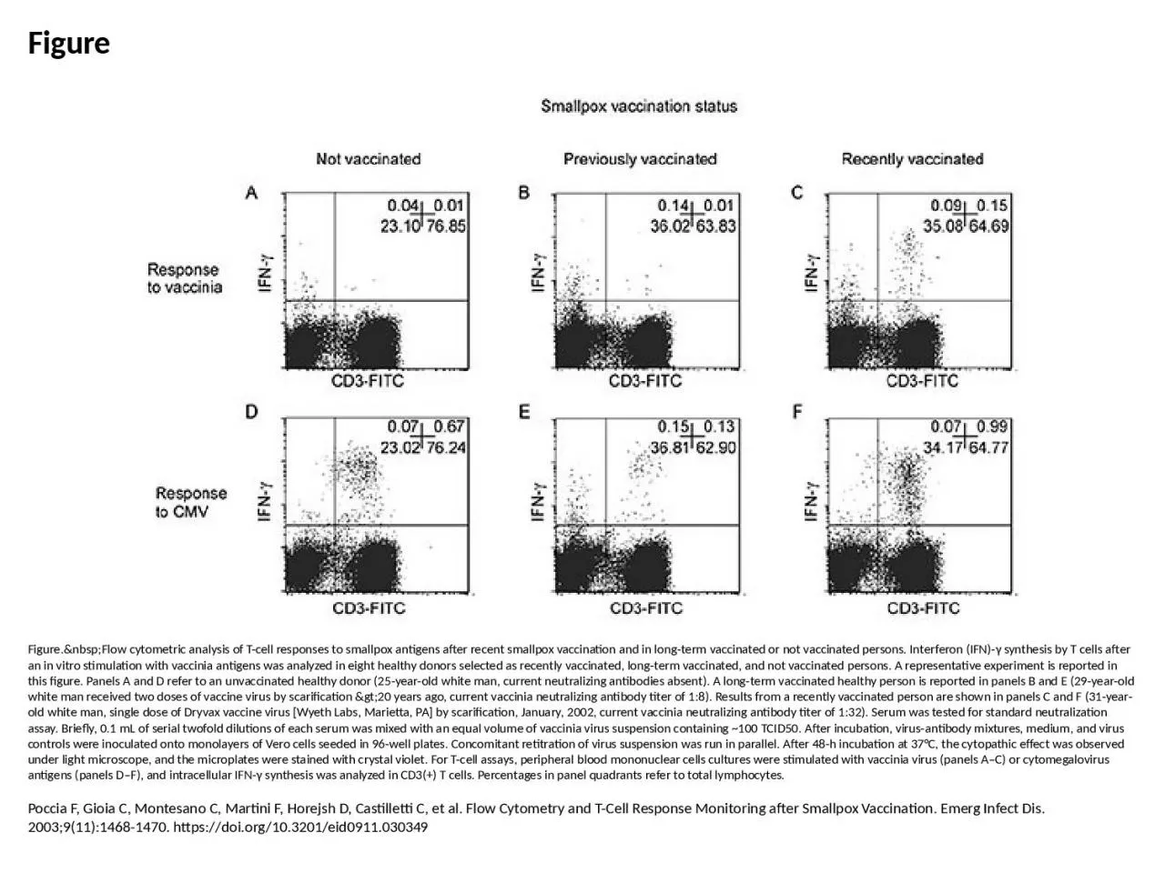
PPT-Figure Figure. Flow cytometric analysis of T-cell responses to smallpox antigens

Author : patricia | Published Date : 2024-01-29

Poccia F Gioia C Montesano C Martini F Horejsh D Castilletti C et al Flow Cytometry and TCell Response Monitoring after Smallpox Vaccination Emerg Infect Dis 200391114681470

Presentation Embed Code

Download Presentation

Download Presentation The PPT/PDF document "Figure Figure. Flow cytometric ..." is the property of its rightful owner. Permission is granted to download and print the materials on this website for personal, non-commercial use only, and to display it on your personal computer provided you do not modify the materials and that you retain all copyright notices contained in the materials. By downloading content from our website, you accept the terms of this agreement.

Figure Figure. Flow cytometric analysis of T-cell responses to smallpox antigens: Transcript


Poccia F Gioia C Montesano C Martini F Horejsh D Castilletti C et al Flow Cytometry and TCell Response Monitoring after Smallpox Vaccination Emerg Infect Dis 200391114681470 httpsdoiorg103201eid0911030349. Figure 23.3. Human Microbiota – Respiratory Tract. The lungs and trachea are usually sterile.. The ciliated mucous lining of the trachea, bronchi, and bronchioles makes up the . mucociliary escalator. Figure 12.1b The functions of cell division: Growth and development. Figure 12.1c The functions of cell division: Tissue renewal. Figure 12.2 Eukaryotic chomosomes. Figure 19.0 Chromatin in a developing salamander ovum. The �ow cytometer was developed in the 1970’s and rapidly be by the HIV pandemic and a plethora of discoveries in hematology, specialized �ow cytometers for use in the clinica Ng M, Lee J, Leong M, Ling A, Tan H, Ooi E. Topographic Changes in SARS Coronavirus–infected Cells during Late Stages of Infection. Emerg Infect Dis. 2004;10(11):1907-1914. https://doi.org/10.3201/eid1011.040195. Jalava K, On SL, Harrington CS, Andersen LP, Hänninen M, Vandamme P. A Cultured Strain of "Helicobacter heilmannii," a Human Gastric Pathogen, Identified as H. bizzozeronii: Evidence for Zoonotic Potential of Helicobacter. Emerg Infect Dis. 2001;7(6):1036-1038. https://doi.org/10.3201/eid0706.010622. Monroe MC, Morzunov SP, Johnson AM, Bowen MD, Artsob H, Yates T, et al. Genetic Diversity and Distribution of Peromyscus-Borne Hantaviruses in North America. Emerg Infect Dis. 1999;5(1):75-86. https://doi.org/10.3201/eid0501.990109. Weinstein RS. Should Remaining Stockpiles of Smallpox Virus (Variola) Be Destroyed?. Emerg Infect Dis. 2011;17(4):681-683. https://doi.org/10.3201/eid1704.101865. Van Delden C, Iglewski BH. Cell-to-Cell Signaling and Pseudomonas aeruginosa Infections. Emerg Infect Dis. 1998;4(4):551-560. https://doi.org/10.3201/eid0404.980405. Takada N, Fujita H, Kawabata H, Takajo I, Sakata A, Takano A, et al. Spotted Fever Group Rickettsia sp. Closely Related to R. japonica, Thailand. Emerg Infect Dis. 2009;15(4):610-611. https://doi.org/10.3201/eid1504.071271. An . antigen. is anything that elicits an adaptive immune response. These are molecules that trigger a specific immune response, eg., components of bacterial cell wall, capsule, pili, flagella, external and internal proteins of viruses, fungi and protozoa, toxins, nonself foreign substances like proteins, nucleoproteins, polysaccharides, some glycoproteins. Consort flow chart summarizing the number of patients screened, enrolled, treated and followed on the study. *one patient was re-assessed for eligibility at recurrence and re-enrolled on the study. Created with BioRender.com.. Kirschner DE, Webb G. Resistance, Remission, and Qualitative Differences in HIV Chemotherapy. Emerg Infect Dis. 1997;3(3):273-283. https://doi.org/10.3201/eid0303.970303. Salzberg SL, Kingsford C, Cattoli G, Spiro DJ, Janies DA, Aly MM, et al. Genome Analysis Linking Recent European and African Influenza (H5N1) Viruses. Emerg Infect Dis. 2007;13(5):713. https://doi.org/10.3201/eid1305.070013. Madariaga MG, Pulvirenti J, Sekosan M, Paddock CD, Zaki SR. Q Fever Endocarditis in HIV-Infected Patient. Emerg Infect Dis. 2004;10(3):501-504. https://doi.org/10.3201/eid1003.030971.

Download Document

Here is the link to download the presentation.
"Figure Figure. Flow cytometric analysis of T-cell responses to smallpox antigens"The content belongs to its owner. You may download and print it for personal use, without modification, and keep all copyright notices. By downloading, you agree to these terms.

Related Documents