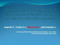
population Ampofo P Nyako EA Blankson P and Konadu A University of Ghana School of Medicine and Dentistry Accra Ghana Korle Bu Teaching Hospital Accra Ghana ID: 779923
Download The PPT/PDF document "Radiographic In-Vivo Determination of th..." is the property of its rightful owner. Permission is granted to download and print the materials on this web site for personal, non-commercial use only, and to display it on your personal computer provided you do not modify the materials and that you retain all copyright notices contained in the materials. By downloading content from our website, you accept the terms of this agreement.
Slide1
Radiographic In-Vivo Determination of the number of Canals in maxillary premolars of a Ghanaian population
Ampofo
P
.,
Nyako
E.A
.,
Blankson
P
.
,
and
Konadu
A
.
University
of Ghana School of Medicine and Dentistry, Accra, Ghana
Korle
-Bu
Teaching Hospital, Accra, Ghana
OUTLINEBackgroundIntroductionObjectives
Materials and Methods
Results
Discussion
References
Slide3Meet Mr. Kpodzro…
Slide4Mr.
Kpodzro
presented at the Emergency Unit with a 4-day history of swelling in the right maxillary region which gradually increased in size.
He was subsequently referred to the Dental department where thorough investigation was done.
Hb-10.8 g/
dL
; WBC- 10.30 x 10⁹
/L; Vitals and other results were grossly normalHe had apparently been seen by a Dentist 3-months prior to presentation where a root-canal was done on the second upper premolar which was confirmed on X-ray.
Slide5X-RAY OF MR. KPODZRO’S TOOTH
Slide6INTRODUCTIONBack to BasicsThe Concept of Reversible
vrs
Irreversible
Pulpistis
Slide7WHAT IS ROOT CANAL THERAPYA sequence of treatment for the infected pulp of a tooth which results in the elimination of infection and the protection of the decontaminated tooth from future
microbial invasion
.
Slide8It is evident
that the root canal varies with size, shape and number of roots in different teeth,
1
it is a known pertinent consideration in
endodontics
. Successful General dental procedures and Root canal therapy in particular requires a thorough knowledge of tooth anatomy and root canal morphology
2,3
Slide9With a variation in the number of canals
4
, the maxillary premolars remain one of the challenging teeth to treat
endodontically
,
A
lso because of the direction and longitudinal depressions of the roots, the various pulp cavity configurations, and the difficulties in visualizing the apical limit by radiographs
5
Slide10The Objective?Many factors contribute to differences in canal morphology4
but largely influenced by ethnic background, age and gender of the populations studied
5,6,7,8
Despite
copious data on several populations worldwide, data is scarce for Ghana.
This study thus describes a clinical survey by use of radiographs to determine the number of canals in maxillary premolars in a Ghanaian population.
Slide11Materials And MethodsFour hundred and two (402) participants were selected from patients who had opted for, and were referred to the Restorative Dentistry Department of the University Ghana School of Medicine and Dentistry for endodontic treatment of maxillary premolar from January 2012 till December 2015
.
Preoperative radiographs were taken for evaluation (root morphology, number of canals, and
periapical
status).
Slide12The teeth that were included in this study were those teeth that required nonsurgical endodontic treatment. The
included teeth were free of root
resorption
, had no calcifications or open apices and no retreatment cases were included in the study, patients who defaulted treatment at any point were also excluded.
Consent
was sought from each patient.
Slide13The teeth included were both clinically and radiographically examined by three specialists with more than 10 years of experience in
endodontics
and all patients operated on by the same certified Dental
Surgeons
The endodontic procedures undertaken were as follows: local anesthesia consisting of 2%
Lidocaine
in 1:100,000 adrenaline was administered by infiltration.
Slide14After the contents of the pulp chamber were removed, a sharp endodontic explorer was used to explore the developmental grooves carefully to locate the orifices of the canals. Copious amount of 2.5% sodium hypochlorite irrigation was used.
Pulp
tissue was extirpated using barbed broaches and the canals were flared with gates
glidden
burs.
Examination
of the floor of the pulp chamber to locate canals orifices was done using 3.5 high resolution magnification loupes (
Azdent). The number of root canals in the maxillary first and second premolars was recorded with correlated folder numbers, age and gender. The data was analyzed
Slide15RESULTSA total of 402 patients were used in the study. 239 of these were females and 163 males, representing 59.45% and 40.55% respectively. Of the total number of teeth used, 160 were maxillary
first premolars while 242 were maxillary second premolars.
The age ranged from 16yrs to 82yrs with a mean age of 40yrs.
Slide16Slide17Slide18DISCUSSIONDetermination of pulp anatomy generally fall within In-vivo (Clinical) or In-vitro studies.
While
in-vitro methods encompass teeth sectioning of extracted or cadaveric teeth, to apply dyes, contrasting media or scanning electron microscopy for analysis,
6,7
Clinical
methods on the other hand uses Anatomy studies, exploration, high resolution CT, Visualization endogram,
Fibreoptic endoscopy, magnetic resonance imaging and radiographs, which our study employs6,8
Slide19While Maxillary first premolars are considered to have two canals, Maxillary second premolar are generally considered to have one canal.
9,10,11,12
In the present study, the latter had one canal in 24.7% of cases. This is at variance with the studies of Green
9
and
Vertucci
et al. 10 in which the maxillary second premolars was reported to have one canal in 72% and 75% of cases respectively.
Slide20Like other anatomical features, it is natural to assume racial and ethnology influences on dental morphology differences. It
has been observed that different populations show distinctive variations of tooth morphology.
18
Our
study’s 95.4% and 75.3% finding for the first and second maxillary premolars respectively
is
much higher than reported in Asian studies in Singapore 20
and Sichuan province, China 21
Slide21In 1969, Weine et al.22,23 provided the first classification of more than one canal system in a single root and used the
mesiobuccal
root of the maxillary first molar as the type specimen.
Vertucci
further studied the canal anatomy extensively establishing eight different classification of pulp anatomy.
24,25
which the authors propose for the curriculum in Ghana
Slide22Type I: A single canal extends from the pulp chamber to the apexType II: Two separate canals leave the pulp chamber and join short of the apex to form one canal
Type III: One canal leaves the pulp chamber and divides into two in the root; the two then merge to exit as one canal
Type IV: Two separate, distinct canals extend from the pulp chamber to the apex
Type V: One canal leaves the pulp chamber and divides short of the apex into two separate, distinct canals with separate apical foramina
Type VI: Two separate canals leave the pulp chamber, merge in the body of the root, and
redivide
short of the apex to exit as two distinct canals
Type VII: One canal leaves the pulp chamber, divides and then rejoins in the body of the root, and finally redivides into two distinct canals short of the apex
Type VIII: Three separate, distinct canals extend from the pulp chamber to the apex
Slide23A LOGICAL EXPLANATION FOR MR KPODZRO
Slide24SUMMARY
This study was done to determine the number of canals in maxillary premolars in a Ghanaian population.
402 participants
were selected from patients who were referred for endodontic treatment of maxillary premolars. These consisted of 239 females (59%) and 163 males (41%).
The
teeth included in the study were examined clinically and
radiographically for the number of root canals
using.95.4% of the maxillary first premolars had two canals while 75.3% of the maxillary second premolars also had two canals. There was no significant association between gender and the number of canals in each premolar.
Slide25CONCLUSIONThe incidence of two canals in maxillary premolars of the Ghanaian population is very high.
Students
and Clinicians should be made aware of this extreme variation so that further inspection and careful interpretation of preoperative radiographs is done for success in endodontic therapy, especially of the maxillary second premolar
.
Slide26“…the end? We’re not at the end, we’re nowhere near the end. We are not at the beginning of the end. If anywhere, then perhaps end of the beginning.”Winston Churchill
Slide27REFERENCE
Ingle, John
Ide
&
Bakland
, Leif K & Health Communication Network 2002,
Endodontics, 5th ed, B C Decker, Hamilton, Ont. ; London
D'Souza R. Development of the pulpodentinal complex. In: Hargreaves K, editor. Seltzer and Bender's Dental Pulp. Chicago, Ill, USA: Quintessence Books; 2002.
Cleghorn
B, Christie W, Dong C. Root and root canal morphology of the human
mandibular
first premolar: A literature review. J
Endod
. 2007;33:509–16. [
PubMed
]
Jayasimha
Raj U,
Mylswamy
S. Root canal morphology of maxillary second premolars in an Indian population. Journal of Conservative Dentistry : JCD. 2010;13(3):148-151. doi:10.4103/0972-0707.71648.
Pecora
JD, Sousa
Neto
MD,
Saquy
PC,
Woelfel
JB. Root form and canal anatomy of maxillary second premolars.
Braz
Dent J. 1992;3:81–5. [
PubMed
]
Author/Editor:
Garg
Nisha
,
Garg
Amit
,
Speciality:Conservative
Dentistry and
Endodontics
Jayasimha
Raj U,
Mylswamy
S. Root canal morphology of maxillary second premolars in an Indian population. J
Conserv
Dent. 2010;13:148–51. [
PMC free article
] [
PubMed
]
Nur
BG, Ok E,
Altunsoy
M,
Aglarci
OS,
Colak
M,
Gungor
E. Evaluation of the root and canal morphology of
mandibular
permanent molars in a south-eastern Turkish population using cone-beam computed tomography.
European Journal of Dentistry
. 2014;8(2):154-159. doi:10.4103/1305-7456.130584
.
Slide289. Green
D. Double canals in single roots. Oral
Surg
Oral Med Oral
Pathol
1973; 35: 689-96.
10. Vertucci
F, Seelig A, Gillis R. Root canal morphology of the human maxillary second premolar. Oral Surg Oral Med Oral Pathol 1974; 58 456-6411. G
.
Cantatore
, E.
Berutti
, and A.
Castellucci
, “Missed anatomy: frequency and clinical impact,” Endodontic Topics, vol. 15, no. 1, pp. 3–31, 2006.
12. L
. J.
Buhrley
, M. J. Barrows, E. A.
BeGole
, and C. S.
Wenckus
, “Effect of magnification on locating the MB2 canal in maxillary molars,” Journal of
Endodontics
, vol. 28,no. 4, pp. 324–327, 2002.
13.
Chima
O. Number of root canals of the maxillary second premolar in Nigerians.
Odonto-stomatol
Trop
1997; 78 : 31-2
14.
Pecora
JD, Sousa
Neto
MD,
Saquy
PC,
Woelfel
JB. In vitro study of root canal anatomy of maxillary second premolars.
Braz
Dent J 1993; 3: 81-5.
15.
Weng
XL, Yu SB, Zhao SL, Wang HG, Mu T, Tang RY
.
Root canal morphology of
permanent maxillary
teeth in the Han nationality in Chinese
Guanzhong
area: A new modified root canal staining technique. J
Endod
2009;35:651-6.
16. Pineda
F,
Kuttler
Y.
Mesiodistal
and
buccolingual
roentogenic
investigations of 7,275 root canals. Oral Surg. 1972;33:101–10. [
PubMed
]
17.
Rwenyonyi
CM,
Kutesa
A,
Muwazi
L,
Buwembo
W. Root and canal morphology of maxillary first premolar teeth in a Ugandan population. Open J
Stomatol
2011;1:7-11.
18. Al-
Nazhan
S, Al-
Daafas
A, Al-
Maflehi
N. Radiographic investigation of
in vivo
endodontically
treated maxillary premolars in a Saudi Arabian sub-population. Saudi
Endod
J 2012;2:1-5.
Slide2919. Ahmad
IA. Root and root canal morphology of Saudi Arabian permanent dentition. Saudi
Endod
J 2015;5:99-106.
20.
Loh
HS. Root morphology of the maxillary first premolar in Singaporeans. Aust
Dent J 1998;43:399-402.21. Deng D, Zhang M, Wu W. The morphology study of root canal system of permanent teeth. Sichuan J Anat 2004;12:226.22. Weine
FS, Healey HJ, Gerstein H,
Evanson
L. Canal configuration in the
mesiobuccal
root of the maxillary first molar and its endodontic significance. Oral
Surg
Oral Med Oral
Pathol
. 1969;28:419–25. [
PubMed
]
23.
Gulabivala
K,
Aung
TH,
Alavi
A, Ng YL. Root and canal morphology of Burmese
mandibular
molars.
Int
Endod
J. 2001;34:359–70. [
PubMed
]
24.
Awawdeh
LA, Al-
Qudah
AA. Root form and canal morphology of
mandibular
premolars in a Jordanian population.
Int
Endod
J. 2008;41:240–8. [
PubMed
]
25.
Vertucci
FJ. Root canal morphology and its relationship to endodontic procedures.
Endod
Topics. 2005;10:3–29
26.
Sert
S,
Bayirli
GS. Evaluation of the root canal configuration of the
mandibular
and maxillary permanent teeth by gender in the Turkish population. J
Endod
. 2004;30:391–8. [
PubMed
]