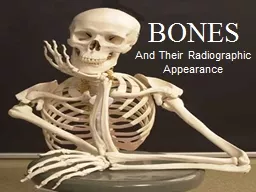
/
 And Their Radiographic Appearance  And Their Radiographic Appearance

And Their Radiographic Appearance - PowerPoint Presentation

mitsue-stanley
mitsue-stanley . @mitsue-stanley
Follow
345 views
Uploaded On 2020-04-04

And Their Radiographic Appearance - PPT Presentation

BONES 22 Bones make up the skull Cranial bones include Occipital one Frontal one Parietal two Temporal two Sphenoid one Ethmoid one Cranial bones surround the brain The Skull ID: 775247

bone nasal maxillary fossa bone nasal maxillary fossa ridge process zygomatic foramen oblique external temporal bones mandibular compact radiopaque

Share:

Link:

Embed:

Download Presentation from below link

Download Presentation The PPT/PDF document " And Their Radiographic Appearance" is the property of its rightful owner. Permission is granted to download and print the materials on this web site for personal, non-commercial use only, and to display it on your personal computer provided you do not modify the materials and that you retain all copyright notices contained in the materials. By downloading content from our website, you accept the terms of this agreement.


Presentation Transcript

Slide1

And Their Radiographic Appearance

BONES

Slide2

22 Bones make up the skull

Cranial bones include:Occipital (one)Frontal (one)Parietal (two)Temporal (two)Sphenoid (one)Ethmoid (one)Cranial bones surround the brain

Slide3

The Skull

Bones:

1 - mandible

2 - maxilla

3 -

zygomatic

4 - nasal bone

5 - frontal bone

6 - sphenoid bone

7 - temporal

8 - occipital bone

9 -

lacrimal

bone

10 -

ethmoid

bone

11 - parietal bone

Slide4

Facial bones include:Mandible (one)Maxilla (two)Zygomatic (two)Lacrimal (two)Nasal (two)Inferior nasal conchal (two)Vomer (one)

Slide5

Terms that describe bone anatomy

Process – general term for any prominenceFossa - depression on the surface of the boneSuture – where two bones joinTuberosity – bony prominance, usually where muscle attaches, ie maxillary tuberosity

Mastoid process

Temporal fossa

suture

Slide6

Terms that describe bone anatomy

Notch – indentation on the edge of a boneRidge – elongated prominenceForamen - opening

Mental foramen

External oblique ridge

Mandibular notch

Slide7

Two types of Bone – compact and cancellous

Cancellous bone (also called spongy bone) makes up center of bonesContains bone marrow spaces (called “trabeculation” on radiographs)More radiolucent than compact bone

trabeculation

Slide8

Compact Bone

Compact bone- forms plates that form outside of bones, linings for alveolus, foramina, etc…Is more radiopaque because of its density

Compact bone

Cancellous bone

Slide9

Compact Bone Lines Alveolus (socket)

It is also called (*and means same thing): Lamina dura (on radiographs only)Cribriform plateCortical boneAlveolar bone proper

Slide10

Lamina dura

(radiopaque lining)

Periodontal ligament space

(radiolucent lining)

Compact bone outlines alveolar crest (when no bone has been lost due to periodontal disease!)

Alveolar crest

(radiopaque)

Slide11

Do you see any compact bone outlining the alveolar crest?

Slide12

Lateral View Identification

Mandible

Maxilla

Zygomatic arch

Condyle

External auditory meatus

Temporal bone

Slide13

Lateral closeup

CondyleArticular eminenceCoronoid processRamusArticular fossaMandibular notchCoronoid notch

6

7

Slide14

Temporomandibular Joint

Mandibular condyle articulates with temporal bone in the articular fossa (also called glenoid fossa, mandibular fossa)Most anterior border of articular fossa is the articular eminanceIf someone opens wide and the condyle slides anterior to the eminance, the person has “lockjaw”

Articular or gleniod fossa

Articular eminance

Slide15

Lateral closeup

DehiscenceFenestrationMental foramenZygomatic process of maxillaAlveolar boneAlveolar crestExternal oblique ridge

7

Slide16

Dehiscence vs Fenestration

Bony defects of unknown causeNeither can be found radiographically, only during surgeryFenestration, defect completely surrounded by boneDehiscence, alveolar crest bone absent

dehiscence

fenestration

Slide17

Bones of the Orbit

In order of appearance

Frontal

Zygomatic

Maxillary

Palatine

Sphenoid

Lacrimal

Ethmoid

Slide18

Maxillary Sinus

Nasal Cavity

Orbit

Slide19

Zygomatic Arch

Commonly called the “cheekbone”Comprised of three bones, temporal, maxilla, zygoma

maxilla

zygoma

temporal

sutures

Slide20

Zygomatic Arch

Maxillary process of zygoma joins with zygomatic process of maxilla

Zygomatic process of

temporal bone joins with temporal process of zygoma

Zygomatic

Arch

Slide21

Radiographically, the zygomatic arch appears as a radiopaque horseshoe shaped structure above maxillary molars (not always seen)

Slide22

Maxillary Sinus – an opening in the maxillary bone, acts as a filter for inhaled air

Location of the sinus

Inside of sinus with bony covering removed

Slide23

Inverted Y

Maxillary sinus meets nasal cavity in area of canineOn radiographs, wall of sinus crosses wall of nasal cavity (both are radiopaque because they are compact bone)Result is the “inverted Y”

Slide24

Inverted Y

Maxillary sinus

Nasal cavity

Inverted Y

Slide25

Floor of maxillary

sinus

(

radiopaque)

Zygomatic Arch

Septa of maxillary sinus (divides cavity)

Slide26

Frontal View Identification

Frontal boneOrbitMental protuberance

Slide27

Skull Identification

Midline sutureAnterior Nasal spineNasal septumInfraorbital foramenLateral fossaSuperior nasal conchae

6

Slide28

Anterior Radiograph

Median palatal suture (radiolucent)Noseline (cartilage)Nasal spine (radiopaque V-shaped prominence)Nasal conchaeNasal septum (elongated, thicker radiopacity)

5

Slide29

Lateral Fossa – a depression between the maxillary cuspid and incisor

Exercise – feel your lateral fossa with your finger

Slide30

Nasal septum

(divides nasal cavity)

Anterior Nasal spine (V-shaped)

Lateral fossa

(Radiolucency inside circle)

Inferior nasal

conchae

Slide31

Mandibular

foramen

Internal Oblique ridge- (slightly inferior to external oblique ridge)

Lingula – a bony projection that partially covers the mandibular foramen

Mental ridge-

see figure 27-56

Iannucci

Slide32

Lingual foramen

Genial tubercles

(muscles attach here)

Submandibular fossa

(depression for submandibular

Salivary gland)

Slide33

Coronoid notch

Coronoid process

External oblique ridge

Mandibular

(sigmoid)

Notch

Ramus

Condyle

Angle of the mandible

Slide34

External

oblique ridge(thicker radiopaque band)

Note

: External/internal ridges often “superimposed” over each other radiographically;therefore difficult to differentiate between the two; external always superior to internaloblique ridge (mylohyoid muscle attachment); internal usually runs below roots of mandibularmolars (see figure 26-62 Haring)

Slide35

Internal on “inside” or lingual

Slide36

A- external oblique ridge

B- internal oblique ridgeC- submandibular fossaD- mandibular canal

B

A

Hyoid bone

Slide37

Mandibular

foramen

Mandibular canal

Submandibular fossa

(large radiolucency

within the circle)

Soft tissue outline-

retromolar

area

Slide38

Mandibular

canal

External

oblique ridge

Slide39

Submandibular fossa

Slide40

Nutrient canals – passageways to

teeth for vessels (arrows on film), often seen around maxillary premolars

Lingual foramen(radiolucency)

Genial tubercles

(Radiopacities)

Slide41

Mental Foramen

Mylohyoid

ridge or internal oblique ridge

(see down by roots of teeth)– actually on lingual of mandible

Slide42

Nasopalatine

orIncisive foramen

Palate

Anterior or Greater

palatine foramen

Median palatine suture

Posterior

or

Lessor

palatine foramen

Hamulus

Slide43

Palatal Radiographs

Median Palatal

suture

Nasopalatine

or Incisive

foramen

Slide44

Sphenoid Bone

A butterfly shaped cranial bone posterior to the palateIt forms part of the orbitIt’s hamulus can sometimes be seen on third molar radiographSome muscles of masticaton attach to Pterygoid plate

Lateral pterygoid plate

Hamulus

Slide45

Temporal bone

A cranial bone that articulates with mandible in its articular fossaOther landmarks include styloid process, mastoid process Forms part of zygomatic arch

Articular

or glenoid fossa(where condyle sits)

Mastoid process

Slide46

Infratemporal Space (skull with mandible removed)

Maxillary

tuberosity

Styloid

process (can sometimes beseen on a panoral)

Articular

or Glenoid fossa

Slide47

Condyle

and glenoid fossa

Hard palate

(horizontal thicker radiopaque line)

Coronoid

process

Slide48

Maxillary tuberosity

Inverted Y

External

oblique ridge

Nasal spine