/
 Accessory Organs to the Digestive Tract  Accessory Organs to the Digestive Tract

Accessory Organs to the Digestive Tract - PowerPoint Presentation

phoebe-click
phoebe-click . @phoebe-click
Follow
348 views
Uploaded On 2020-04-05

Accessory Organs to the Digestive Tract - PPT Presentation

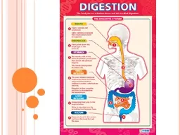
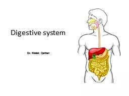
Pages 473476 and 494497 Teeth mechanical digestion through mastication Salivary glands parotid submandibular sublingual Secrete saliva a bicarbonate rich juice chemical digestion via secretions of enzymes ID: 775882

liver bile blood duct liver bile blood duct glucose pearson education 2015 duodenum hepatic cholesterol gallbladder digestion organs colon

Share:

Link:

Embed:

Download Presentation from below link

Download Presentation The PPT/PDF document " Accessory Organs to the Digestive Tract" is the property of its rightful owner. Permission is granted to download and print the materials on this web site for personal, non-commercial use only, and to display it on your personal computer provided you do not modify the materials and that you retain all copyright notices contained in the materials. By downloading content from our website, you accept the terms of this agreement.


Presentation Transcript

Slide1

Accessory Organs to the Digestive Tract

Pages 473-476 and 494-497

Slide2

Teeth – mechanical digestion through masticationSalivary glands – parotid, submandibular, sublingualSecrete saliva, a bicarbonate rich juicechemical digestion via secretions of enzymesSalivary amylase begins starch digestionSaliva also contains lysozyme and antibodies to inhibit bacteriaPancreasLiverGallbladder

© 2015 Pearson Education, Inc.

Accessory Digestive Organs

Slide3

Figure 14.1 The human digestive system: Alimentary canal and accessory organs.

Mouth (oral cavity)

Tongue

Esophagus

Parotid gland

Sublingual gland

Submandibular

gland

Salivary glands

Pharynx

Stomach

Pancreas

Transverse colon

Descending colon

Ascending colon

Cecum

Sigmoid colon

Rectum

Appendix

Anal canal

Anus

Small

intestine

Duodenum

Jejunum

Ileum

Liver

Gallbladder

Large intestine

Slide4

Produces digestive enzymes that break down all categories of foodreleases into the duodenum via the hepatopancreatic ampullaAlkaline (bicarbonate) fluid with these enzymes neutralizes the acidic chymeProduces hormones: (negative feedback loops)Insulin – reduces blood glucose Glucagon – raises blood glucose

© 2015 Pearson Education, Inc.

Pancreas

Slide5

largest gland in the body; consists of four lobescan regenerate if part is damaged/removedsuspended from the diaphragm and abdominal wall by the falciform ligamentConnected to the gallbladder via the common hepatic duct

© 2015 Pearson Education, Inc.

Liver

Slide6

the body’s key metabolic organAfter the GI tract, blood travels to liver via the hepatic portal circulationRoles in digestion:Manufactures bile and blood clotting proteinsDetoxifies drugs and alcoholDegrades hormonesProduces cholesterol:Low Density Lipoproteins: transport cholesterol/lipids to the cells; large amounts circulating can build up in vessel lumen (BAD GUYS)High Density Lipoproetins: transport cholesterol from the cells to the liver for disposal in bile (GOOD GUYS)

© 2015 Pearson Education, Inc.

The Central Role of the Liver in Metabolism

Slide7

The liver manages blood glucose levels through:Glycogenesis: “Building glycogen”Glucose is removed from the blood (via insulin)Converted to glycogen and stored in the liverGlycogenolysis: “Splitting glycogen”Glucose is released into the blood (via glucagon)Glycogen is “split” and put back into bloodGluconeogenesis: “Building glycogen from other sources”The Liver can make glucose using fats and amino acids

The Liver and Blood Glucose

Slide8

produced by the livera yellow-green, watery solution containing:Bile salts and bile pigments (mostly bilirubin from the breakdown of hemoglobin)Cholesterol, phospholipids, and electrolytesleaves the liver via the common hepatic ductenters duodenum through the bile ductFunction: emulsify fatsChemically breaks down large fat globules

© 2015 Pearson Education, Inc.

Bile

Slide9

Sac found in shallow fossa of liverWhen no digestion is occurring: bile backs up the cystic duct for storage in the gallbladderWater is removed at this time to concentrate itGallstonescrystallized cholesterol that can cause blockagesResult from removal of too much waterProlonged storage in gallbladder

© 2015 Pearson Education, Inc.

Gallbladder

Slide10

Figure 14.6 The duodenum of the small intestine and related organs.

Right and left

hepatic ducts

from liver

Cystic duct

Common hepatic duct

Bile duct and sphincter

Accessory pancreatic duct

Pancreas

Jejunum

Main pancreatic duct and sphincter

Duodenum

Hepatopancreatic

ampulla and sphincter

Duodenal

papilla

Gallbladder