/
 Using Karyotypes To Diagnose Genetic Disorders  Using Karyotypes To Diagnose Genetic Disorders

Using Karyotypes To Diagnose Genetic Disorders - PowerPoint Presentation

phoebe-click
phoebe-click . @phoebe-click
Follow
343 views
Uploaded On 2020-04-02

Using Karyotypes To Diagnose Genetic Disorders - PPT Presentation

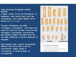
A regular human cell has 46 chromosomes 44 autosomes which come in pairs and 2 sex chromosomes which specify gender XX for female and XY for male The pairs of autosomes are called homologous chromosomes One of each pair came from mom and the other came from dad Homologous chromosomes ha ID: 774783

gene traits chromosomes genes gene traits chromosomes genes recessive chromosome autosomal inheritance dominant linked dominance sex disease disorders incomplete

Share:

Link:

Embed:

Download Presentation from below link

Download Presentation The PPT/PDF document " Using Karyotypes To Diagnose Genetic Di..." is the property of its rightful owner. Permission is granted to download and print the materials on this web site for personal, non-commercial use only, and to display it on your personal computer provided you do not modify the materials and that you retain all copyright notices contained in the materials. By downloading content from our website, you accept the terms of this agreement.


Presentation Transcript

Slide1

Using Karyotypes To Diagnose Genetic DisordersA regular human cell has 46 chromosomes: 44 autosomes, which come in pairs, and 2 sex chromosomes, which specify gender (XX for female and XY for male).The pairs of autosomes are called "homologous chromosomes." One of each pair came from mom and the other came from dad. Homologous chromosomes have all of the same genes arranged in the same order, but with slight differences in the DNA sequences of the genes.What happens when a person has something different, such as too many or too few chromosomes, missing pieces of chromosomes, or mixed up pieces of chromosomes?                                                     

Using

Karyotypes To Diagnose Genetic DisordersA regular human cell has 46 chromosomes: 44 autosomes, which come in pairs, and 2 sex chromosomes, which specify gender (XX for female and XY for male).The pairs of autosomes are called "homologous chromosomes." One of each pair came from mom and the other came from dad. Homologous chromosomes have all of the same genes arranged in the same order, but with slight differences in the DNA sequences of the genes.What happens when a person has something different, such as too many or too few chromosomes, missing pieces of chromosomes, or mixed up pieces of chromosomes?

Using Karyotypes To Diagnose Genetic Disorders

Slide2

Slide3

So how are genes passed on from parent to child?

Genes - sections of chromosomal DNAOne set of chromosomes is inherited from each parentTherefore, for each pair of genes, one is inherited from a person’s mother, and one from their father Sometimes the genes, or chromosomes they are on, have defects which are passed to offspring

Chromosome

Gene

Homologous Chromosome pair

Slide4

Classification of genetic disorders

Male

Mutations

in single genes

Whole chromosomes or sections messed up

Chromosome number imbalance

Due to nondisjunction during meiosis

ex.

Patau

(Trisomy 13), Down syndrome (Trisomy 21)

Single Gene Disorders

Chromosome disorders

Slide5

Caused by a mutant allele for a gene.Mutant allele may be the dominant or recessive one

The three common types of single gene disorders are :Autosomal recessiveAutosomal dominantX-linked

Single gene disorders

Slide6

Examples of Autosomal recessive diseases

Tay SachsCystic fibrosis Phenylketonuria (PKU) Sickle Cell disease

Autosomal

recessive

inheritance

Must have Homozygous Recessive Genotype

Slide7

Fig. 1.2 ©Scion Publishing Ltd Photos (a) and (b) courtesy of Dr Tim David

Tay-Sachs 1 – 27 Ashkanazi Jews, Lousiana Cajuns and French Canadians – HIGHER FREQUENCY1- 250 in other population Heterozygotes have Selective Survival Advantage)Problems with brain cell membrane chemical build up causes blindness and seizures and early death

Slide8

Fig. 1.2 ©Scion Publishing Ltd Photos (a) and (b) courtesy of Dr Tim David

Cystic fibrosis

Most common genetic disease among CaucasiansFaulty cell membrane transport protein

Slide9

Fig. 1.2 ©Scion Publishing Ltd Photos (a) and (b) courtesy of Dr Tim David

Phenylketonuria “PKU”Lacks enzyme to breakdown amino acid phenylalanine which builds up in brain

Slide10

Ltd

Sickle cell disease

Slide11

Parents

AUTOSOMAL RECESSIVE INHERITANCE

Parent who are carriers for the same autosomal recessive condition have one copy of the usual form of the gene and one copy of an

altered gene

of the particular pair

Slide12

Parents

Sperm/Eggs

AUTOSOMAL RECESSIVE INHERITANCE

A parent who is a carrier passes on either the usual gene

or the

altered gene

into the eggs or sperm

The other parent who is also a carrier for the same condition passes on either the usual gene or the

altered gene

into his/her eggs or sperm

Slide13

Parents

AUTOSOMAL RECESSIVE INHERITANCE

Affected

Unaffected (carrier)

Unaffected

Sperm/Eggs

Unaffected (carrier)

Slide14

D – No disease d- disease

Slide15

Examples of Autosomal Dominant Disorders

Achondroplasia (Dwarfism)Huntington disease

Autosomal dominant

inheritance

Homo dominant or Heterozygous

Slide16

Achondroplasia Dwarfism

Slide17

Huntingtons chorea

Slide18

Parents

Gametes

Autosomal dominant inheritance

Slide19

Parents

Gametes

Autosomal dominant inheritance

Affected

Affected

Unaffected

At

conception

Slide20

RecessiveHomozygotes must have two copies of the altered gene to be affected

DominantThese individuals are called Heterozygotes with one copy of the altered gene they are affected

X-linked recessiveMales with an altered gene on the X-chromosome are always affected

Male

Slide21

Slide22

Examples of Autosomal recessive diseases

Tay SachsCystic fibrosis Phenylketonuria (PKU) Sickle Cell disease

Autosomal

recessive

inheritance

Must have Homozygous Recessive Genotype

Slide23

Examples of Autosomal Dominant Disorders

Achondroplasia (Dwarfism)Huntington disease

Autosomal dominant

inheritance

Homo dominant or Heterozygous

Slide24

Sex-Linked Traits

X

chromosome

Y

chromosome

Slide25

Slide26

What are Sex Linked Traits?

Most traits we inherit are located on our

autosomes

.

BUT

Some traits are determined by genes located on the

sex chromosomes

.

These traits are called

sex linked traits

Slide27

The X chromosome is larger in size and has many more genes than the Y chromosome.Those traits with genes on the X sexchromosome are called X-linked traits

X

chromosome

Y

chromosome

Slide28

Gene Linkage

Sex-linked traits were discovered in 1910 by Thomas Hunt Morgan who studied inheritance in fruit flies (Drosophila).

Slide29

Mendel’s Law of Independent Assortment applies to

chromosomes

which are assorted independently

in meiosis

.

Genes

located close

together are

inherited

together.

Slide30

Slide31

Sex linked Inheritance is different in males and females

Since males only have one X chromosome they can only receive one gene for these traits.

The gene is on the X chromosome they inherit from their

mother

The Y chromosome, they got from their

father

doesn’t have a copy of the gene

Slide32

What he gets is what he is!!!

Males express all X-linked genes –There’s no second allele to mask the effects of the only one he inherits on his X chromosomes

Slide33

Males inherit one X chromosome

2 Possible Genotypes:

XᴿY or

Y

Slide34

Females inherit two X chromosomes

3 Possible Genotypes:

XᴿXᴿ

XᴿXʳ

Slide35

Sex-Linked Traits –

1. Color Blindness – recessive disorder

2. Hemophilia – blood clotting disorder

3. Baldness – recessive trait

4. Muscular dystrophy – recessive disorder

Slide36

Beyond Mendel’s Principle of Dominance

Incomplete Dominance

Codominance

Multiple Alleles

Polygenic Traits

Slide37

Incomplete dominance

Dominant allele doesn’t fully mask the expression of recessive allele.

A “blending” of both dominant and recessive alleles is seen as an intermediate (

inbetween

) phenotype in heterozygotes.

Slide38

Slide39

Slide40

Slide41

Slide42

Slide43

Slide44

Incomplete Dominance vs. Codominance

Incomplete dominance – Heterozygote’s traits are a blend of the two alleles

Ex. Red X White flowers > Pink flowers

Codominance – Both alleles for gene are equally strong and are both seen

Ex. Red x White feathers > Both colors seen

Slide45

With

incomplete dominance

, a cross between organisms with two different phenotypes produces offspring with a third phenotype that is a

blending

of the parental traits

.

With

codominance

, a cross between organisms with two different phenotypes produces offspring with a third phenotype in which both of the

parental traits appear together.

Slide46

Which inheritance pattern does each cross represent? Codominance or Incomplete Dominance

12/1/2015

X

=

100%

X

=

100%

Slide47

Multiple Alleles

Many genes come in more than just 2 forms – there can be many alleles for one gene

The gene for blood type comes in 3 different allele forms

I

A

I

B

i

Slide48

Slide49

Polygenic Traits

More than one gene determines a traitUsually the cause in traits with a lot of variation (height, skin color, hair color)

Slide50

Slide51

Phenotype results from the interaction of genes and environmental influences

Slide52


Eye

color is determined by many different

genes


Polygenic trait

2

. Height and skin color comes in many forms on a continuum


Polygenic trait

3. A

hoo

can have curly hair, spiked hair or a mix of curly and spiked


Codominance – both traits seen

4. A horse has both red and white hairs making them look

pinkish (roan).


Codominance – both traits seen

5. A puppy inherits a gray coat from its black coat dad and white

mom

Incomplete dominance – blended traits


6

. Snapdragons homozygous red crossed with pure white make

pink

Incomplete dominance – blended traits