/
How to properly diagnose invasive aspergillosis? How to properly diagnose invasive aspergillosis?

How to properly diagnose invasive aspergillosis? - PowerPoint Presentation

callie
callie . @callie
Follow
344 views
Uploaded On 2022-05-18

How to properly diagnose invasive aspergillosis? - PPT Presentation

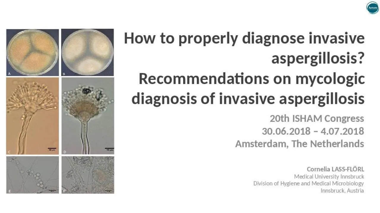
Recommendations on mycologic diagnosis of invasive aspergillosis Cornelia LASSFLÖRL Medical University Innsbruck Division of Hygiene and Medical Microbiology Innsbruck Austria 20th ISHAM ID: 911844

diagnose pcr specificity sensitivity pcr diagnose sensitivity specificity patients blood aspergillus bal samples test 2018 positive invasive sor cmi

Share:

Link:

Embed:

Download Presentation from below link

Download Presentation The PPT/PDF document "How to properly diagnose invasive asperg..." is the property of its rightful owner. Permission is granted to download and print the materials on this web site for personal, non-commercial use only, and to display it on your personal computer provided you do not modify the materials and that you retain all copyright notices contained in the materials. By downloading content from our website, you accept the terms of this agreement.


Presentation Transcript

Slide1

How to properly diagnose invasive aspergillosis?Recommendations on mycologic diagnosis of invasive aspergillosis

Cornelia LASS-FLÖRLMedical University InnsbruckDivision of Hygiene and Medical MicrobiologyInnsbruck, Austria

20th ISHAM Congress30.06.2018 – 4.07.2018Amsterdam, The Netherlands

Slide2

Disclosure of speaker's interests

No (potential) conflict of interests

Slide3

Slide4

Hard factsyeasts:

endogenous microflorafungi can be both colonizers and pathogensthe finding of organisms from sputum, or GI does not necessarily indicate infectionclinical manifestations are non-specific direct examination or cultures from sterile sites are golden standard

conventional diagnostic tests insensitive, positive latepatients with disseminated candidasis may have negative blood cultures vigilance is required in the interpretation of superficial cultures , antigen tests, PCR screening, presence of antibodies and/or metabolites

Slide5

The patientConventional testsAntigen tests

Molecular based testsDiagnosisImaging

& clinicLab parameters

Slide6

The human specimen

superficial cultures

sputum

, TS, BAL

biopsy

fine needle-aspiration

commensale

flora

sterile

Infection

blood

Slide7

Microscopy & Culture „must have“

Slide8

PopulationIntention

InterventionSoR

QoECommentAnyTo identify fungal elements in histological sections and stainsHistological examinationGomori's methenamine silver stain Periodic acid–Schiff

A

III

Histopathology is an essential investigation

Inability

to definitively distinguish other filamentous fungi

GMS

: removes cellular background; more sensitive to hyphal elements

PAS

: advantage of counter stain to check cellular detail

Any

To identify fungal elements in histological sections and stains

Fluorescent dyes:

Calcofluor

white™,

Uvitex

2B,

Blancophor

A

II

Not specific to

Aspergillus

but high sensitivity and the micromorphology may provide information on the fungal class (e.g.

Aspergillus

: typically dichotomous and septate,

Mucorales

:

pauci

-septate and 90° angle branching, yeast: budding)

Rapid

turnaround time

Broad

applicability

May

be applied to frozen sections, paraffin-embedded tissue

Any

To identify fungal elements in histological sections and stains

ImmunohistochemistryMonoclonal antibody WF-AF-1 or EB-A1In situ hybridization

B

II

Have the potential to provide genus- and species-specific dataCommercially available monoclonal antibodiesWF-AF-1 is specific for Aspergillus fumigatus, Aspergillus

flavus, and Aspergillus niger

Time consuming and not broadly available

Any

To identify fungal elements in fresh clinical specimens (e.g. BAL)

Application of fluorescent dyes

Calcofluor white™ or Uvitex 2B or Blancophor™AII

Essential investigationNot specific for

Aspergillus speciesHigh

sensitivityRapid turn-around timeBroad applicabilityNo species identification but the micromorphology may provide information on the fungal class (e.g.

Aspergillus: typically dichotomous and septate, Mucorales

: pauci-septate and 90° angle branching, yeast: budding)

BAL, bronchoalveolar lavage; CNS, central nervous system; GMS, Gomori's methenamine silver stain; HE,

haematoxylin-eosin; PAS, Periodic acid–Schiff; QoE, Quality of evidence;

SoR, Strength of recommendation

Microscopic examinations

Ullmann AJ et al. CMI 2018;24:e1

Slide9

Slide10

Slide11

Slide12

Slide13

PopulationIntentionIntervention

SoRQoE

CommentAnyTo achieve a homogeneous sample of viscous samples such as sputumLiquefaction using a mucolytic agent, e.g. Pancreatin®, Sputolysin®, or using sonication and 1,4-dithiothreitolA

III

Essential investigation

High-volume

sputum culture (entire sample) shown to significantly increase recovery

Any

To achieve optimal recovery of

Aspergillus

from BAL by centrifugation and investigation of the sediment

Centrifugation of BALs or bronchial aspirates

A

III

Essential investigation

Isolation

of

Aspergillus

dependent on volume cultured

Abbreviations: BAL,

bronchoalveolar

lavage; PCR, polymerase chain reaction;

QoE

, Quality of evidence;

SoR

, Strength of recommendation

Sample

selection

and

pre-analytical

respiratory

sample

treatment

Ullmann AJ et al. CMI 2018;24:e1

Slide14

PopulationIntentionIntervention

SoR

QoECommentAnyPrimary isolation from deep sites samples (e.g. biopsies, CSF)Culture on SDA, BHI agar, PDA at 30°C and 37°C for 72 h

A

III

Blood inhibits

conidiation

;

BHI

can help to recover some isolates; isolation of several colonies or isolation of the same fungus from a repeat specimen enhance significance

Primary isolation from non-sterile samples, e.g. sputum, respiratory aspirates

Culture on SDA, BHI agar, PDA with gentamicin plus chloramphenicol at 30°C and 37°C for 72 h

A

III

High-volume sputum culture (entire sample) shown to significantly increase recovery; quantitative cultures are not discriminative for infection or colonization

Identification of species complex

Macroscopic and microscopic examination from primary cultures

A

II

Colony

colour

,

conidium

size, shape and

septation

.

Colour

of conidia and conidiophore and

conidiogenesis

(tease or tape mounts are preferred); expertise needed for interpretation

Thermotolerance

test (growth at 50°C for species confirmation of

A. fumigatus

)

Identification of species complex (and species identification of

A. fumigatus

specifically)

Culture on identification media at 25–30°C, 37°C and 50°C (2% MEA and

Czapek

-Dox Agar) and microscopic examination

A

II

Identification at species level

MALDI-TOF MS identification

B

II

In-house databases are often used to improve identification rates

Identification at species level

Sequencing of ITS, β-tubulin and calmodulinA

III

Not necessary in organisms with typical growth, but in cases of atypical growth

To study outbreaks

Microsatellite and CSP analysis

C

II

To study outbreaks (which in general may comprise more than one genotype)

B

II

To study colonization patterns

Culture

and

Aspergillus species identificationBHI, brain–heart infusion; CSF, cerebrospinal fluid; CSP, cell surface protein; ITS, internal transcribed spacer; MALDI-TOF MS, matrix-assisted laser desorption/ionization time-of-flight mass

spectometry identification; MEA, malt extract agar; PDA, potato dextrose agar; QoE, Quality of evidence; SDA, Sabouraud dextrose agar; SoR, Strength of recommendationUllmann AJ et al. CMI 2018;24:e1

Slide15

Serology and/or PCRs are „add on tests“

Slide16

Copyright 2011 Rainer Poulet

Slide17

PopulationIntentionIntervention

SoRQoE

CommentPatients with prolonged neutropenia or allogeneic stem cell transplantation recipients not on mould-active prophylaxisProspective screening for IAGM in blooda

A

I

Highest test accuracy requiring two consecutive samples with an ODI ≥0.5 or retesting the same sample

Prospective

monitoring should be combined with HRCT and clinical evaluation

Patients with prolonged neutropenic or allogeneic stem cell transplantation recipients

on

mould

active prophylaxis

Prospective screening for IA

GM in

blood

a

D

II

Low prevalence of IA in this setting with consequently low PPV of blood GM test

Prophylaxis

may have a negative impact on sensitivity of the test or the low yield may be due to decreased incidence of IA

Patients with a

haematological

malignancy

To diagnose IA

GM in

blood

a

Significantly

lower sensitivity in non-neutropenic patients

Neutropenic

patients

A

II

Non-neutropenic

patients

B

II

ICU patients

To diagnose IA

GM in

blood

a

C

II

Better performance in neutropenic than in non-neutropenic patients

Solid organ recipients

To diagnose IA

GM in

blood

a

C

II

Low sensitivity, good specificity

Most

data for lung SOT

Any other patient

To diagnose IA

GM in

blood

a

C

II

Piperacillin/

tazobactam may no longer be responsible for false-positive results according to recent studiesCross-reactivity in case of histoplasmosis, fusariosis, talaromycosis (formerly: penicilliosis)False-positive results reported due to ingestion of ice-pops, transfusions, antibiotics, Plasmalyt® infusionCancer patients

To monitor treatment

GM in

blood

a

A

II

Galactomannan

testing

in

blood

samples

Abbreviations: GM, galactomannan; IA, invasive aspergillosis; ICU, intensive care unit; ODI, optical density index; PPV, positive predictive value;

QoE

, Quality of evidence;

SoR

, Strength of recommendation; SOT, solid organ transplantation.

a

Serum or plasma.

Ullmann AJ et al. CMI 2018;24:e1

Slide18

PopulationIntention

InterventionSoR

QoECommentAnyTo diagnose pulmonary IATo apply GM test on BAL fluidA

II

Diagnostic assay

GM

in BAL is a good tool to diagnose, optimal cut-off to positivity 0.5 to 1.0

Any

To diagnose cerebral IA

To apply GM test on cerebrospinal fluid

B

II

No validated cut-off

Any

To detect GM in tissue

To apply GM test on lung biopsies

B

II

Using a cut-off 0.5 resulted in a sensitivity of 90 % and a specificity of 95%; specimens need to be sliced, precondition for doing so is that sufficient material is available; dilution in isotonic saline

Galactomannan

testing

in

samples

other

than

blood

BAL,

bronchoalveolar

lavage; GM, galactomannan; IA, invasive aspergillosis;

QoE

, Quality of evidence;

SoR

, Strength of recommendation.

Ullmann AJ et al. CMI 2018;24:e1

Slide19

PopulationIntentionIntervention

SoRQoE

CommentMixed population: adult ICU, haematological disorders, SOTTo diagnose IFDDiagnostic assayCII

Five different assaysOverall sensitivity of 77% and specificity of 85%

Specificity

limits its value in this setting

Screening assays

C

II

Two or more consecutive samples: sensitivity: 65%; specificity: 93%

Studies

included once to thrice weekly. Varies with assay and cut-off: Wako assay sensitivity: 40%–97%, specificity: 51%–99%

Adult

haematological

malignancy and HSCT

To diagnose IFD

Diagnostic assay

C

II

Overall sensitivity: 50%–70%, specificity: 91%–99%

ICU—mixed adult immunocompromised patients (haematology, SOT, cancer, immunosuppressive therapy, liver failure, HIV)

To diagnose IA

Diagnostic assay

C

II

Overall sensitivity: 78%–85%, specificity: 36%–75%, NPV: 85%–92%

Specificity

increased at higher cut-off values

ICU—mixed adult population: SOT, liver failure, immunosuppressed

Screening assays

C

III

Sensitivity: 91%, specificity: 58%, PPV: 25%, NPV: 98%.

Positive

mean of 5.6 days before positive

mould

culture

High

false-positive rate in early ICU admission

Adult

haematological

malignancy and HSCT

To diagnose IA

Diagnostic assay

C

II

Overall sensitivity: 57%–76%, specificity: 95%–97%

Screening assays

C

IIOverall sensitivity: 46%, specificity: 97%Confirmation with GM increases specificityData suggest BDG is unsuitable for ruling out diagnosis of IA

β

-D-

glucan assaysBDG, β-d-glucan test; GM, galactomannan; HSCT, haematopoietic

stem cell transplantation; IA, invasive aspergillosis; ICU, intensive care unit; IFD, invasive fungal disease; NPV, negative predictive value; PPV, positive predictive value; QoE

, Quality of evidence; SoR, Strength of recommendation; SOT, solid organ transplantationUllmann AJ et al. CMI 2018;24:e1

Slide20

PopulationIntention

InterventionSoR

QoECommentHaematological malignancy and solid organ transplantTo diagnose IALFD applied on BAL samplesB

II

Retrospective study. Sensitivity and specificity of BAL LFD tests for probable IPA were 100% and 81% (PPV 71%, NPV 100%), five patients with possible IPA had positive LFD, no proven IA

Haematopoietic stem cell transplantation

To diagnose IA

LFD applied on serum samples

B

II

Prospective screening in 101 patients undergoing allogeneic HSCT

Immunocompromised patients

To diagnose IA

LFD applied on BAL samples

B

II

Retrospective study. Sensitivities for LFD, GM, BDG and PCR were between 70% and 88%. Combined GM (cut-off >1.0 OD) with LFD increased the sensitivity to 94%, while combined GM (cut-off >1.0 OD) with PCR resulted in 100% sensitivity (specificity for probable/proven IPA 95%–98%).

Lateral

flow

device

antigen

test

for

invasive

aspergillosis

BAL,

bronchoalveolar

lavage; BDG,

β-

D-glucan test; GM, galactomannan; HSCT,

haematopoietic

stem cell transplantation; IA, invasive aspergillosis; IFD, invasive fungal diseases; LFD, lateral device flow; NPV, negative predictive value; PCR, polymerase chain reaction; PPV, positive predictive value;

QoE

, Quality of evidence;

SoR

, Strength of recommendation

Ullmann AJ et al. CMI 2018;24:e1

Slide21

PopulationIntention

InterventionSoR

QoECommentPatients undergoing allogeneic stem cell transplantation recipients not on mould-active prophylaxisTo diagnose IABAL PCRB

II

Patients with pulmonary infiltrates and haematological malignancies and prolonged neutropenia

To diagnose IA

BAL PCR

B

II

Methodically different in-house assays, better performance in patients without antifungal treatment, PCR and galactomannan: increases specificity

ICU patients, mixed populations

To diagnose IA

BAL PCR

B

II

Commercially available

Aspergillus

PCR assays with good performance data

Patients with

haematological

malignancies

To diagnose CNS aspergillosis or meningitis

CSF PCR

B

II

113 CSF samples from 55 immunocompromised patients sensitivity 100%, specificity 93% (retrospective)

PCR on

bronchoalveolar

lavage

or

cerebrospinal

fluid

BAL,

bronchoalveolar

lavage; CNS, central nervous system; CSF, cerebrospinal fluid; IA, invasive aspergillosis; ICU, intensive care unit;

QoE

, Quality of evidence;

SoR

, Strength of recommendation

Ullmann AJ et al. CMI 2018;24:e1

Slide22

PopulationIntentionIntervention

SoRQoE

CommentPatients with haematological malignanciesTo diagnose IAPCR on blood samplesBII

Meta-analysis: 16 studies PCR single positive test: Sensitivity: 88%, specificity: 75%; PCR two consecutive positive tests: Sensitivity: 75%, specificity: 87%

To diagnose IA

PCR on serum samples

97% of protocols detected threshold of 10 genomes/mL serum volume >0.5 mL, elution volume <100 

μL

, sensitivity: 86%; specificity: 94%

To diagnose IA

PCR on whole blood samples

First blood PCR assay to be compatible with EAPCRI recommendations, fever driven: Sensitivity: 92%, specificity: 95%, negative PCR result to be used to rule out IA

Haematopoietic

stem cell transplantation

To diagnose IA

Prospective screening PCR on whole blood samples

B

II

Combination of serum and whole blood superior

To diagnose IA

Prospective screening PCR on blood samples

B

II

Addition of GM and PCR monitoring provides greater accuracy, PPV 50%–80%, NPV 80%–90%

To diagnose IA

PCR and GM in BAL

A

II

PCR on

whole

blood

,

serum

or

plasma

BAL,

bronchoalveolar

lavage; EAPCRI, European

Aspergillus

PCR Initiative; GM, galactomannan; IA, invasive aspergillosis; NPV, negative predictive value; PCR, polymerase chain reaction; PPV, positive predictive value;

QoE

, Quality of evidence;

SoR

, Strength of recommendation

Ullmann AJ et al. CMI 2018;24:e1

Slide23

PopulationIntention

InterventionSoR

QoECommentBiopsy with visible hyphaeTo detect and specify a fungusBroad-range PCR

A

II

High sensitivity (>90%) and high specificity (99%); various molecular-based techniques available

Biopsy with no visible hyphae

To detect and specify a fungus

Broad-range PCR

C

II

Sensitivity (57%) and specificity (96%); ability to distinguish other fungi; performance only in addition to other tests

Biopsy with visible hyphae

To detect and specify a fungus

Broad-range PCR on wax-embedded specimens

A

II

TaKaRa

DEXPAT kit and

QIAamp

DNA mini kit detected fewer than 10 conidia/sample

Any

To detect and specify a fungus

Fresh tissue samples

B

II

Aspergillus

PCR performance analysis yielded sensitivity/specificity rates of 86%/100% (79 patients, retrospective study)

Molecular

diagnostics

on

biopsies

QoE

, Quality of evidence;

SoR

, Strength of recommendation

Ullmann AJ et al. CMI 2018;24:e1

Slide24

PopulationIntention

InterventionSoR

QoECommentAnyTo diagnose IAAspergillus-specific antibodies by EIA: Serion (Germany), Omega (France), Bio-Rad (France), Dynamiker (China)

C

II

Antibodies take a mean of 11 days to develop after onset of illness; detectable in 29% to 100% of patients during course of acute IA

Precipitating antibodies by agar gel double diffusion (Microgen Ltd. UK) or counter-immuno-electrophoresis

C

III

Consider

false-negative

results

due to

hypogammaglobulinaemia

Agglutinating antibodies by indirect

haemagglutination

(

EliTech

/

Fumouze

, France)

C

II

Specific immunoglobulins to

Aspergillus

by

ImmunoCap

®

C

III

EIA, enzyme immunoassay; IA, invasive aspergillosis;

QoE

, Quality of evidence;

SoR

, Strength of recommendation

Antibody-

based

diagnosis

of invasive

aspergillosisUllmann AJ et al. CMI 2018;24:e1

Slide25

PopulationIntention

InterventionSoR

QoECommentAll clinically relevant Aspergillus isolates (in patient groups or regions with known azole resistance)Identify azole resistanceReference MIC testing

A

II

In situations where rapid testing is available

Clinically relevant

Aspergillus

isolates in patient groups with high prevalence of azole resistance or patients unresponsive to treatment

Identify isolates with intrinsic resistance

Species identification to complex level

A

III

Some species are intrinsically resistant—e.g.

A. 

calidoustus

(azole resistant)

and A.

terreus

(

AmB

resistant)

Clinically relevant

A. fumigatus

isolates

Identify azole-resistant

A. fumigatus

Routine azole agar screening

B

III

Identifies resistant colonies that require MIC-testing

All isolates –resistance surveillance

Determine the local epidemiology of azole resistance

Periodical reference MIC testing of

A. fumigatus

complex

A

II

Test at least 100 isolates

Azole-resistant isolates

Determine nature and trends in Cyp51A mutation distribution

Cyp51A-gene mutation analysis

A

II

Test resistant isolates from surveillance survey

Indications

for

testing

for

azole resistance in clinical

Aspergillus isolates

AmB, Amphotericin B; MIC, minimum inhibitory concentration; QoE, Quality of evidence; SoR, Strength of recommendation

Ullmann AJ et al. CMI 2018;24:e1

Slide26

„Puzzle

diagnosis

Microscopic

Examination

Culture

Serology

tests

Clinical

features

CT

Slide27

Important rules Educate your doctors to give you the „best clinical specimens“ Choose tests according your „local epidemiology“ and „patients‘ symptoms & history“Culture and microscopic examinations: must have! Define indirect tests as an „add on“ and have „assay variabilities“ in mind

Be aware of the pro & consNo test covers all fungi!

Slide28

Thank

you

for your attention!