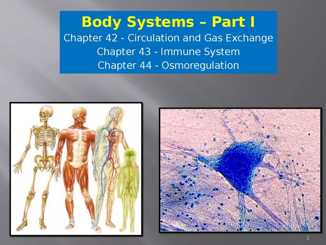
/
Body Systems – Part I Chapter 42 - Circulation and Gas Exchange Body Systems – Part I Chapter 42 - Circulation and Gas Exchange

Body Systems – Part I Chapter 42 - Circulation and Gas Exchange - PowerPoint Presentation

phoebe
phoebe . @phoebe
Follow
27 views
Uploaded On 2024-02-03

Body Systems – Part I Chapter 42 - Circulation and Gas Exchange - PPT Presentation

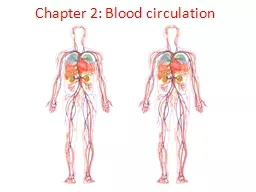
Chapter 43 Immune System Chapter 44 Osmoregulation 1 Chapter 42 Circulation and Gas Exchange 2 Circulation in Animals Diffusion not sufficient for transport of substances oxygen carbon dioxide glucose etc because it is too slow therefore transport systems that can move substances i ID: 1044711

cells blood body water blood cells water body system antibodies oxygen heat heart cell transport lungs air antigen fluid

Share:

Link:

Embed:

Download Presentation from below link

Download Presentation The PPT/PDF document "Body Systems – Part I Chapter 42 - Cir..." is the property of its rightful owner. Permission is granted to download and print the materials on this web site for personal, non-commercial use only, and to display it on your personal computer provided you do not modify the materials and that you retain all copyright notices contained in the materials. By downloading content from our website, you accept the terms of this agreement.


Presentation Transcript

1. Body Systems – Part IChapter 42 - Circulation and Gas ExchangeChapter 43 - Immune SystemChapter 44 - Osmoregulation1

2. Chapter 42 Circulation and Gas Exchange2

3. Circulation in AnimalsDiffusion not sufficient for transport of substances (oxygen, carbon dioxide, glucose, etc) because it is too slow; therefore transport systems that can move substances in bulk are necessaryEx. Heart pumps blood to all parts of the body and the blood carries oxygen to all the body cellsInverts = have either a gastrovascular cavity OR a circ. System for transportOpen and Closed Circ. SystemsThree Basic components: 1. Circ. Fluid (blood)2. Tubes (blood vessels)3. Pump (heart)2 chambered heart (fish) vs. 3 chambered heart (amphibian) vs. 4 chambered heart (mammal)3

4. OPEN Circ. Systems-Blood and interstitial fluid are combined and called hemolymph (NO DISTINCTION between the two)-Heart pumps hemolymph into sinuses, which are spaces that surround the organs-Hemolymph NOT confined to blood vessels- The heart draws hemolymph into the circulatory system through pores called ostiaCLOSED Circ. Systems-Blood is distinct from interstitial fluid; confined to blood vessels-Materials are exchanged by diffusion between blood and interstitial fluid that bathes the cells4

5. Atrium (atria = pl.)  receives blood; thinner walls (collection)Ventricles  pumps blood; thicker, muscular walls for pumpingBlood vesselsArteries – carry blood away from heart (oxygenated); towards capillariesVeins – carry blood toward the heart (non-oxygenated); from capillariesCapillaries – microscopic vessels with thin, porous walls; good for diffusionAdaptations of Cardiovascular SystemArteryVein5

6. Circuit of Blood Flow Systemic Circulation = branch of circulatory system that supplies all body organs then returns oxygen-poor blood back to atrium (pumps to body)Pulmocutaneous Circulation = leads capillaries to the gas-exchange organs (lungs, skin, etc) where the blood picks up oxygen and then returns to the LEFT atrium; from here pumped to systemic circulation (to rest of body) (pumps to lungs)Combination of these two things is called double circulationRIGHT Atria  RIGHT Ventricle  LUNGS  LEFT Atria  LEFT Ventricle  BODY6

7. Fish, amphibians, mammals, and birdsFish  two main chambers (one atrium, one ventricle); TWO CHAMBERED HEART; single circuit (systemic circulation)Path of Blood: Pumped from ventricle  gills (exchange CO2 for O2)  capillary beds (oxygenates rest of body)  veins  atriumAmphibians  three main chambers (two atria, one ventricle); THREE CHAMBERED HEART; double circulation (both systemic and pulmocutaneous circulation)7

8. Mammals and Birds four chambers (two atria, two ventricles); FOUR CHAMBERED HEART; double circulation- Evolved to support the endothermic way of lifePumping cycle of Mammalian Heart: Blood from body gets brought back to RA via 2 vena cavaBlood goes from RA to RV where it gets pumped to the LUNGS via pulmonary arteryOxygenated blood comes back to heart to the LA via the pulmonary veinBlood goes from LA to LV to get pumped to the rest of the body via the aortaMammals and Birds  4 Chambered Hearts8Superior vena cava (anterior vena cava) = brings oxygen poor blood from the head, neck and forelimbs back to the heartInferior vena cava (posterior vena cava) = brings oxygen poor blood from the trunk and hind limbs back to the heart

9. Cardiac CycleRegulated by cardiac cycle-Systole = contraction phase-Diastole = relaxation phase-Cardiac output = amt of blood pumped per minute; regulated by two things: -Heart rate (beats/min)-Stroke volume (amt pumped by LV)9

10. Valves of the HeartAV (Atrioventricular) Valve  between each atrium and ventricle; keeps blood from flowing back into the atriaSemilunar Valve  at the two exits of the heart (where pulmonary artery leaves the RV and where the aorta leaves the LV); prevents the blood from flowing back into the ventriclesPacemaker of the heart  Sinoatrial (SA) node = sets the rate and timing at which all the cardiac muscles contract (found in the right atrium)-Makes sure the atria contract in unison and then, once they are emptied, the ventricles all contract in unison -Uses electrical currents to maintain timing Heart Murmur = defect in one for the valves; blood can leak back inYOU NEED TO KNOW THE PARTS OF THE HEART!! Understand this picture and the blood flow in the heart!10

11. Structural Differences in arteries, veins, and capillaries (correlate with FUNCTION)-ARTERIES  THICKEST; three layers of tissue; innermost layer = endothelium (flattened cells; minimize resistance to blood flow; good for diffusion); thick middle and outer layers for strength when pumping blood-VEINS  three layers of tissue; thinner middle and outer layers than arteries; lower velocity and lower pressure…so do not need to be so thick-CAPILLARIES  only the endothelium (one layer); facilitates exchange of substances; the velocity of the blood flow is the LOWEST in the capillaries11

12. Blood Pressure-Highest in arteries during times of contraction (systole); lower in veins-Stress raises blood pressure b/c it constricts blood vessels-BP is determined by both cardiac output AND peripheral resistance (the impedance of blood flow by the arterioles)-When being measured, the first number is the systolic pressure (contraction) and the second number is the diastolic number (relaxation)Average BP of an adult = 120/7012

13. Lymphatic SystemThe lymph system is a series of vessels and organs called lymph nodes that is separate from the circulatory system that returns fluid and proteins to the blood; it also helps defend the body against infectionLymph = fluid in the lymph system (blood, proteins, etc – similar makeup as interstitial fluid)13

14. BLOODPLASMA-Composition = 90% water and inorganic salts -Acts as a buffer against pH changes, helps maintain osmotic balance, and contributes to the blood’s viscosity-The kidney maintains plasma electrolytes at specific -Blood is connective tissue with cells suspended in plasma (liquid matrix)-When taking blood, you can separate out the cellular parts and the plasma concentrationsCellular Components: RBC’sWBC’sPlateletsFunction of the Plasma: BufferViscosity of BloodEscorts for lipidsAntibodiesClotting factors14

15. Two major classes: red blood cells (RBC’S) and white blood cells (WBC’s)Red Blood Cells (ERYTHROCYTES)-Function = transport oxygen-Most numerous-Large SA for diffusion-Lack nuclei (therefore more space for hemoglobin, about 250 million per cell)Red Blood Cells-Hemoglobin = iron-containing protein that transports oxygen-Lack mitochondria and make ATP via anaerobic metabolism-Lifespan = about 3-4 months (phagocytic cells in liver and spleen destroy them)- Produced in the bone marrow15

16. White Blood Cells (LEUKOCYTES)-Function = fight infection-Spend most of their time outside the circ. system in the interstitial fluid and the lymphatic system-5 types: -Monocytes (phagocyte)-Neutrophils (phagocyte)-Lymphocytes (B and T cells)-Basophils-EosinophilsWBC attacking bacterium16

17. Platelets - Function = clotting Fragments of cells Lack nucleiBlood ClottingBlood contains a sealant that clots the blood; it is always present in its inactive form called fibrinogen. When the blood needs to clot, the enzyme thrombin converts fibrinogen to the active form FIBRIN (which forms the main part of the blood clot)All the cellular elements (RBCs, WBCs, and platelets) in blood are made from multipotent stem cells (potential to differentiate into any type of blood cell) in the red marrow of bonesScab = external clotBruise = internal clot17

18. Cardiovascular Diseases Heart attack (also called Myocardial infarction) = death of cardiac muscle tissue; resulting from blockage of one or more coronary arteries (arteries that supply oxygen-rich blood to the heart)Stroke = death of nervous tissue in the brain; due to blockage/rupture of arteries in the head; the effect of the stroke depends on the extent and location of the damaged brain tissue*Leading cause of Death in the US*18

19. -Hypertension = high blood pressure; promotes atherosclerosis and increases risk of heart attack and stroke; controlled by diet, exercise, and healthy life style-These diseases are partially genetic; also due to lifestyle-CholesterolLDL’s (low-density lipoprotein) = “bad cholesterol”; forms plaquesHDL’s (high-density lipoprotein) = “good cholesterol”; looks for excess cholesterol and returns it to the liver (decreases cholesterol)-Atherosclerosis = plaques develop on the arteries and narrow their diameter-Arteriosclerosis = hardening of the arteries (calcification of plaques); a form of atherosclerosis19

20. Gas Exchange in Animals (Respiratory System)-Respiratory medium = source of oxygen (aquatic animals = water; terrestrial animals = air)…more oxygen available from air than water-Respiratory surface = part of animal where gases are exchanged; CO2 and O2 diffuse here: -Usually thin and have large surface areas-Must be moist-Some animals use entire outer skin as a respiratory surface (ex. Earthworm)-Most common respiratory organs = gills, tracheae, lungsThe warmer and saltier the water, the LESS oxygen it can hold20

21. Gills-Outfoldings of the body surface that are suspended in water-HIGH surface area-Problems with getting oxygen from water: -Oxygen concentration in water are low (as compared to air)-The warmer and saltier the water, the less dissolved oxygen it can holdVentilation  process that increases the flow of water over the gills to maximize oxygen consumption21

22. Countercurrent Exchange  efficient process of oxygen being transferred from the water to the blood; MAXIMIZES DIFFUSION!-The blood travels through the capillaries the opposite way of how the water is flowing over the gills-As blood moves through the gill capillary, it becomes more and more loaded with oxygen, but it simultaneously encounters water with even higher oxygen concentrations because the water is just beginning its passage over the gills; THIS MEANS THAT ALONG THE ENTIRE LENGTH OF THE CAPILLARY, THERE IS A DIFFUSION GRADIENT FAVORING THE TRANSFER OF OXYGEN FROM THE WATER TO THE BLOOD!Because of the efficiency of the countercurrent exchange system, the gills can remove 80% of the oxygen dissolved in the water passing over the respiratory surfaceMaximizes DIFFUSION!22

23. Tracheal Systems-Found in insects-Made up of air tubes that go throughout the body; largest tubes (tracheae) open to the outside-Tracheoles (smaller tubes) take oxygen to the individual body cellsTrachea are reinforced with rings of chitin so they don’t collapse23

24. Lungs-Circulatory system transports gases between lungs and rest of body-Amphibians  small lungs, also rely on diffusion (through skin for example)-Birds, Mammals, and most Reptiles  rely entirely on lungs for gas exchangeGenerally the size of the lungs correlates with the animals metabolic rate24

25. -Located in thoracic cavity-Lungs have a spongy texture with a honeycombed/moist epithelium that acts as the respiratory surfaceMammalian Respiratory System-Path of air: -Nostrils  nasal cavity  pharynx (path where air and food crosses)  larynx (voicebox/ upper part of resp. tract)  trachea (windpipe)  lungs-glottis = opening of the windpipe; when open, we can breathe-epiglottis = flap; covers the glottis when food is there so it doesn’t go “down the wrong pipe”25

26. Process that ventilates lungs is called BREATHINGNegative pressure breathing  process by which mammals ventilate their lungs; PULL air into lungs instead of PUSHING itPositive pressure breathing  how frogs ventilate their lungs; PUSH air into their lungs instead of PULLING it (like gulping air)Trachea forks into two bronchi, one leading to each lung-Each of these branch into finer and finer tubes called bronchioles-Bronchioles dead end into air sacs called alveoli, which is where gas exchange occurs (blood does NOT go into alveoli…this is just where gas exchange happens)26

27. Diaphragm -Inhalation = diaphragm contracts; moves down; lung volume increased (MORE SPACE FOR AIR)  volume in thoracic cavity increases-Exhalation = diaphragm relaxes; moves up; lung volume reduced (NEEDS LESS SPACE)  volume in thoracic cavity decreasesTidal volume  volume of air animal inhales and exhales with each breath; would differ if someone was at rest vs. exercisingVital capacity  maximum tidal volumeResidual volume  amount of air that remains in the lungs even after we forcefully exhale as much as we can (can never deflate the alveoli completely)When we get old, lungs lose their resilience and the newly inhaled air is mixed with old oxygen-depleted air; this increases the residual volume and decreases the tidal volume = limits effectiveness of gas exchange!27

28. Control centers in brain regulate rate and depth of breathing-Automatic mechanisms regulate our breathing; coordinates respiratory system with cardiovascular system-Breathing Control centers are located in two part of the brain = medulla oblongata and the pons-Sets the basic breathing rhythm-Uses negative feedback so the lungs don’t over-expand (STRETCH SENSORS)-Works by monitoring level of CO2 in blood and regulates breathing accordingly-CO2 reacts with H20 to form carbonic acid, which lowers pH-When the Medulla recognizes a lower pH, it increases the depth and rate of breathing28

29. -Hyperventilating = tricks the breathing center; messes up CO2 to O2 ratio-Breathing center responds to a variety of nervous and chemical signals and adjusts the rate and depth of breathing to meet the changing demands of the bodyHyperventilation29

30. Respiratory Pigments-Major problem: low solubility of oxygen in water-Most animals use special proteins called respiratory pigments that bind oxygen and circulate with the blood; the pigments attract the oxygen molecules-This greatly increases the amount of oxygen that can be carried in the blood-Hemocyanin  found in the hemolymph of arthropods and many mollusks; has copper as its O2 binding component-Hemoglobin  resp. pigment used by almost all vertebrates-Found in red blood cells-Made of 4 subunits; each with an iron center-Iron binds O2; so each hemoglobin molecule can carry 4 molecules of O2-Hemoglobin’s conformation is sensitive; ex. Drop in pH lowers the affinity of hemoglobin for O230

31. CO2 Transport-Hemoglobin also helps transport CO2 and helps in buffering-70% of CO2 is transported in the blood in the form of bicarbonate ions (HCO3), which diffuses into the plasma31

32. Chapter 43 – The Body’s Defenses32

33. The Body’s Defenses -INNATE (Nonspecific) IMMUNITY : response that does not discriminate one infectious agent from another-ADAPTIVE (Specific) IMMUNITY : responds in specific ways to particular microorganisms-3 lines of defense: -First line: external (epithelial tissue; ex. skin and mucous membranes)- NONSPECIFIC-Second line : internal (phagocytic cells and antimicrobial proteins indiscriminately attack invaders) - NONSPECIFIC-Third line : internal (immune system) – SPECIFIC33(MACROPHAGES)

34. Nonspecific Defenses Against Infection-External nonspecific defense-Skin and mucous membrane is first line of defense; it inhibits microbes from entering the body-Skin has a pH of 3 to 5 (acidic enough to kill some microbes)-Secretions (saliva, tears, mucous) contain antimicrobial proteins-Contains lysozymes, an enzyme that digests cell walls of bacteria-Mucus traps microbes 34

35. Monocytes-5% of wbc’s-Circulate in blood then migrate into tissue where they form macrophages (“big eaters”); very large-Some migrate throughout the body and some stay in certain tissues; phagocytic-Internal nonspecific defense ® depends on phagocytosis (ingestion of invading organisms by white blood cells); uses phagocytes, NK cells, inflammation, and antimicrobial proteins  NONSPECIFIC IMMUNE RESPONSES: MACROPHAGES ENGULFING BACTERIA/ VIRUSESNeutrophils-60-70% of all wbc’s-Damaged cells release signals that attract neutrophils; they then attack and engulf the invaders-Self-destruct; lifespan = few daysEosinophils-1.5% of wbc’s-Defend against larger parasites (ex. Blood flukes)-Position against invader and secrete destructive enzymesNatural Killer (NK) cells-Destroy virus-infected body cells (don’t attack the microorganism directly)35

36. -Interferons: limit cell-to-cell spread of viruses-Chemicals secreted by virus-infected cells-Diffuses to neighboring cells and induces them to make other chemicals that hinder viral reproductionAntimicrobial Proteins -Nonspecific defense: can attack microbes directly OR impede reproduction-Complement System: a biochemical cascade that helps, or “complements”, the ability of antibodies to clear pathogens from an organism; it is part of the immune system; it consists of a number of small proteins found in the blood (generally synthesized by the liver), that normally circulate as inactive precursors (pro-proteins) ; the complement system may amplify the inflammatory response, enhance phagocytosis, or directly lyse pathogens; the complement system is activated by the onset of the immune response or by surface antigens on the microorganisms or other foreign cells 36

37. Inflammatory Response-Blood clotting marks beginning of repair process (blocks spread of microbes)-Phagocytes migrate to area of infection within the hour-Chemokines – chemicals that attract phagocytes to area of injuryCaused by damage to a tissueIncreased blood supply to area to bring phagocytic cells- Initiated by chemical signals (both from microorganism itself OR released from the body)-When the basophils are injured, they release histamine which triggers dilation and increased permeability of nearby capillaries - Histamine: causes the inflammatory response; main effect: dilates blood vessels-Characteristic redness, heat, fever, edema- Cytokines (signaling molecules)  increase local blood supply and cause the characteristic redness and heat of inflammation37

38. How SPECIFIC Immunity Arises-Lymphocytes = key of immune system; provide specificity and diversity; originate from pluripotent stem cells in the bone marrow or liver of a developing fetus; two main types: -B lymphocytes (B cells) – cells remain in bone marrow and become B cells; produce antibodies (HUMORAL IMMUNITY; extracellular pathogens and antibodies)-T lymphocytes (T cells) – cells migrate from the bone marrow to the thymus (gland above the heart) and develop into T cells (CELL-MEDIATED IMMUNITY; intracellular pathogens)-Lymphocytes are concentrated in the spleen, lymph nodes, and lymphatic tissue; circulate throughout the blood and lymph-Antigen = foreign molecule that elicits a response from a lymphocyte; causes antibodies to be made (stands for ANTIbody GENerating foreign macromolecule)38

39. -Epitope  part of the ANTIGEN where the antibody binds to- One antigen may have several epitopes-Antibodies make up the group immunoglobulins (proteins)-Antibodies usually have 2 identical antigen-binding sites specific for the epitope that provoked its productionStructure of an Antibody-Two identical heavy chains and two identical light chains linked together by disulfide bridges in a Y-shape-At the tips of the Y, there are variable regions (V) …they vary from antibody to antibody. -The rest of the antibody is made up of constant regions (C) …they vary little from antibody to antibody-Five different classes of antibodies; based on differences in the C regions of the heavy chainsAntibody Structure/ Function 39

40. MHC- Major Histocompatibility Complex-Two main types of T cells (each responds to either MHC I or MHC II):-Cytotoxic T cells (Tc) – bind to MHC I molecules; kill infected cells-Helper T cells (TH) – bind to MHC II molecules-Antigen-Presenting Cells (APC’s) ® cells that ingest bacteria and viruses and then destroy them; then they present signals to the Helper T Cells, which can activate the humoral and cell mediated immunity-MHC is a family of genes that encodes the glycoproteins in cell membranes-MHC molecules vary from person to person (helps with allowing some survivors for an epidemic – some may be immune to microorganism)- Determines “self” from “non-self”; it is also involved in tissue rejection (transplants)40

41. -B cells and T cells recognize specific antigens by their antigen receptors (on the plasma membrane)-When activated by a microorganism binding to it, Lymphocytes form 2 clones (it divides and differentiates): -Effector cells ® addresses the antigen immediately; can form plasma cells - Effector cells of B cells = Plasma cells  secrete antibodies - Effector cells of T cells = helper T cells and cytotoxic T cells-Memory cells ® long lived; stay around in case of a secondary “attack”-So even if only a few lymphocytes are activated, thousands of cells result specific for that antigen (because of the cloning)-Primary Immune Response: first time a body is exposed to an antigen; 10-17 days to get maximum response (effector B and T cells made)-Secondary Immune Response: second time a person is exposed to an antigen; response is faster; antibodies produced more quickly; these antibodies usually have a higher affinity for the antigen then the original ones did ® this is called Immunological Memory41

42. -Humoral immunity ® B cells activated; defend against free bacteria, toxins, and viruses present in body fluids; Plasma cells secrete antibodies-Cell-mediated immunity ® T cells used; active against viruses and bacteria within infected body cells; cytotoxic T cellsCytokines ® molecules secreted by activated TH cells that stimulate other lymphocytes42

43. The APC engulfs the pathogen, and presents a piece of the protein to the Helper T cell, which activates the Tc and B cellsThe cytotoxic T cell has a molecule called perforin that it injects into infected cells that cause the cell to lyse (pokes holes in the cell membrane) so it can not reproduce and cause further harm to the organism. 43

44. How do antibodies work? -Antibody binds to the antigen to form the antigen-antibody complex -Neutralization  binding blocks the activity of the antigen-Opsonization  bound antibodies enhance attachment of macrophages (yields phagocytosis)-Agglutination  clumping of bacteria or viruses; occurs b/c each antibody has at least two antigen binding sites; these large complexes are phagocytosed by macrophages-Complement Fixation  activation of the complement system (serum proteins are activated); yields lysis of many types of viruses and pathogenic cells44

45. What are the steps of a SPECIFIC Immune response?? (to a FREE pathogen – not a virus-infected cell)Antigen engulfed by a macrophage (APC)APC presents antigen and it is recognized by the Helper-T cellHelper-T cell activates B cellsB cells (plasma cells) secrete antibodiesAntibodies recognize antigen and destroy itMemory B cells and Helper-T cells are madeKNOW THESE STEPS!!45

46. First Exposure vs. Second Exposure to antigenYour body can respond MUCH faster and make MANY more antibodies the SECOND time you are exposed to an antigen. This is due to your memory cells and immunological memory. MAKE SURE YOU UNDERSTAND THIS PICTURE!46

47. What happens if your humoral immunity is compromised??When you are initially exposed to a pathogen, you would NOT make antibodies or memory B cells (because you have no humoral immunity)When you are exposed a SECOND time, the response would NOT be faster (like it would in a normal person) because you have no memory cellsSo… what WOULD you be able to do to fight something off? You could still use the cell-mediated part of your immune system (T-cells) and also your non-specific immunity (macrophages)47

48. Immunity in Health and Disease-Active Immunity  you create the antibodies yourself; can be acquired naturally or from vaccination; can make the antibodies again if you are exposed a second time-Passive Immunity  antibodies are transferred from one person to another (ex. Antibodies of a pregnant woman cross the placenta into the fetus; breast milk)-Lasts only as long as those antibodies last (weeks to months)- If a baby is nursing and is exposed to a bacteria that the mom had been previously exposed to, hew would be fine because of the antibodies being passed though the breastmilk. -The immune systems ability to distinguish self from non-self limits blood transfusion and tissue transplantation-Placenta plays an important role in fetus development because a woman’s body DOES NOT reject the fetus as a foreign substance48

49. -Blood Typing-A blood makes antibodies to B-like epitopes-B blood makes antibodies to A-like epitopes-O blood makes antibodies to both A and B-like epitopes-No memory cells with blood cells, so each occurrence is like a primary response-Rh factor (problem if mom is Rh neg and fetus is Rh pos if small amounts of blood cross the placenta – unlike how the ABO antibodies cannot!)The Rh blood group system is the clinically most important blood group system besides ABO. Individuals either have, or do not have, the "Rh factor" on the surface of their red blood cells.If someone is exposed to the “wrong” type of blood, the antibodies will bind to the “new” blood and clump around it  for example if a person with O blood was given a transfusion with type B, the anti-B antibodies in his body would clump around the transfused red blood cells49Blood Transfusions

50. -Allergies are exaggerated responses to certain environmental antigens-Histamines are released when these antigens bind; histamine causes dilation and increased permeability of the small blood vessels; this causes allergy symptoms (runny nose, watery eyes, etc.)-Antihistamines decrease allergy symptoms by blocking receptors for histamine; you would take this if you were allergic to flowers (for example) and were exposed to them-Anaphylactic shock  life-threatening reaction to injected or ingested allergens (ex. bee stings, penicillin, peanuts, fish); triggers abrupt dilation of peripheral blood vessels causing a quick drop in blood pressure; death may occur in a few minutesPeanuts50

51. Autoimmune and Immunodeficiency Diseases-Immunodeficiency Diseases-Either the humoral or the cell-mediated immune systems fail (sometimes both!)-Working on gene therapy’s to fix-Cancers (ex. Hodgkin’s disease), AIDS function as immunodeficiency diseases-AIDS-Immunodeficiency disease caused by a virus-Susceptible to opportunistic diseases (those that take advantage of weakened immune system)-Caused by HIV; most lethal pathogen ever encountered-Evolution of drug-resistant strains of HIV is a problem, so they use a combination of drugs-Autoimmune Diseases-Loses tolerance of self and turns against your own cells (ex. lupus, arthritis, MS)-Caused by some failure in immune regulation51

52. Chapter 44 – Osmoregulation and Excretion

53. -Homeostasis  regulating the internal environment-Thermoregulation  regulating the internal temperature-Osmoregulation  regulation of solute balance and gain and loss of water-Excretion  getting rid of nitrogen containing wasteOverview of Homeostasis:-Regulators  regulates internal environment when external environment changes-Conformers  allow internal environment to vary slightly with external changes-Some will conform even if it causes death-Many organisms are in the middle of the range (regulate in some situations, conform in others)-Maintaining homeostasis is very energetically expensiveKey Terms

54. Water Balance and Waste Disposal-Osmoregulation  management of body’s water content and solute composition-Main goal = maintain composition of cytoplasm-Composition of interstitial fluid is controlled by managing the comp of blood (ex. Kidneys)-Transport Epithelia  layers of epithelial cells that regulate solute movement; control which direction and how much solute moves-Ensures solutes must move through a semi-permeable membrane

55. Isoosmotic, Hypoosmotic, Hyperosmotic SolutionsIsoosmotic  solutions on both sides of the membrane have the same osmolarity; no NET movement across the membraneHypoosmotic  LESS solute in the solution, MORE solute in the cell; cell swellsHyperosmotic  MORE solute in the solution, LESS solute in the cell; cell shrinks

56. -Osmoconformer  isoosmotic to surroundings; marine organisms; water must have a stable composition-Osmoregulator  not isoosmotic to environment, so control internal osmolarity; enables animals to live in varied environments (land/ freshwater); costs energy! (active transport)-Stenohaline  animals that can’t tolerate sharp changes in external osmolarity (both osmoconformers and osmoregulators)-Euryhaline  animals that can tolerate sharp externals changes in osmolarityOsmoregulators vs. OsmoconformersFor example: if you put an organism that NORMALLY lives in salt water into FRESH water and it could NOT osmoregulate, it would be HYPERTONIC to the environment and gain too much water and die

57. Maintaining water balance in freshwater:-PROBLEM = gain water by osmosis and lose salt by diffusion-Contractile vacuoles-Excrete large amounts of very dilute urine-Actively uptake salt from surroundings-Living in temporary water (ex. Puddle)-Anhydrobiosis  invertebrates lose almost all water and survive in dormant state when environment dries up; when water is added, they come back to lifeMaintaining Osmotic Balance on Land:-Adaptations to reduce water loss = waxy cuticle, body coverings that prevent water loss, nocturnal, eat/drink moist foods, using metabolic water that is produced in cellular respiration- Avoiding dessication

58. Maintaining water balance in the sea:-Most marine invertebrates = osmoconformers-Most marine vertebrates = osmoregulators-Constantly losing water by osmosis (water saltier than internal environments)-Drink large amounts of seawater and external salt is secreted by active transport out of the gills-Very little urine is produced = water conservation-Sharks  kidneys help remove salt; also excreted by rectal gland or lost in feces-Bodies are actually hypoosmotic to ocean; therefore, water diffuses in = produce dilute urine!

59. Transport Epithelia- In most animals, osmotic regulation and metabolic waste disposal rely on transport epithelia to move specific solutes in controlled amounts in specific directions.- In most animals, transport epithelia are arranged into complex tubular networks with extensive surface area.- Some transport epithelia directly face the outside environment, while others line channels connected to the outside by an opening on the body surface.- Transport epithelia in excretory organs often have the dual functions of maintaining water balance and disposing of metabolic wastes.

60. -Ammonia  TOXIC!-Usually converted to a less toxic form; SOLUBLE in water-Animals that secrete ammonia need access to lots of water (common in aquatic species; it diffuses out into the water)-Urea  made by mammals, most adult amphibians, marine fishes and turtles-Less toxic than ammonia; SOLUBLE in water; primary waste product in humans-Produced by liver  combines ammonia with carbon dioxide-Disadvantage: body uses energy to produce it-Nitrogenous waste correlates with phylogeny and habitat-Waste type impacts water balance-Uric Acid  made by land snails, insects, birds, reptiles-Nontoxic; NON-SOLUBLE in water -More expensive to produce than urea (made from ammonia); minimal water loss when secreted; semi-solid paste (think bird poop)-Urea or Uric Acid….how to decide?-Mode of Reproduction  reptile/bird eggs = waste cannot diffuse out; uric acid precipitates out of solution and can be stored as harmless solid in the egg until the baby hatches, urea won’t

61.

62. Excretory Systems-Production of Urine-2 step process -Body fluid (blood, hemolymph, etc) is collected (involves filtration; Usually nonselective-Selective reabsorption-Absorb valuable solutes (glucose, salts, etc)-Nonvaluable materials are left in the filtrate

63. Filtration/ Filtrate/ ReabsorptionAlthough excretory systems are diverse, nearly all produce urine in a process that involves several steps.First, body fluid (blood, coelomic fluid, or hemolymph) is collected.Filtration  solutions go through selectively permeable membranes consisting of a single layer of transport epithelium, driven by hydrostatic pressure.Filtrate  a solution of water and small solutes, such as salts, sugars, amino acids, and nitrogenous wastes that gets filtered outIt is important to recover useful molecules from the filtrate and return them to the body fluids.Reabsorption  excretory systems specifically transport materials into or out of the filtrate through this processValuable solutes—including glucose, certain salts, vitamins, hormones, and amino acids—are reabsorbed by ACTIVE TRANSPORT in excretory systems. The processed filtrate containing nitrogenous wastes is excreted as urine.

64. Excretory Systems= Tubular Theme-Protonephridia: Flame bulb system (function in osmoregulation)-Found in flatworms (Platyhelminthes)-Series of dead end tubules throughout body; capped by flame bulbs with cilia; cilia wave water and solutes through (filtration) and move urine out through the nephridiopores-Tubules can reabsorb solutes-Metanephridia-Found in annelids; each segment has a pair-Fluid enters through nephrostome (internal opening; ciliated funnel) from the coelom; goes through collecting tubule which includes a storage bladder and exits to the outside via the nephridiopore (external opening) -Has internal openings that collect fluids (storage bladder)-Balance excessive water uptake by making dilute urine

65. -Malpighian Tubules-Found in insects and terrestrial arthropods-Remove nitrogenous waste-Outfoldings of the digestive tract; absorb waste from the hemolymph and empties into the lumen; water is reabsorbed and nearly-dry wasted is excreted-Vertebrate Kidneys-Built of tubules and associated with capillaries

66. Functional Unit of Kidney= Nephron-Mammals have a pair of kidneys; 10cm long; supplied with blood by renal artery and renal vein-Urine exits each kidney via the ureter and empties into the urinary bladder which releases urine via urethra during urination (which is controlled by sphincter muscles)-Structure of a kidney-Outer part  Renal cortex-Inner part  Renal medulla-Both renal cortex and renal medulla are associated with many excretory tubules-NEPHRON = Functional Unit-Single long tube-Ball of capillaries (glomerulus) surrounded by Bowman’s capsule

67. -Filtration of Blood occurs as blood pressure forces fluid from the blood in the glomerulus into the lumen of Bowman’s capsule-Pathway of the filtrate: -Three regions of the Nephron(1) Proximal tubule (2) Loop of Henle (descending/ ascending limbs) (3) Distal Tubule COLLECTING DUCT = gets emptied into renal pelvis which gets emptied by ureter

68. -Cortical Nephrons  shorter loops of Henle; confined to renal cortex; make up 80% of mammalian nephrons-Juxtamedullary Nephrons  longer loops of Henle that extend into renal medulla; 20% of mammalian nephronsNephrons lined by transport epithelium-Afferent arteriole  supplies blood to nephrons; branch of renal artery-Vasa recta  capillary system that serves the loop of Henle

69.

70. From Blood Filtrate to Urine-Proximal Tubule-Secretion/reabsorption alter composition of filtrate-Reabsorb 90% of buffer bicarbonate = IMPORTANT!-Most important function: reabsorb water and NaCl-Descending Loop of Henle-Transport epithelium-Very permeable to: water-NOT very permeable to: salts and solutes-Therefore, osmolarity of interstitial fluid becomes increasingly greater as it gets towards the inner medulla (bottom of the loop is most concentrated!) (because water flows out = filtrate becomes more concentrated)-Look at the picture in the book!Understand this picture!

71. -Ascending Loop of Henle-Transport epithelium-Very permeable to: NaCl-NOT very permeable to: water-Therefore, loses salt so filtrate becomes more dilute-Distal Tube-Regulates [K+] and [NaCl] of body fluid by varying amount of K+ that is secretedpH regulation-Important in secretion and reabsorption-Collecting duct-Carries filtrate through medulla to the renal pelvis-Water is constantly being absorbed because the interstitial fluid is more concentrated, so water diffuses out  therefore filtrate becomes more concentrated

72. -Mammalian Kidney’s ability to conserve water = key terrestrial adaptation-Different permeabilities in the sides of the loop of Henle maximize reabsorption-Most salt is collected in interior of kidney (inner medulla) -From the cortex to the inner medulla, the interstitial fluid increases in osmolarity; the two solutes that contribute to this gradient are: NaCl and Urea-NaCl diffuses out of the ascending loop-Urea diffuses out of the collecting duct (although most remains in the duct and is excreted)-Therefore, osmotic gradient between the cortex (low concentration) and medulla (high concentration)

73. -ADH – Antidiuretic Hormone  amplifies water reabsorption-Regulates water balance-Made by the hypothalamus-Stored and released by pituitary gland-Osmoreceptor cells in hypothalamus monitor blood-Low water (ex. Sweating) = increase ADH into blood stream which gets to kidney; transport epithelium is then made more permeable to water to reabsorb as much as possible-Negative Feedback = once levels return to normal, less ADH is secreted-Also works vice versa if too much water in blood-Alcohol = inhibits release of ADH; therefore causes excessive water loss and dehydration-Kidney uses much ATP (ACTIVE TRANSPORT of NaCl out of ascending loop higher up)- Juxtamedullary nephrons (long loops) conserve much water-Kidney functions are regulated by: -Nervous System-HORMONAL CONTROLS

74. -JGA – Juxtaglomerular Apparatus  located near afferent arteriole that supplies blood to the glomerulus-Monitors blood pressure and blood volumeAngiotensinogen  Angiotensin II(plasma protein) (enz = renin) (peptide that fns as a hormone)-Angiotensin II  increases BP by constricting arterioles-Also stimulates adrenal glands to release aldosterone (hormone) which causes nephrons distal tubules to reabsorb more Na+ and water  which increases blood volume-ADH and RAAS (Renin-Angiotensin-Aldosterone-System) usually work together-ADH alone = lower Na+ in blood-RAAS = helps maintain [Na+]-ANF – Atrial Natriuretic Factor-Opposes RAAS; inhibits release of renin and inhibits NaCl reabsorption-ANF decreases BP and blood volume

75.

76. Effect of beverages on Urine ProductionWhat would happen if you drank: Salt water  produce little urine because your body would try to retain as much water as possible to balance out the solutePlain (fresh) water  produce a normal amount of urineAlcohol  produce a LOT of urine because alcohol inhibits ADH so you release a lot of water (urinate a lot) and can become dehydrated76

77. This next stuff is not in your notes, but important to go over for your general knowledge and for the test; use these slides for your notes

78. Thermoregulation Thermoregulation  regulation of internal temperature; four processes account for heat loss and gain-Conduction  heat exchange due to direct contact-Water more effective in conducting heat than air-Convection  heat exchange due to movement of air/liquid past a surface-Ex. Heat loss of an animal due to a breeze-Radiation  emission of electromagnetic waves; can transfer heat between objects that aren’t in direct contact-Ex. Animal absorbing heat from radiating sun-Evaporation  removal of heat from the surface of a liquid-Ex. Evaporative cooling

79. -ECTOTHERMS  low metabolic rate; does not generate enough heat to affect body temp; cannot perform rigorous activities for a long amount of time; relies on environment to heat/cool (lay in sun if hot/ burrow in ground or go in water if cold); have a DECREASED metabolic rate in colder temps and a HIGHER metabolic rate in warmer temps; shows a 1:1 ratio with body temperature and air temperature (see graph on next slide)-ENDOTHERMS  high metabolic rate; generates enough heat to warm body; typically body temp stays the same regardless of the external air temperature-Advantages: -High and stable body temperature-Elaborate circulatory and respiratory systems-High levels of aerobic metabolism; can perform rigorous activities longer than ectotherms-Disadvantages: - VERY EXPENSIVE energetically- Need to consume more food to support metabolic rateEndothermEctotherm

80. Endotherms vs. EctothermsUnderstand these lines!!!80

81. Behaviors/Adjustments that balance heat gain/loss  can adjust the RATE of heat exchange:-Insulation (fat, feathers)-Adaptations in circulatory system: -Vasodilation  increase diameter of superficial blood vessels (vessels near the surface of skin); warms skin; cools body b/c heat radiates-Vasoconstriction  decreases diameter of superficial blood vessels; warms body b/c minimal heat loss

82. Feedback Mechanisms in Thermoregulation-Hypothalamus  part of brain that controls thermoregulation; thermostat of body; this signals when to activate heat loss/gain mechanisms

83. MORE BEHAVIORS/ADJUSTMENTS THAT BALANCE HEAT GAIN/LOSS: -Countercurrent Heat Exchanger  special arrangement of blood vessels that helps trap the heat in the core of the body; the heat goes towards the blood returning to the body (not the blood going towards extremities); minimizes heat loss to environment-Cooling by Evaporative Heat Loss-Behavioral Responses  shade/ sun; hibernation; migration-Endotherms only: Change the rate of metabolic heat production (make more heat in colder temps)

84. -Torpor  physiological state where activity and metabolism are low-Saves energy-Hibernation  long term torpor-Body temperature decreases-Metabolic rate is 100’s of times lower than “normal”-Energy is obtained from body tissue-Various types of torpor (Ex. Belding’s ground squirrel – wakes up every two weeks)-Estivation  summer torpor-Daily torpor  sleeping at night/day; controlled by internal clockGodless Killing Machine

85. Q-10 EffectQ10 effect  rates of enzyme-mediated reactions increase 2-3 times for every 10 temperature increaseWe will practice some of these problems with a worksheet later…

86. HomeostasisInsulin  released by pancreas into the blood; tells the body cells and liver to take up glucose and DECREASES BLOOD SUGARGlucagon  pancreas releases this and liver and body cells break down glycogen and release glucose to blood to INCREASE BLOOD SUGARHomeostasis is the body’s attempt to maintain a relatively constant internal environment (ex. pH, temperature, blood sugar levels, etc). They maintain a variable (such as body temperature) at or near a particular set point. Fluctuations above/below that set point serve as a stimulus for action.