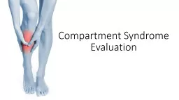
Acute Joint Dislocation Saleh WaslAllah Alharbi Professor KSU Objectives Compartment Syndrome CS To explain the pathophysiology of CS To identify patients at risk To be able to diagnose and manage CS ID: 1044460
Download Presentation The PPT/PDF document "Orthopedic Emergencies Compartment Syndr..." is the property of its rightful owner. Permission is granted to download and print the materials on this web site for personal, non-commercial use only, and to display it on your personal computer provided you do not modify the materials and that you retain all copyright notices contained in the materials. By downloading content from our website, you accept the terms of this agreement.
1. Orthopedic EmergenciesCompartment SyndromeAcute Joint DislocationSaleh WaslAllah AlharbiProfessorKSU
2. ObjectivesCompartment Syndrome (CS)To explain the pathophysiology of CS.To identify patients at risk.To be able to diagnose and manage CS.To be able to describe the complications of CS.
3. CSWhat is compartment?
4. CSWhat is compartment?مقصورة,غرفة, حيز
5. CSNormal blood flow is impaired.Artery- arteriole- capillary- venule- vein.Tissue perfusion failing.
6. CS Hypoxia
7. CSBP 120/80 + - 10Tissue pressure should be less than diastolic pressure by 30 mm Hg.
8. CSDefinition:Compartment syndrome develops when there is excessive, sustained increase of local tissue pressure in a closed compartment.
9. CSRisk Factors(edema)Elevated tissue pressureTense tissues Impaired diffusion / hypoxiaCell damageMore swelling , more hypoxiaVicious circle
10.
11. CSLocal causes:Trauma (crush, fracture open/closed)InjectionBleedingProlong vascular occlusion (reperfusion inj)BurnsVenomous biteIV extravasationPost opBandages
12. CSGeneral causes:HypotensionHead injury
13. CSDiagnosisEarly Pain out of proportion to injury Pain with stretching fingers / toes Risk factors High index of suspicion Measurement of compartment
14.
15. DiagnosisLate Numbness, parasthesia, weakness, Paralysis Pulseless Tooooo Late
16. DiagnosisS/S Pallor Altered perfusion Diminished pulses or pulselessness Altered capillary refill Palpable fullness or tenseness of a compartment, the forgotten "P" Altered sensibility Pain on passive muscle stretch
17. CSManagementInitial ( undeveloped) CS Remove any bandages/ cast/ brace … Maintain normal BP Keep limb at heart level Regular close monitoring (15-30 min) Avoid sedation, nerve block ( pt feedback)
18. CSManagementFully developed CS Above plus Diuretics to flush kidneys Urgent surgical decompression (Fasciotomy)
19. CS
20. CS
21. CSFasciotomy Decompress all compartments Allows muscles to expand Thus, Reduction compartment pressure Stops further damage Should be done very early If too late, shouldn`t be done
22. CSFasciotomy Debridement of all necrotic tissue Second and third debridement needed Skin closure/graft after few days
23. CSFasciotomy Indications: 6 hours of ischemia significant tissue injury Worsening limb condition Developed clinical evidence of CS In doubt
24. CSComplications:Myonecrosis-----Myoglobinuria----kidney tubular damageLimb contractures/paralysis/sensation loss
25. CSComplications:- Leg: Anterior compartment (foot drop) Deep post compartment (clawed toes/anesthesia sole) Volar compartment (acute Volkman’s ischemia/contracture)
26. CS
27. Acute Joint DislocationAJDObjectives To describe mechanisms of joint stability To be able to diagnose AJD To know general principles of management To describe possible complications in major joints (shoulder,hip,knee)
28. AJD
29. AJDJoint stability:Bony stability Shape of bone ends (ball and socket/flat)Soft tissues Dynamic stabilizers: Tendons/muscles Static Stabilizers: ligaments/mensci/labrum
30.
31. Hinge joint
32. Condylar
33. Pivot
34. Plane
35. Saddle
36. Ball and socket
37. StabilityComplex synergy leading to FUNCTIONAL stability
38. AJDHigher energy is needed to dislocate a bony stable joint than a joint with mainly soft tissue stability.Example: Hip and Shoulder
39. AJDDislocation of major joint is associated with other injuries.
40. AJDRisk Major trauma victims Athletes Connective tissue disease patients
41. AJDWhen a joint is strained: it may sprain it may fracture it may dislocate it may fracture and dislocate
42. AJDSome joints dislocate in one or two directions depending on the force,,, (hip)Others may dislocate in different directions (shoulder)
43. AJDA joint dislocation is described in reference to the distal segment (shoulder dislocation)
44. Damage to the labrum Bankart’s lesion, and capsule.Damage to the head of humerus.
45. Knee dislocation
46. Knee dislocation
47. S/SHistory of traumaPain and pt is holding limbInability to use limbDeformity loss of contourShorteningMalalignmentMalrotationCheck NV status and CS
48. DiagnosisHistory and physical examX ray urgent ( no delay) (special views)
49. AJDManagement principles:Exclude other injuriesPain controlUrgent reductionCheck stabilityCheck NV after reductionXray post reductionProtect the jointRehabilitationLook for late complications
50. AJDManagement:Better with anesthesia. WHYUrgent Closed reduction firstIf fail open reduction
51. AJDComplicationsEarlyNV injuryCSFracturesOsteochondral lesion/fractureHeterotopic calcification
52. AJDComplicationsLateStiffnessChronic instabilityAVN/ avascular necrosisArthrosis
53. AJDSpecial considrations:Hip jointPost dislocation is commonestDashboard injury with hip flexedSciatic nerve injury commonLate AVNAn orthopedic emergency
54. AJDSpecial consideration:Shoulder dislocationCommonAnterior is more commonPt with seizures prone to posterior dislocationMay cause chronic instabilityChances of axillary nerve injury
55. AJDSpecial consideration:Knee dislocationHigh energy traumaThree ligaments or morePopl artery injury (serious emergency)Peroneal nerve injuryFracture/CSRequire additional reconstructive surgeryPost reduction arteiogram
56.