/
Preliminary Exam Department of Electrical and Computer Engineering Preliminary Exam Department of Electrical and Computer Engineering

Preliminary Exam Department of Electrical and Computer Engineering - PowerPoint Presentation

reese
reese . @reese
Follow
342 views
Uploaded On 2022-02-16

Preliminary Exam Department of Electrical and Computer Engineering - PPT Presentation

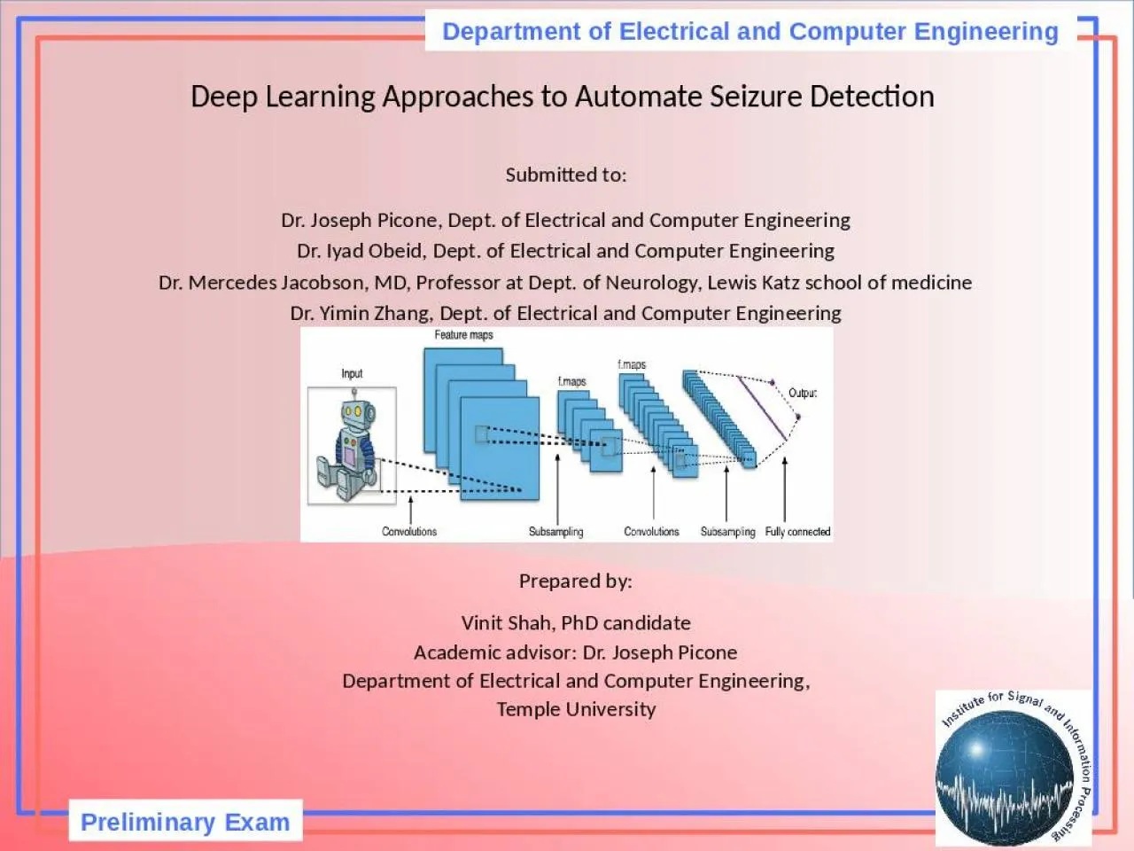
Deep Learning Approaches to Automate Seizure Detection Submitted to Dr Joseph Picone Dept of Electrical and Computer Engineering Dr Iyad Obeid Dept of Electrical and Computer Engineering ID: 909450

dcnn seizure eeg qeeg seizure dcnn qeeg eeg seizures filters svm detection neural hour convolutional cnn tools electrical filter

Share:

Link:

Embed:

Download Presentation from below link

Download Presentation The PPT/PDF document "Preliminary Exam Department of Electrica..." is the property of its rightful owner. Permission is granted to download and print the materials on this web site for personal, non-commercial use only, and to display it on your personal computer provided you do not modify the materials and that you retain all copyright notices contained in the materials. By downloading content from our website, you accept the terms of this agreement.


Presentation Transcript

Slide1

Preliminary Exam

Department of Electrical and Computer Engineering

Deep Learning Approaches to Automate Seizure Detection

Submitted to:

Dr. Joseph

Picone

, Dept. of Electrical and Computer Engineering

Dr.

Iyad Obeid, Dept. of Electrical and Computer EngineeringDr. Mercedes Jacobson, MD, Professor at Dept. of Neurology, Lewis Katz school of medicineDr. Yimin Zhang, Dept. of Electrical and Computer Engineering

Prepared by:

Vinit Shah, PhD candidate

Academic advisor: Dr. Joseph

Picone

Department of Electrical and Computer Engineering,

Temple University

Slide2

Outline

Advances in Neurology community and QEEG tools

Intra-patient variation in seizure morphologySupport Vector Machine (SVM)Convolutional Neural Network (CNN)

Doubly Convolutional Neural Network (DCNN)

Slide3

Common issue in neurology community

Epileptic seizures affects approximately 1% of the world’s population. (Annegers

, 1997)Scalp Electroencephalogram (EEG) monitoring is a non-invasive and convenient method to assess electrical activity from brain.Assessment of long-term monitoring (LTM) EEGs is tedious, time consuming and susceptible to missing events-of-interest such as seizures.

Slide4

qEEG tools

Quantitative EEG (qEEG) is the analysis of the digitized EEG. Various transformation techniques have been applied using DSP for visual interpretation of EEGs to assist and even augment our understanding of the EEG and brain function.DSP algorithms such as Fourier and Wavelet analysis have been applied to develop qEEG

/cEEG slides such as envelop trend (aEEG), color density spectral array (CDSA), rhythmicity spectrogram, asymmetry index, etc..

Slide5

Background

Slide6

Seizure

Slide7

Seizure offset

Slide8

What if ?

Patterns during Periodic/Rhythmic discharges or low amplitude status epilepticus would seem confusing.

Slide9

Example of 1-hour QEEG panel

Slide10

Experiment at Emory University

Total of 18 ANCS certified Neurophysiologists participated.

15 epochs were selected containing 126 seizures

9 Neurologists created the gold standard seizure data by reviewing raw EEGs.

Remaining 9 reviewed it using two methods:

QEEG + raw EEG

QEEG only

Automatic seizure detection algorithm (

Persyst

Inc.) was run on this data.

1 min. and 2.5 min. variation was allowed for detection of seizure onset.

Allowed margins for Seizure identification

Q

QR

SzD

Sensitivity

FA

Sensitivity

FA

Sensitivity

FA

1 min. Onset variation

51%

1/hour

63%

0.5/hour

25%

0.07/hour

2.5 min. Onset variation

67%

1/hour

68%

0.5/hour

27%

0.07/hour

Slide11

Results

Prolonged

Low

Amplitude

Seizures

Brief

Low

Amplitude

Seizures

Slide12

q

EEG tools Vs. SzD

From comparison between implementation of QEEG tools Vs. SzD (automatic seizure detection algorithm). QEEG seems more reliable and its common practice in hospitals recently.On the other hand, reviewing QEEG slide’s could have less temporal resolution and it is prone to missing brief and/or slowly evolving seizures.

SzD’s sensitivity is 26.2% to 26.7%.

Slide13

Intra-patient varying seizure morphologies

Example: 1

Slide14

Example: 2

Intra-patient varying seizure morphologies

Slide15

Preprocessing EEG signals

EEG signal for P number of channels can be defined as:

The nonlinear energy operator (NLEO):

[n] =

[n – 1]

[n – 2] -

[n]

[n – 3]

Frequency-weighted in left half of the window is subtracted from that of the right half of the window:[n] =

Threshold value for boundaries can be defined as:

T[n] =


= [

[n]…..

[n]]


Slide16

Pictorially….

Slide17

Support Vector Machines

SVM is a discriminative classifier which constructs a hyperplane (or set of hyperplanes) in a higher dimensional space.

To classify datapoints

, SVM uses the sign of the decision function:f(x) =

For standard two class SVM classification, solution to following formula is needed:


Slide18

Multiclass SVM classification

In case of multiclass classification, the task specific separating hyper-plane is defined as:

And optimization problem becomes:

Once optimized, the multitask

and

can be obtained from w of the standard SVM.


Slide19

Binary class hyperplane vs. Multiclass hyperplane

Let’s throw out task-specific component; then classification function becomes:

f(x) =


Slide20

Results on CHBMIT seizure database

Slide21

CNNs are special kind of neural network for processing data that has a grid like topology.

Typical CNN Layer:

Let’s move on to Deep learning (CNN)

Slide22

Best features to recognize output image ?

Whether it’s X shaped or not…?

Intuitively we can see three of them here.

Slide23

For first Feature patch

Convolution step to generate feature maps

Similarly for all three features

Slide24

Pooling

2

filter size with stride value set at 2.


Pooling layers reduce the spatial size and number of parameters associated with the network.

Slide25

Using non-linear detector function such as

ReLU (in our case), sigmoid, etc..

Detector stage

Note that our output array has become sparse

Slide26

Note that multiple layers help reduce dimensionality.

Complex layer

Eventually, flattening the data where all the weights are shared with the following layers (Fully connected layers)

Slide27

Backpropagation through stochastic gradient descent method can find out perfect weights for each layer and eventually features by reducing total error.

How did we get features in the first place ?

Slide28

Input image

is a real-valued 3D-tensor, where c is the number of channels; w, h are the width and height.

Let’s define convolution operation as

becomes output image after convolution.


CNN continued

The spatial dimension of the output image

without zero padding remains

+ z – 1 and

, respectively.


Slide29

Zero padding is necessary in DCNN to preserve the shape of the filter for further convolution.

Doubly Convolutional Neural Network (DCNN)

and

are the input and output image, respectively.

are a set of

meta-filters, with filter size

,

.


Slide30

K-translation correlation between two convolutional filter within a same layer.

Main ingredient of DCNN

In CNNs, learned filters are slightly translated version of each other.

By considering correlation between filters inside meta-filters identifies maximally correlated filters.

Slide31

DCNN results on CIFAR-10, CIFAR-100 and ImageNet

Results of CNN variants Vs. DCNN on CIFAR database with and without data augmentation

Results of CNN variants Vs. DCNN on ImageNet

Slide32

NLEO segmentation can help us define the size of meta filter that we are interested in for DCNN operation.

DCNN should be able to learn in more detail the dependency (correlation) between channels for artifact identification.

How to implement DCNN for seizure detection

Slide33

Summary and future work

Although,

qEEG tools are reliable and becoming pervasive to detect most seizures, they are still not efficient to detect brief, slowly evolving or low amplitude seizures. Also,

qEEG tools in conjunction with raw EEG are preferable to detect accurate onset and to reduce false positives.In Neuro-ICU and EMU environment, there is a strong need of automated seizure detection tool which can show significantly low FPR.

Multitask learning approach to train classifiers for various seizure types is a good approach to avoid over-training on most common seizure types for SVM.

DCNN generates meta-filters which looks for highly correlated filters inside the primary patch and then convolution takes place again with such output filters. This additional layer yields highly correlated feature maps then regular CNN approach.The useful approaches to be extracted (for artifact reduction) from these papers should be:

NLEO operator for adaptive segmentation

Applying this Segments as a meta filter size in DCNN approach

.However, the question about detecting seizure with slow evolution and low amplitude remains unanswered.

Slide34

Brief Bibliography

Annegers

, J. F. (1997). The treatment of epilepsy: Principle and practice. Baltimore: Williams and Wilkins.Van

Esbroeck, A., Smith, L., Syed, Z., Singh, S., & Karam, Z. (2016). Multi-task seizure detection: addressing intra-patient variation in seizure morphologies. Machine Learning

, 102(3), 309–321.Shoeb, A., Kharbouch, A., Soegaard

, J., Schachter, S., & Guttag, J. (2011). An algorithm for detecting seizure termination in scalp EEG. In Proceedings of the Annual International Conference of the IEEE Engineering in Medicine and Biology Society, EMBS (pp. 1443–1446).

Goodfellow, I., Bengio, Y., &

Courville, A. (2017). Deep Learning (1st ed.). Cambridge, MA, USA: MIT Press.Zhai, S., Cheng, Y., Zhang, Z. (Mark), & Lu, W. (2016). Doubly Convolutional Neural Networks. In

NIPS (pp. 1082–1090).Haider, H. A., Esteller, R., Hahn, C. D., Westover, M. B., Halford, J. J., Lee, J. W., … Pargeon, K. (2016). Sensitivity of quantitative EEG for seizure identification in the intensive care unit. Neurology, 87(9), 935–44.Brandon, R. (August 18, 2016). How do Convolutional Neural Networks work? Retrieved from: http://brohrer.github.io/how_convolutional_neural_networks_work.html

Slide35

Thank You !