/
Brain Aneurysm Brain Aneurysm

Brain Aneurysm - PDF document

sadie
sadie . @sadie
Follow
342 views
Uploaded On 2022-08-22

Brain Aneurysm - PPT Presentation

What is a brain aneurysm 
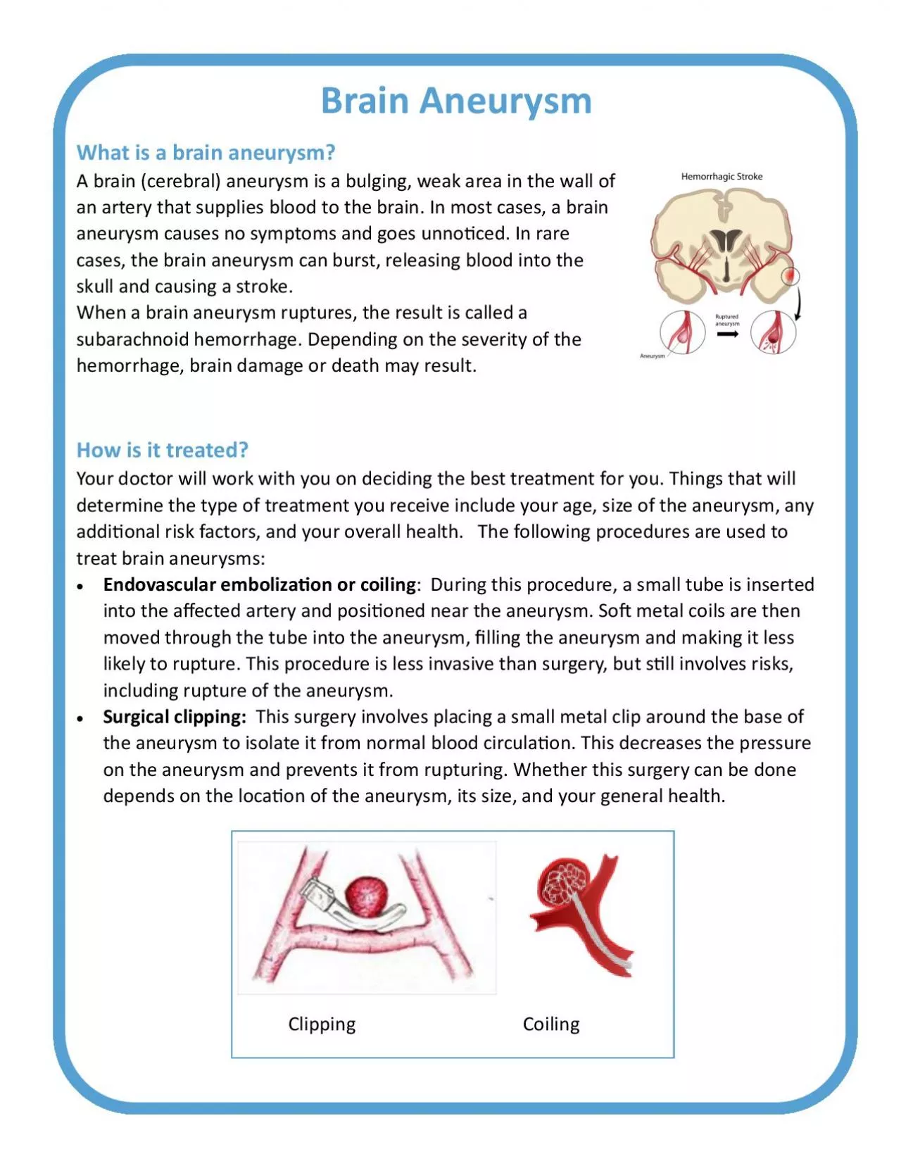
A brain cerebral aneurysm is a bulging weak area in the wall of an artery that supplies blood to the brain In most cases a brain aneurysm causes no symptoms and goes u ID: 939699

aneurysm brain x0066006c x00740069 brain aneurysm x00740069 x0066006c uid blood sah x00660074 pressure treatment called result vasospasm hemorrhage hydrocephalus

Share:

Link:

Embed:

Download Presentation from below link

Download Pdf The PPT/PDF document "Brain Aneurysm" is the property of its rightful owner. Permission is granted to download and print the materials on this web site for personal, non-commercial use only, and to display it on your personal computer provided you do not modify the materials and that you retain all copyright notices contained in the materials. By downloading content from our website, you accept the terms of this agreement.


Presentation Transcript

Brain Aneurysm What is a brain aneurysm? A brain (cerebral) aneurysm is a bulging, weak area in the wall of an artery that supplies blood to the brain. In most cases, a brain aneurysm causes no symptoms and goes unno�ced. In rare cases, the brain aneurysm can burst, releasing blood into the skull and causing a stroke. When a brain aneurysm ruptures, the result is called a subarachnoid hemorrhage. Depending on the severity of the hemorrhage, brain damage or death may result. How is it treated? Your doctor will work with you on deciding the best treatment for you. Things that will determine the type of treatment you receive include your age, size of the aneurysm, any addi�onal risk factors, and your overall health. The following procedures are used to treat brain aneurysms:  Endovascular emboliza�on or coiling : During this procedure, a small tube is inserted into the a�ected artery and posi�oned near the aneurysm. So� metal coils are then moved through the tube into the aneurysm, �lling the aneurysm and making it less likely to rupture. This procedure is less invasive than surgery, but s�ll involves risks, including rupture of the aneurysm.  Surgical clipping: This surgery involves placing a small metal clip around the base of the aneurysm to isolate it from normal blood circula�on. This decreases the pressure on the aneurysm and prevents it from rupturing. Whether this surgery can be done depends on the loca�on of the aneurysm, its size, and your general health. Clipping Coiling Subarachnoid Hemorrhage (SAH) Subarachnoid hemorrhage is usually caused by an aneurysm that ruptures or bursts. Your treatment a�er a SAH includes hospitaliza�on, intensive care and close monitoring of the pressure in your brain, neu

rologic status and vital signs. You may also need treatment to prevent the aneurysm from bleeding again. We will also monitor you for other condi�ons that may result from your SAH, including: Vasospasm or narrowing of the arteries  This may occur up to 21 days a�er an aneurysm bleeds, but the risk decreases a�er 14 days. You may need to stay in the ICU during these �rst 14 days. Vasospasm comes from irrita�on of the blood vessels as a result of your SAH. Vasospasm causes the blood vessels to clamp down, and decreases the �ow of blood through them. This condi�on can cause other strokes and can be fatal if not treated.  During your stay, you will require close monitoring, frequent neurologic checks and will require treatment if vasospasm occurs. Some addi�onal things you may need during your stay include medica�on called nimodipine, transcranial dopplers and IV �uids. Hydrocephalus or �uid buildup in the brain  Some�mes the blood in your brain a�er a SAH can block the pathways that allow the cerebral spinal �uid (CSF) to circulate. CSF is a �uid in your brain that cushions and protects the brain.  The blockage of CSF a�er a SAH can cause a buildup of �uid in spaces in your brain called ventricles. This is called hydrocephalus. Hydrocephalus can cause increased pressure in your brain that can lead to damage.  To treat hydrocephalus, you may need a device to moni- tor brain pressure and drain o� extra brain �uid. This device is called an external ventricular drain (EVD). An EVD is a catheter that is placed by a neurosurgeon into the ventricle or �uid space in the brain. This allows for drainage of the extra �uid and monitoring of your brain pressure.