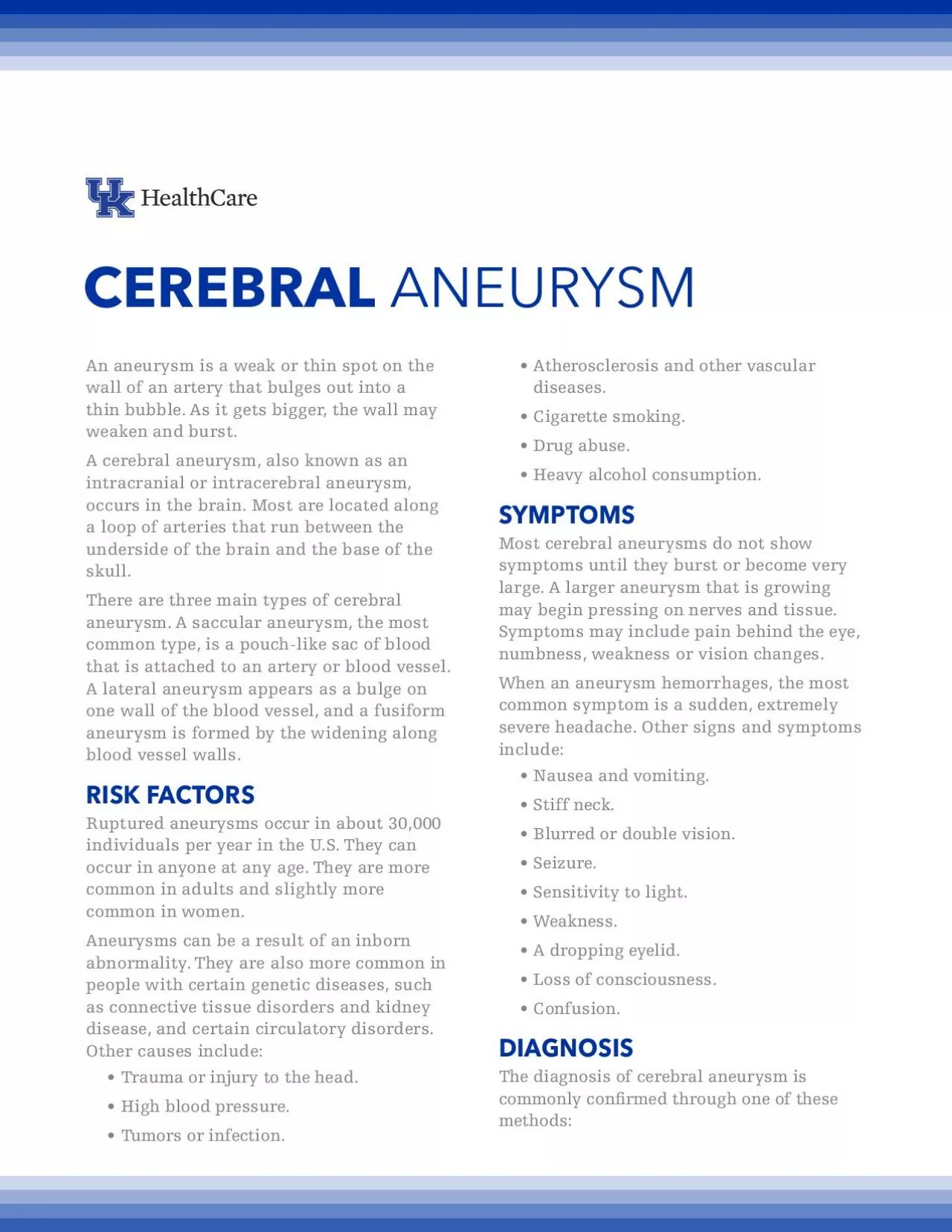
/
CEREBRAL ANEURYSMAn aneurysm is a weak or thin spot on the wall of an CEREBRAL ANEURYSMAn aneurysm is a weak or thin spot on the wall of an

CEREBRAL ANEURYSMAn aneurysm is a weak or thin spot on the wall of an - PDF document

williams
williams . @williams
Follow
342 views
Uploaded On 2022-08-19

CEREBRAL ANEURYSMAn aneurysm is a weak or thin spot on the wall of an - PPT Presentation

Angiography This is a dye test used to analyze arteries or veins An intracerebral angiogram can identify changes in an artery or vein like an aneurysm A 30exible catheter through the body A sma ID: 938178

cerebral aneurysm blood artery aneurysm cerebral artery blood common brain symptoms wall aneurysms kentucky stroke bleeding procedure vessel small

Share:

Link:

Embed:

Download Presentation from below link

Download Pdf The PPT/PDF document "CEREBRAL ANEURYSMAn aneurysm is a weak o..." is the property of its rightful owner. Permission is granted to download and print the materials on this web site for personal, non-commercial use only, and to display it on your personal computer provided you do not modify the materials and that you retain all copyright notices contained in the materials. By downloading content from our website, you accept the terms of this agreement.


Presentation Transcript

CEREBRAL ANEURYSMAn aneurysm is a weak or thin spot on the wall of an artery that bulges out into a thin bubble. As it gets bigger, the wall may weaken and burst. A cerebral aneurysm, also known as an intracranial or intracerebral aneurysm, occurs in the brain. Most are located along a loop of arteries that run between the underside of the brain and the base of the There are three main types of cerebral aneurysm. A saccular aneurysm, the most common type, is a pouch-like sac of blood that is attached to an artery or blood vessel. A lateral aneurysm appears as a bulge on one wall of the blood vessel, and a fusiform aneurysm is formed by the widening along blood vessel walls.RISK FACTORSRuptured aneurysms occur in about 30,000 individuals per year in the U.S. They can occur in anyone at any age. They are more common in adults and slightly more common in women.Aneurysms can be a result of an inborn abnormality. They are also more common in people with certain genetic diseases, such as connective tissue disorders and kidney disease, and certain circulatory disorders. Trauma or injury to the head.Tumors or infection.Atherosclerosis and other vascular Drug abuse.Heavy alcohol consumption.SYMPTOMSMost cerebral aneurysms do not show symptoms until they burst or become very large. A larger aneurysm that is growing may begin pressing on nerves and tissue. Symptoms may include pain behind the eye, numbness, weakness or vision changes. When an aneurysm hemorrhages, the most common symptom is a sudden, extremely severe headache. Other signs and symptoms Nausea and vomiting.Stiff neck.Sensitivity to light.Weakness.A dropping eyelid.DIAGNOSISThe diagnosis of cerebral aneurysm is commonly conrmed through one of these Angiography: This is a dye test used to analyze arteries or veins. An intracerebral angiogram can identify changes in an artery or vein like an aneurysm. A exible catheter through the body. A small amount of contrast dye is used, and a series of x-rays are tak

en, noting any changes.Computed tomography (CT): This is a fast, painless, noninvasive diagnostic tool that can reveal the presence of a cerebral aneurysm brain. Magnetic resonance imaging (MRI): This uses computer-generated radio waves and a powerful magnetic eld to produce images of the brain. It can show the size and shape bleeding in the brain. MRI is painless and noninvasive.Cerebrospinal uid analysis: This is where a small amount of uid is removed by a spinal needle and tested to detect any bleeding or brain hemorrhage. TREATMENTHealthcare professionals can choose from a range of treatments for cerebral aneurysm. Medications are aimed at relieving symptoms and managing complications. Physical, speech or occupational therapy may be needed to relearn skills. Two common treatment options for ruptured Microvascular clipping: This involves cutting off the ow of blood to the aneurysm. A neurosurgeon will place a small, metal clip on the aneurysm, halting blood supply. The clip prevents the risk of future bleeding.A related procedure is an occlusion, in which the entire artery is clamped off. This is performed when the aneurysm has damaged the artery.Endovascular embolization: This procedure is an alternative to surgery, in which the doctor inserts a catheter into an artery and threads it to the site of the aneurysm. The procedure effectively destroys the aneurysm and may have to be performed more than once.The Kentucky Neuroscience Institute (KNI)integrates the expertise of the University of Kentucky’s neurology and neurosurgery clinicians and researchers. KNI is a referral center dedicated to providing comprehensive care to our patients with the most complex For appointments or more information, call https://ukhealthcare.uky.edu/kentucky-neuroscience-instituteNational Stroke Association’s by developing compelling education and programs focused on prevention, treatment, rehabilitation and support for all impacted by stroke. www.stroke.org