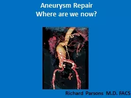
Richard Parsons MD FACS Endovascular treatment of aortic disease Anatomy Indications for repair of AAA Size gt than 5cm Expansion greater than 024 cmyear Symptomatic aneurysm ID: 914406
Download Presentation The PPT/PDF document "Aneurysm Repair Where are we now?" is the property of its rightful owner. Permission is granted to download and print the materials on this web site for personal, non-commercial use only, and to display it on your personal computer provided you do not modify the materials and that you retain all copyright notices contained in the materials. By downloading content from our website, you accept the terms of this agreement.
Slide1
Aneurysm RepairWhere are we now?
Richard Parsons M.D. FACS
Slide2Endovascular treatment of aortic disease
Slide3Anatomy
Slide4Indications for repair of AAASize > than 5cm Expansion greater than 0.2-.4 cm/year
Symptomatic aneurysm Rupture
Slide5Endovascular stent graft repair of Abdominal Aortic Aneurysm(EVAR)First performed in the US in 1994 Has become the most common way to repair AAA 90+% at Abington Hospital
From 2014-2016 we have performed 83 EVARs Length of stay is usually 1 night
Slide6Endovascular repair of Abdominal Aortic Aneurysm
Slide7Technically challenging features of endovascular aneurysm repair
Inverted funnel
Slide8Technically challenging features of endovascular repair
Thrombus
thrombus
neck
Accessory renal artery
Angulated neck
Slide9Complications of repairRenal failure Colonic ischemia
Aortic ruptureEndoleaks
Slide10Bowel ischemia
Bowel ischemia
Colon ischemia
Renal ischemia
Slide11CAUTION—Investigational device. Limited by Federal (or United States) law to investigational use.
Juxtarenal:
AAA
Slide12Post treatment leaks
Slide13Endoleak classification types I-V
Slide14Type I endoleak
Slide15Type II endoleak from IMA retrograde flow
Slide16Thoracic aneurysm
Slide17Endovascular thoracic aortic aneurysm repair
Slide18Slide19Endovascular vs open Thoracic Aneurysm Repair
Repair when 6cm or greater Less painful Shorter length of stayLess morbidity and mortality
Slide20Complications of TAA repairEndoleakGraft migration
Stent fracture Delayed ruptureInfectionParaplegia
Slide21Best case scenario rare !Left subclavian artery not involved
Does not extend below diaphragmSpinal ischemia risk diminished
LSCA
Slide22Risk factors for paraplegiaLong thoracic segment coveragePrevious abdominal aortic repair
Intra or postoperative hypotension
Slide23Slide24Mechanisms to decrease paraplegia riskAvoid hypotension
Stage thoracic and abdominal repair3-6 months apartCSF catheter drainage to decrease spinal cord pressures to be below 10mmEvoked potential monitoring using balloon occlusion
Temporarily creating an endoleak that is later closed
Slide25Thoracic dissection
Slide26Treatment of type B dissection90 % can be treated with BP control and pain medication
Continued pain or aortic rupture requires immediate repairInvasive treatment is reserved for nonperfused vascular bedsMesenteric
Renal
Lower extremity
Late aneurysmal degeneration
Slide27Treatment strategiesOpen fenestrationEndovascular fenestration
Proximal endograft placement to open the true lumen and close the false lumen
Slide28Risks of treatmentAortic ruptureStroke
Spinal cord ischemia( paraplegia)Ischemia of branch vessels( renal, mesenteric,extremeties)
Slide29Dissection Endovascular Stents
STABLE I Trial Enrollment
83 pts. enrolled
US and OUS centers
CAUTION—Investigational device. Limited by Federal (or United States) law to investigational use.
Slide30Remodeling of the aorta after dissection flap is closed
Slide31Treatment of asymptomatic aortic dissectionPrevention of late aneurysm dilatationAortic remodeling occurs in 90% of treated patients
Only 70% of untreated patients remodeled 30% have aneurysmal dilatationUnclear if treatment of all asymptomatic dissections is justified
Slide32Slide33Traumatic aortic dissectionHigh speed deceleration injuryThe aorta is tethered at the ligamentum arteriosum dissection occurs just distal to subclavian
Wide mediastinum on chest X-rayCTA confirms dissection
Slide34Slide35Thank you!
Richard Parsons MD