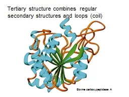
/
Example of Tertiary and Quaternary Structure of Protein Example of Tertiary and Quaternary Structure of Protein

Example of Tertiary and Quaternary Structure of Protein - PowerPoint Presentation

scarlett
scarlett . @scarlett
Follow
345 views
Uploaded On 2022-06-18

Example of Tertiary and Quaternary Structure of Protein - PPT Presentation

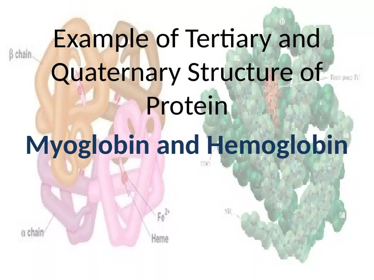
Myoglobin and Hemoglobin Myoglobin Was the first protein the complete tertiary structure was determined by Xtray crystallography Has 8 α helical region and no β pleated Hydrogen binding stabilize the ID: 920336

heme bind oxygen affinity bind heme affinity oxygen bpg group structure c02 release molecule lungs tissues hemoglobin bound protein

Share:

Link:

Embed:

Download Presentation from below link

Download Presentation The PPT/PDF document "Example of Tertiary and Quaternary Struc..." is the property of its rightful owner. Permission is granted to download and print the materials on this web site for personal, non-commercial use only, and to display it on your personal computer provided you do not modify the materials and that you retain all copyright notices contained in the materials. By downloading content from our website, you accept the terms of this agreement.


Presentation Transcript

Slide1

Example of Tertiary and Quaternary Structure of Protein

Myoglobin

and Hemoglobin

Slide2

Myoglobin

Was the first protein the complete tertiary structure was determined by X-tray

crystallographyHas 8 α-helical region and no β-pleatedHydrogen binding stabilize the α-helical regionConsist of a single polypeptide chain of 153 a.acid residue and includes prosthetic group- one heme groupStore oxygen as reserve against oxygen deprivation

Slide3

Hemoglobin

Example of quaternary structure of protein

Consist 4 polypeptide chain -4 subunit- tetramerEach subunit consist one heme group (the same found in myoglobin)The chain interact with each other through noncovalent interaction – electrostatic interaction, hydrogen bonds, and hydrophobic interaction any changes in structure of protein- will cause drastic changes to its property this condition is called allostery

Slide4

Hemoglobin

An

allosteric proteinTetramer, 4 polypeptide chains (α2β2) - 2α-chains and 2β-chains – nothing to do with αhelix and βsheet- its just a greek nameBind O2 in lungs and transport it to cellsTransport C02 and H+ from tissue to lungs

The same

heme

group in

mb

and

hbCyanide and carbon monoxide kill because they disrupt the physiologic function of hemoglobin2,3- biphosphoglycerate (BPG) promotes the efficient release of 02

Slide5

Heme

Group

Mb and Hb contain heme – a prosthetic groupResponsible to bind to 02Consist of heterocyclic organic ring (porphyrin) and iron atom (Fe2+)Oxidation of Fe 2t to Fe3+ destroy their biologic activityFe has 6 coordination sites that can form complexation bondsFour are occupied by the N atomsFree heme

can bind CO 25,000 times than 02 – how Mb and

Hb

overcome this problem?

Slide6

Structure of

heme

group in Mb and HBThe perfect orientation for CO binding is when all 3 atoms (Fe, C and O) perpendicular to the to the plane of hemeMb and Hb create hindered environment- do not allow O2 to bind at the required orientation- less affinityThe fifth coordination is occupied by Histidine residue F8The O2 is bound at the 6th coordination site of iron

Slide7

heme

group

The second histidine His E7 – not bound to the heme, but acts a gate to open and closes as oxygen enter the hydrophobic pocketE7 inhibit O2 to bind to perpendicularly to heme The presence of His E7 – will force CO to bind at the 120 angle – make it lose it affinity to heme

Slide8
Oxygen saturation in Mb and

Hb

One molecule of Mb- can bind one molecule 02

HB (4 molecule)- can bind 4 02O2 bind to HB thru positive cooperativity – when one O2 is bound, it become easier for the next to bindDissociation of one O2 from oxygenated Hb will make the dissociation of 02 from other subunits easier

Slide9
Different form of HB

Hb

is bound to 02-

oxyhemoglobin – relaxed (R state)Without 02 – deoxyhb – tense (T) stateIf Fe2+ is oxidized to Fe3+ - unable to bind 02- methemoglobinC0 and NO have higher affinity for heme FE2+ than 02- toxicity

Slide10

Oxygen-saturation curve

Myoglobin

is showing hyperbolic curve – easily saturated by increment of O2 pressure Hb-sigmoidal curve – under the same pressure where Mb already near to saturation, Hb is still ‘struggling’ to catch 02.But, once one 02 bind to the molecule – more will bind to it-cooperativity- increase in saturationSame condition for dissociation of O2Hb will release 02 easily in tissues compare to MB-thus make it a good 02 transporter

Slide11
Bohr Effect

Hb

also transport CO2 and H+ from tissues to lungs

When H+ and C02 bind to Hb- affect the affinity of Hb for oxygen – by altering the 3D structureThe effect of H+ - Bohr EffectNot occur in Mb

Slide12

Bohr effect

↑[H+] – protonation of N terminal in Hb

Create a salt bridgeLow affinity of Hb to O2Metabolically active tissues need more 02- they generate more C02 and H+ which causes hemoglobin to release its 02C02 produced in metabolism are in the form of H2CO3→ HCO3- and H+HC03- is transported to lungs and combined with H+→ C02 – exhaledThis process allow fine tuning Ph and level of C02 and 02

Slide13

2,3

Biphosphoglycerate

(BPG)An intermediate compound found in glucose metabolism pathwayBind to T state of Hb to stabilize Hb and make it less affinity towards 02- will release 02 to celle:g: animal is quickly transported to mount side at altitude 4500m where the P02 is lower – delivery of 02 to tissue reducedAfter few hours at high altitude-[BPG] in blood increase- decrease affinity of Hb to 02-delivery of O2 to tissues is restored The situation is reversed when the animal is returned to the sea level

Slide14

2,3

Biphosphoglycerate

(BPG)BPG also play role in supplying growing fetus with oxygenFetus must extract oxygen from its mother’s blood- Fetal Hb (HbF) must have higher affinity than the maternal Hb (HbA) for 02HbF has α2γ2 subunitThis s/u has lower affinity towards BPG - higher affinity to O2 compare to HbA