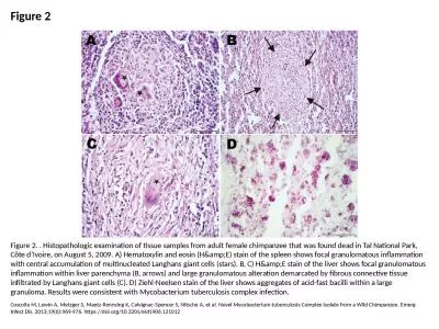
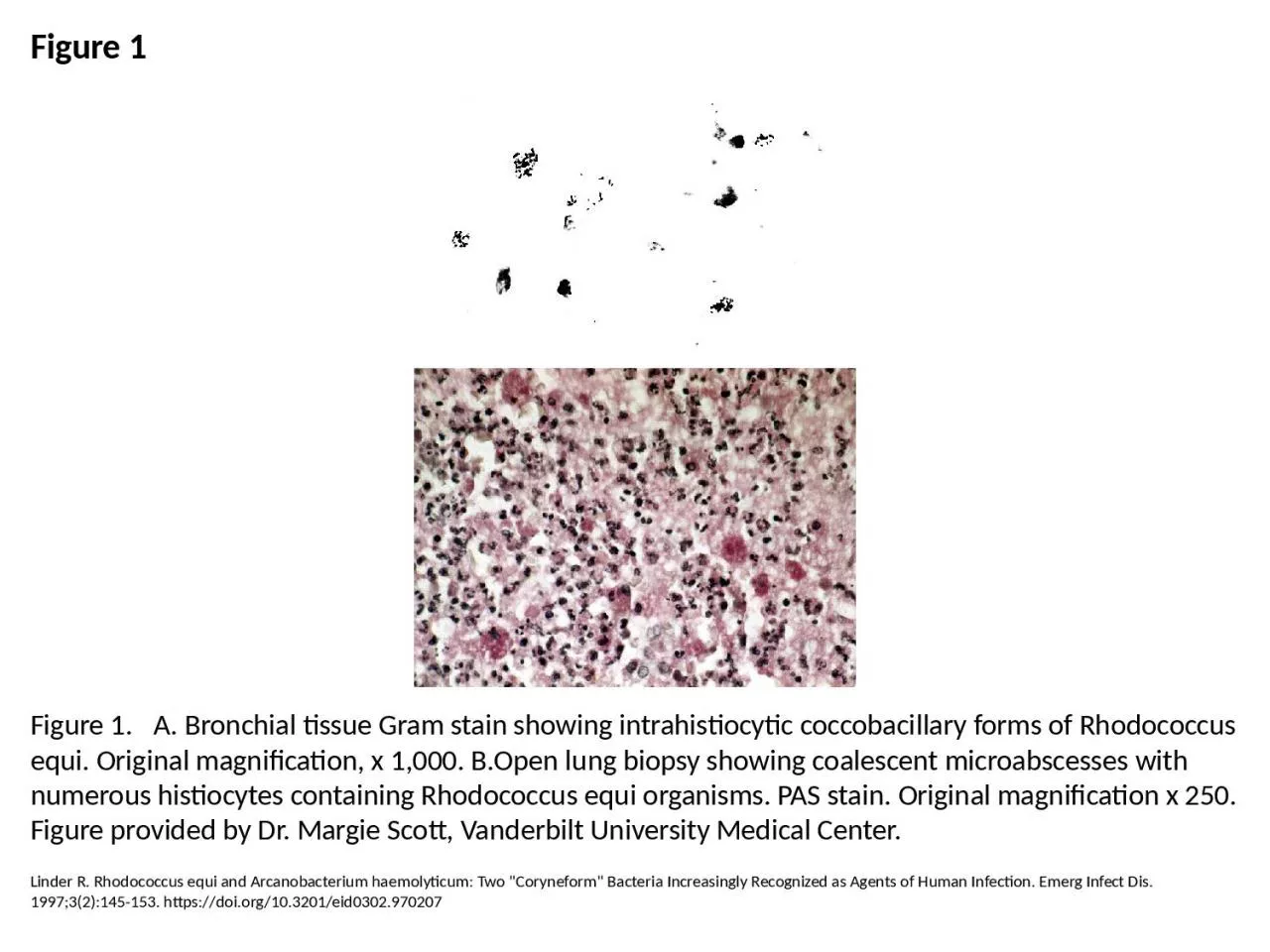
PPT-Figure 1 Figure 1. A. Bronchial tissue Gram stain showing intrahistiocytic coccobacillary

Author : scarlett | Published Date : 2023-07-18

Linder R Rhodococcus equi and Arcanobacterium haemolyticum Two Coryneform Bacteria Increasingly Recognized as Agents of Human Infection Emerg Infect Dis 199732145153

Presentation Embed Code

Download Presentation

Download Presentation The PPT/PDF document "Figure 1 Figure 1. A. Bronchial tissue..." is the property of its rightful owner. Permission is granted to download and print the materials on this website for personal, non-commercial use only, and to display it on your personal computer provided you do not modify the materials and that you retain all copyright notices contained in the materials. By downloading content from our website, you accept the terms of this agreement.

Figure 1 Figure 1. A. Bronchial tissue Gram stain showing intrahistiocytic coccobacillary: Transcript


Linder R Rhodococcus equi and Arcanobacterium haemolyticum Two Coryneform Bacteria Increasingly Recognized as Agents of Human Infection Emerg Infect Dis 199732145153 httpsdoiorg103201eid0302970207. Cheng . Zhang. Thurs 1 Dec 11. MM Tutorial. Gram staining. Fixed film (heat kill bacteria etc...). Methyl violet . Lugol’s. /gram’s iodine. Decolourise with acetone. Methyl red .  . STAIN 2 (counter stain): pink-red colour. Gram stain . Ziehl. . Neelsen. stain. . Requires the use of at least 3 chemical reagents.. Primary Stain. : . stain all the cells.. Decolorizing agent: . may or may not remove the primary stain from the entire the cell or only from certain cell structure. . Goals and Objectives. :. Exercise . 37. : Morphological Study of Unknown Bacterium. Each person make . smears of assigned organism: one from plate, one from broth (review smears Exercise 11). Gram stain (Exercise 15) and record results: Gram reaction, . Gram stain . Ziehl. . Neelsen. stain. . Requires the use of at least 3 chemical reagents.. Primary Stain. : . stain all the cells.. Decolorizing agent: . may or may not remove the primary stain from the entire the cell or only from certain cell structure. . Differentiate bacteria into two large groups (the Gram Positive and the Gram negative).. Gram status is important in medicine; the presence or absence of a cell wall will change the bacterium's susceptibility to some antibiotics. Eastern Journal of Medicine 4 1 39-41 1999 Traumatic bronchial rupture YALINKAYA 1 BLCLER U2Department of Thoracic Surgery1 School of Medicine Yznc Yl University Van Van Military Hospital2 Van Key wor . . . . Dr.oruba. Khalid abbas. ACTINOMYCETES. In this lecture , we shall discuss 3 genera of actinomycetes: . Actinomyces, Nocardia. , and . Streptomyces.. . These organisms have been shown to be . & Mycobacterium. Bacterial shape. Bacilli. Cocci. Gram +ve. Gram –ve. Gram +ve. Neisseria spp.. Staphylococcus. Streptococcus. Gram -ve. Enterobacteriaceae. Spore forming:. 1. Bacillus spp. (aerobic). 1 The slide is washed in water; excess water is removed using a blotting paper, dried in air and heat fixed before observing under microscope. Those bacteria that hold on to primary dye-iodine comple Coscolla M, Lewin A, Metzger S, Maetz-Rennsing K, Calvignac-Spencer S, Nitsche A, et al. Novel Mycobacterium tuberculosis Complex Isolate from a Wild Chimpanzee. Emerg Infect Dis. 2013;19(6):969-976. https://doi.org/10.3201/eid1906.121012. Harris JR, Blaney DD, Lindsley MD, Zaki SR, Paddock CD, Drew CP, et al. Blastomycosis in Man after Kinkajou Bite. Emerg Infect Dis. 2011;17(2):268-270. https://doi.org/10.3201/eid1702.101046. Brent AJ, Matthews PC, Dance DA, Pitt TL, Handy R. Misdiagnosing Melioidosis. Emerg Infect Dis. 2007;13(2):349. https://doi.org/10.3201/eid1302.061290. Lab # 6. NEISSERIA SPECIES. Contains many species; 2 are human pathogens. Neisseria . gonorrhoea. (gonococcus). Neisseria . meningitidis. (. meningococcus. ). Other species are normal flora that inhabit the upper respiratory tract of humans.. refractile. body within the vegetative cells. Procedure: (fig.10) . 1- Prepare a fixed smear.. 2- Flood the smear with 5% malachite green and let the stain react for 1 minute .Using your . bunsen. burner, heat the top of the stained slide until steam rises but do not let it boil. This step is performed by passing the burner back and over the slide for about 5 minutes..

Download Document

Here is the link to download the presentation.
"Figure 1 Figure 1. A. Bronchial tissue Gram stain showing intrahistiocytic coccobacillary"The content belongs to its owner. You may download and print it for personal use, without modification, and keep all copyright notices. By downloading, you agree to these terms.

Related Documents