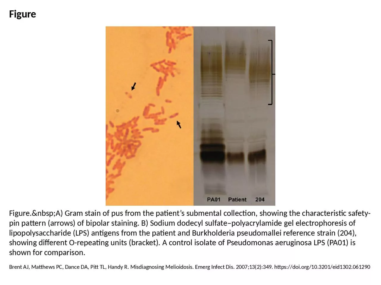
PPT-Figure Figure. A) Gram stain of pus from the patient’s submental collection,
Author : taylor | Published Date : 2023-07-19
Brent AJ Matthews PC Dance DA Pitt TL Handy R Misdiagnosing Melioidosis Emerg Infect Dis 2007132349 httpsdoiorg103201eid1302061290
Presentation Embed Code
Download Presentation
Download Presentation The PPT/PDF document "Figure Figure. A) Gram stain of..." is the property of its rightful owner. Permission is granted to download and print the materials on this website for personal, non-commercial use only, and to display it on your personal computer provided you do not modify the materials and that you retain all copyright notices contained in the materials. By downloading content from our website, you accept the terms of this agreement.
Figure Figure. A) Gram stain of pus from the patient’s submental collection,: Transcript
Brent AJ Matthews PC Dance DA Pitt TL Handy R Misdiagnosing Melioidosis Emerg Infect Dis 2007132349 httpsdoiorg103201eid1302061290. jjdendexjp eue CAC Cafed u u w Cafed Appende GND 02 D 2014 81 J D u Devepen ew Jn e uan Jn u n anenane nan age n anenane 77 Appved 21 n Revn Unde Devepen a Revn Jn u w pendng ange Jn pu e ndaed deeed pendng devepen evn f upeedng puan Cange n Leu f R Cheng . Zhang. Thurs 1 Dec 11. MM Tutorial. Gram staining. Fixed film (heat kill bacteria etc...). Methyl violet . Lugol’s. /gram’s iodine. Decolourise with acetone. Methyl red . . STAIN 2 (counter stain): pink-red colour. Gram stain . Ziehl. . Neelsen. stain. . Requires the use of at least 3 chemical reagents.. Primary Stain. : . stain all the cells.. Decolorizing agent: . may or may not remove the primary stain from the entire the cell or only from certain cell structure. . Biology and Biotechnology department. Differential . stain separate bacteria into group . . Gram . Stain :. Differentiate bacteria into 2 groups:. Gram . positive bacteria . Gram . negative bacteria . .. The presence of color gives the cells significant contrast so are much more visible. . Different stains have different affinities for different organisms, or different parts of . organisms. They are used to differentiate different types of organisms or to view specific parts of organisms. Leslie Teachout. MT (ASCP), CIC. Riverton Memorial and Lander Regional Hospitals. Infection Prevention. Objectives. Discover and discuss the importance of the infection preventionist and microbiology working as a team.. 11/14/2012. Background. Adipocyte physiology. Hyperplasia occurs after a critical mass has been reached. Liposuction reduces # of adipocytes, regardless of their size. Should provide long lasting results. Only use oil on 100x objective lens. Never get it onto any of the other objectives! . Never use the coarse adjustment with the 40x and 100x objectives.. . Ex. . 3: Staining Techniques - Gram Stain, part 1. Differentiate bacteria into two large groups (the Gram Positive and the Gram negative).. Gram status is important in medicine; the presence or absence of a cell wall will change the bacterium's susceptibility to some antibiotics. Differentiate bacteria into two large groups (the Gram Positive and the Gram negative).. Gram status is important in medicine; the presence or absence of a cell wall will change the bacterium's susceptibility to some antibiotics.. . By. Dr. . Shnyar. Hamid Qadir. Light Microscopy. Direct examination of stained or unstained preparations by . light (bright-field) microscopy. is particularly useful for detection of bacteria, fungi, and parasites.. Linder R. Rhodococcus equi and Arcanobacterium haemolyticum: Two "Coryneform" Bacteria Increasingly Recognized as Agents of Human Infection. Emerg Infect Dis. 1997;3(2):145-153. https://doi.org/10.3201/eid0302.970207. Song D, Kang B, Lee C, Saif LJ, Ha G, Kang D, et al. Transmission of Avian Influenza Virus (H3N2) to Dogs. Emerg Infect Dis. 2008;14(5):741-746. https://doi.org/10.3201/eid1405.071471. Gram Positive. Gram Negative. Gram-Positive bacteria. Gram-Negative bacteria. Cell Wall. A single-layered, smooth cell wall. A double-layered, wavy cell-wall. Cell Wall thickness. The thickness of the cell wall is 20 to 80 .
Download Document
Here is the link to download the presentation.
"Figure Figure. A) Gram stain of pus from the patient’s submental collection,"The content belongs to its owner. You may download and print it for personal use, without modification, and keep all copyright notices. By downloading, you agree to these terms.
Related Documents