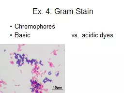
/
Gram Stain Differential stain (Hans Christian Gram, a Danish doctor ). He developed a Gram Stain Differential stain (Hans Christian Gram, a Danish doctor ). He developed a

Gram Stain Differential stain (Hans Christian Gram, a Danish doctor ). He developed a - PowerPoint Presentation

stefany-barnette
stefany-barnette . @stefany-barnette
Follow
349 views
Uploaded On 2019-06-25

Gram Stain Differential stain (Hans Christian Gram, a Danish doctor ). He developed a - PPT Presentation

Differentiate bacteria into two large groups the Gram Positive and the Gram negative Gram status is important in medicine the presence or absence of a cell wall will change the bacteriums susceptibility to some antibiotics ID: 760270

Share:

Link:

Embed:

Download Presentation from below link

Download Presentation The PPT/PDF document "Gram Stain Differential stain (Hans Chri..." is the property of its rightful owner. Permission is granted to download and print the materials on this web site for personal, non-commercial use only, and to display it on your personal computer provided you do not modify the materials and that you retain all copyright notices contained in the materials. By downloading content from our website, you accept the terms of this agreement.


Presentation Transcript

Slide1

Slide2

Gram Stain

Differential stain (Hans Christian Gram, a Danish doctor ). He developed a new method to stain bacteria so they can be visible in specimen samples.Differentiate bacteria into two large groups (the Gram Positive and the Gram negative).Gram status is important in medicine; the presence or absence of a cell wall will change the bacterium's susceptibility to some antibiotics.

Slide3

Almost all bacteria are described by their Gram stain characteristics.Based on differences of Cell wall structures

Slide4

Gram Positive & Negative Bacteria

Slide5

Slide6

Slide7

Reagents for Gram Stain

Crystal Violet (purple).Primary stain; positive stainStains cell wall purpleIodineMordantCombines with primary stain to form an insoluble complex that gets trapped in bacterial cell wall

Slide8

EthanolDecolorizerCV-I complex washed out of Gram negative organisms because it cannot be trapped by lippopolysaccharide layer; flows right through outer membrane

Safranin (pink)CounterstainSimple positive stain that provides contrasting dye for decolorized cells (Gram negative)Stains all cells, but only the negative ones actually appear pink.

Gram +

Gram –

Primary stain:

Crystal violet

Purple

Purple

Mordant:

Iodine

Purple

Purple

Decolorizing agent:

Alcohol-acetone

Purple

Colorless

Counterstain:

Safranin

Purple

Pink

Slide9

Gram Staining Procedure

After air drying and heat fixation.

Slide10

Slide11

Slide12

Slide13

Errors During Staining

Never ever used old culture

.

Never ever used sample for patient take antibiotic.

Time of Decolorizer:

Over: G + see as G -.

Low: G- see as G +.

Time of fixation:

Over: G + see as G -.

Low: no sample on slide.

Slide14

Acid-fast stain (Ziehl-Neelsen stain)

The acid-fast stain is another

differential staining

method.

In this case, the target cells are usually members of the genus

Mycobacterium

.

The cell walls of these bacteria contain an unusually high concentration of waxy lipids, thus making conventional simple stains and Gram stains useless.

The genus

Mycobacterium

contains two important human pathogens,

M. tuberculosis

and

M. leprae

,

which cause tuberculosis and leprosy, respectively.

Slide15

The reagents used are

Ziehl

Neelsen

carbolfuchsin, acid alcohol, and methylene blue.

Acid-fast bacilli will be bright red after staining.

Slide16

Acid-fast stains are useful in identification of acid-fast bacilli (AFB)and rapid, preliminary diagnosis of tuberculosis (with greater than 90% predictive value from sputum samples).

It also can be performed on patient samples to track the progress of antibiotic therapy and determine their degree of contagiousness.

Slide17

Acid Fast Reagents

Carbolfuchsin (red), a phenolic stain: is the primary stain in the acid-fast test. It is soluble in the lipids of the mycobacterial cell wall.

Heating the specimen, or adding a wetting agent such as

Tergitol

, increases the penetration of the carbolfuchsin

.

Following application of the carbolfuchsin, the specimen is cooled and decolorized with a solution of 3% hydrochloric acid and 95% ethanol

(

acid-alcohol

).

Slide18

Since carbolfuchsin is more soluble in waxy cell lipids than in acid-alcohol, the acid-alcohol removes the carbolfuchsin from non-acid-fast organisms, but not from acid-fast organisms.

Following decolorization, the sample is counterstained with methylene blue which Cannot penetrate mycolic acid; provides contrast to non acid fast cells.

Slide19

Procedures

Prepare a smear organism and a on glass slides.

Allow the slides to air dry, and then heat fix the organisms.

Apply enough of carbolfuchsin with

Tergitol

to cover the bacteria. Allow it to set for five minutes

.

(Alternate) If

Tergitol

is not available, apply enough carbolfuchsin to cover the bacteria. Place the slide on a pre-warmed hot plate set on low for 8 minutes.

Do not allow the stain to evaporate or Boil

. Add additional stain, if necessary. Remove the slide and allow it to cool.

Slide20

Apply enough methylene blue to cover the bacteria. Allow it to set for 30 sec. Gently rinse the slide with water.Blot (don't wipe) the slide dry with bibulous paper. Allow the slide to air dry.Examine the slide under oil immersion. Positive organisms will appear pink or red; Negative organisms will appear blue.

Rinse the slide with acid-alcohol (15-20 sec), drop by drop, just until the alcohol runs clear.

Gently rinse the slide with water.

Slide21

Slide22

Slide23

Slide24

Note

The

reddish-pink

color and “cording” (sticking together in long ropy masses) of the

Mycobacterium

cells due to the excess lipids of the cell wall

Slide25