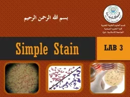
/
Laboratory diagnosis Smear preparation, Simple  and Gram stains Laboratory diagnosis Smear preparation, Simple  and Gram stains

Laboratory diagnosis Smear preparation, Simple and Gram stains - PowerPoint Presentation

jones
jones . @jones
Follow
0 views
Uploaded On 2024-03-13

Laboratory diagnosis Smear preparation, Simple and Gram stains - PPT Presentation

5292023 1 Practical Bacteriology lab 2 By Asst Prof Shiamaa AlSalihy Learning objectives 5292023 2 After this lab you must be able to Identify laboratory diagnosis steps Understand staining techniques ID: 1048105

gram stain sample collection stain gram collection sample stained staining csf blood urine swab acid diagnosis antibiotic fluid steps

Share:

Link:

Embed:

Download Presentation from below link

Download Presentation The PPT/PDF document "Laboratory diagnosis Smear preparation, ..." is the property of its rightful owner. Permission is granted to download and print the materials on this web site for personal, non-commercial use only, and to display it on your personal computer provided you do not modify the materials and that you retain all copyright notices contained in the materials. By downloading content from our website, you accept the terms of this agreement.


Presentation Transcript

1. Laboratory diagnosisSmear preparation, Simple and Gram stains5/29/20231Practical Bacteriologylab. 2By:Asst. Prof. Shiama'a Al-Salihy

2. Learning objectives:5/29/20232After this lab, you must be able to:Identify laboratory diagnosis steps.Understand staining techniquesIllustrate the purpose of each stainInterpret laboratory tests results

3. Laboratory diagnosis steps:Specimen: according to type of infectionMicroscopic examination : stained or wetCulture and antibiotic susceptibility test: inoculation of M.Os in or/and on culture media, and testing their sensitivity to antibiotic.Biochemical tests: catalase, coagulase, sugar fermentation tests…etc.Serological and immunological testMolecular tests

4.

5. Sample collection (specimens): Urine ,blood, stool ,sputum , pus, CSF, plural fluid ,peritoneal fluid, different types of swabs.

6. A. Urine sample:Collection: Mid stream urine (M.S.U.) sample to avoid contamination with normal flora (E.coli), Staphylococcus saprophyticus.The sample of urine must be no delay more than 1-2hrs.B. Stool sample:In sterile container or by rectal swab. The collected stool should be processed within 2hrs or inoculated in transport mediaurine collection container

7. C. Swabs: Very important methods for collect the following:Urethral swabs: for urethritis diagnosis in men and vaginal swabThroat swab: culture on chocolate agarNasopharyngeal swabEar swab.Eye swab.wound swab.

8. D. Sputum:Material from lower RTI or ( a mixture of bronchial secretion and inflammatory exudates).Collection steps: Make the collection in a disposables wide mouth screw-capped plastic containerHomogenize the sample.Collect the sample before antibiotic therapy is began.Sputum collection container

9. E. Cerebrospinal fluid (CSF):The examination of CSF is an essential step in the diagnosis of bacterial and fungal meningitis. Normal CSF is sterile, clear, with no RBCs.Collection: 5-10 ml of CSF should be collected in 2 sterile tube by lumber puncture

10.

11. F. Blood :Samples of blood should be taken before antibiotic administrationskin antisepsis: Iodine, Povidone –Iodine, Alcohol 70% or Chlorhexidine10 ml from adult, 2 - 5 ml from children, 1-2 ml from infants and neonates

12. Microscopic examination: Smear: A thin film of material containing M.Os which is spread over the surface of slide (stained or wet)- Wet preparation are used to demonstrate:Blood cells and microbes in fluid specimens such as urine, feces, CSFCysts, eggs or parasite in feces.Fungi in skin.Protozoa in blood and tissues.- Stained smear: if bacteria are seen in gram stained smear, their shape, size, arrangement and reaction should be observed.

13. Steps of preparation smear

14. Staining procedure can also be classified into:Simple staining: It is the use of single basic dye to color the bacterial organism. Such as methylene blue, crystal violet, safranin.Differential staining: uses more than one dyes and it is also used to differentiate the organisms into one of two groups such as gram stain and acid fast stain. Also it can give the shape of bacteriaStructural stain: such as capsule stain, endospore and flagella

15. Methods of simple staining

16. Gram stain It is a differential stain uses two or more types of dye to differentiate between organism according to their response to these dye Gram Stain Gram positive: Appears violet after Gram’s stainb) Gram negative: Appears red after Gram’s stain

17. Gram stain procedure

18.

19. Ziehl-Neelsen stain (Acid fast Bacilli)To stain Mycobacterium species especially M. tuberculosis High lipid content- make decolorization very difficultPrinciple:Acid fast (resist): once the bacteria stained with primary dye- difficult to decolorize with acid due to high contain of lipid substances within their cell walls called mycolic acids. Basic carbol fuchsin in combination with phenol (the staining solution) penetrates the cell wall and stain the bacilli bright red. they resist decolorization with strong acid.

20. Mycobacterium tuberculosisunder microscope stained with AFB stainSteps of AFB stain

21. Albert StainUsed for staining and demonstrated of Corynebacterium diphtheriae, They are thin gram positive bacilli which resemble Chinese letters when stained by simple stain.In albert stain another characteristic is presence of metachromatic granules (RNA or polymetaphosphate) the granules are bluish-purple by methylene blue and dark purple with albert stain

22. CapsuleEndospore

23. Thank you