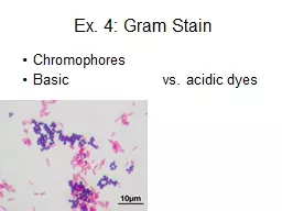
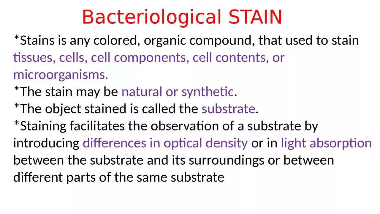
tissues cells cell components cell contents or microorganisms The stain may be natural or synthetic The object stained is called the substrate Staining facilitates the observation of a substrate by introducing ID: 926503
Download Presentation The PPT/PDF document "Bacteriological STAIN *Stains is any col..." is the property of its rightful owner. Permission is granted to download and print the materials on this web site for personal, non-commercial use only, and to display it on your personal computer provided you do not modify the materials and that you retain all copyright notices contained in the materials. By downloading content from our website, you accept the terms of this agreement.
Slide1
Bacteriological STAIN
*Stains is any colored, organic compound, that used to stain
tissues, cells, cell components, cell contents, or microorganisms.
*The stain may be
natural or synthetic
.
*The object stained is called the
substrate
.
*Staining facilitates the observation of a substrate by introducing
differences in optical density
or in
light absorption
between the substrate and its surroundings or between different parts of the same substrate
Slide2ACIDIC
:
Have negative charge so they bind to positively charged cell structures like some proteins.Ex: Nigrosine, Picric acid, Eosin, Acid fuschin, India ink BASIC: Have Positive charge so bind to negatively charged molecules in cell like cell wall.Ex: Crystal Violet, Methylene Blue, Safranin , basic fuschin.NEUTRAL: Both positively and negatively charged imparts different colors to different components.Ex: Geimsa’s stain, Leishman’s stain, Wright’s stain.
According to nature of stain
Slide3Bacterial smear
Slide4Slide5Classification of stains according to the purpose of use
1-Simple stains:
Crystal violet, Methylene blue and Carbolfuchsin.
Slide6Results:
All cells have the same color
Morphology – cocci/spherical / rod.Arrangement – clusters/chains.
Slide72-Differential stains:
*more than one stains are involved in the staining proses and stains react differently with different cell types.
Gram staining
Slide8Gram
Staining
Procedure
Slide9(CV-I) complex
Slide10Gram staining principles
-
Slide11Results for gram stain G –
ve
and G +ve
Slide12Factors affecting the efficiency of Gram staining process
1- Bacterial smear thickness
2- Smear fixation 3- Concentration and freshness of reagents may affect the quality of the stain4- The nature and age of the bacterial culture5- The washing waterFalse results G -ve may appear as G +veG+ ve may appear as G-ve
Slide13Acid-fast organisms are characterized by wax-like, nearly impermeable cell walls; they contain mycolic acid and large amounts of fatty acids, waxes, and complex lipids
Acid-fast stain
Slide14PRINCIPLE OF
ZIEHL-NEELSEN STAIN
Carbolfuchsin, is lipid-soluble and contains phenol, which helps the stain penetrate the cell wall**
Slide151
2
3
4
5
6
7
Slide16How the Acid fast
bacteria appear
Slide17SPECIAL
STAINS 1- Capsules Stain (Negative stain)
Nigrosine
India ink
Slide18Spores Stain
Flagella Stain
Leifson’s stain Malachite green