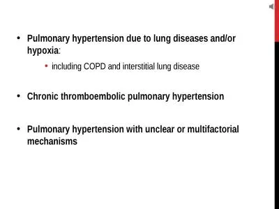
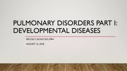
Nicole Cacho Do MPH August 16 2018 Objectives Discuss Following Developmental Diseases of Neonatal Lung Pulmonary Dysplasia Congenital Diaphragmatic Hernia Capillary Alveolar Dysplasia Congenital Pulmonary ID: 775410
Download Presentation The PPT/PDF document " Pulmonary Disorders Part I: Development..." is the property of its rightful owner. Permission is granted to download and print the materials on this web site for personal, non-commercial use only, and to display it on your personal computer provided you do not modify the materials and that you retain all copyright notices contained in the materials. By downloading content from our website, you accept the terms of this agreement.
Slide1
Pulmonary Disorders Part I: Developmental Diseases
Nicole Cacho Do, MPH
August 16, 2018
Slide2Objectives
Discuss Following Developmental Diseases of Neonatal Lung:
Pulmonary Dysplasia
Congenital Diaphragmatic Hernia
Capillary Alveolar Dysplasia
Congenital Pulmonary
Lymphangiectasia
Chylothorax
Congenital Cystic Pulmonary Malformations
Slide3Pulmonary Dysgenesis
Agenesis-total absence of pulmonary parenchyma, its supporting vasculature and bronchi after the bifurcation
Aplasia-subset of agenesis
Hypoplasia-underdeveloped lungs
Slide4Pulmonary Agenesis
Complete
agenesis is usually unilateral, although in some cases there has been dysgenesis of the second lung as
well
Rare
Likely vascular origin (dorsal aortic arch)
Associated with other congenital malformations, considered a subset of VACTERL sequence or
Goldenhar
syndrome
Malformations are ipsilateral to the pulmonary defect
Slide5Pulmonary agenesis: Diagnosis
Antenatal ultrasound: mediastinal shift (to affected side) in the absence of diaphragmatic hernia
Antenatal echo: absence of pulmonary artery or one of the branches
MRI: confirm diagnosis, evaluate size of remaining lungs and look for other malformations
Slide6Pulmonary Agenesis: CXR
Radiopaque
hemithorax and medial shift to
affected side
Jentzsch 2014
Slide7Pulmonary Aplasia
Presence of blind-ending, rudimentary bronchus without associated lung parenchyma or pulmonary vasculature
Slide8Pulmonary hypoplasia
Incomplete development of lung parenchyma leading to decreased number of distal airways, alveoli and associated pulmonary vessels.
Primary is intrinsic failure of normal lung development
Secondary can be from oligohydramnios, space occupying lesions, or absence or abnormal diaphragmatic activity from neuromuscular disorders
1/3 of patients with oligohydramnios have pulmonary hypoplasia
High mortality rate
Slide9Pulmonary hypoplasia: Diagnosis
Measuring lung-to-body ratio
Only captures babies with lethal hypoplasia
Pathologic specimens
show low ratio of lung to body weight
low DNA content
decreased radial alveolar count
peripheral bronchioles and pulmonary arterioles are decreased in number
a
rterioles hypertrophied medial smooth muscle which predisposes to PPHN
Slide10Pulmonary hypoplasia: Treatment
Inhaled nitric oxide
Gentle ventilation
Retrospective
cohort
of 151
matched-pairs
of 22-29
weekers
with
hypoplasia
.
iNO
did
not
improve
survival
prior
to
NICU
discharge
or transfer
JAMA
Pediatr
.
2018
Jul
2;172(7):
e180761
Slide11Congenital Diaphragmatic Hernia: Epidemiology
Developmental defect during formation of diaphragm and abdominal contents herniate into thoracic cavityClassified by anatomic region affectedPosterior lateral (Bochdalek) 70%Anterior (Morgagni) 25%Central 5%Incidence 1/2000 to 1/3000Left sided is most common (85%)
Slide12Congenital Diaphragmatic Hernia: Anomalies
Isolated or associated with other congenital anomalies (40-60%)
CDH Study Group reported a 28% incidence of severe malformations (major cardiac, syndromal, and chromosomal)
Importance of evaluation of associated malformations of these patients
Donnai
-Barrow (hearing, vision, brain,
omphalocele
)
Fryns
(abnormal facies, fingers/toes, cleft) and Pallister-Killian mosaic (
hypotonia
, course facies, severe develop delay) mosaic
Slide13Congenital Diaphragmatic Hernia: Syndromes
Holder et al. 2007, Genetic
Factors in Congenital Diaphragmatic Hernia
Slide14Congenital Diaphragmatic Hernia: Pathophysiology
Pulmonary insufficiency and PPHN (secondary to pulmonary hypoplasia, low # alveoli, and airway and vascular muscular hypertrophy associated with compression by abdominal contents)
Severity related to degree of lung hypoplasia
Size of defect
presence of liver in chest
how early in gestation abdominal contents were displaced
Slide15Congenital Diaphragmatic Hernia: Outcomes
2007
Systematic
R
eview
by Logan et al found that ~60% cases diagnosed antenatal
Antenatal diagnosis is associate with poor prognosis
Infants with a prenatal diagnosis have better outcomes if born at a tertiary center
Predictors based on assessment of the contralateral lung
Lung area to head circumference ratio (LHR) between 22 and 28 weeks gestation
Liver in chest (most predictive)
Estimated fetal lung volume by MRI
Slide16Congenital Diaphragmatic Hernia: OUtcomes
Corrected gestational age LHR=observed LHR/expected ratio
Fetal Corrected LHR (%)Survival (%)<15015-252026-35, or 36-45 liver up30-60>45, or 36-45 liver down>75
CDH registry n=184 with Left CDH 22-28 weeks
Slide17Congenital Diaphragmatic Hernia: Index of Suspicion
Baby with severe respiratory distress, cyanosis, scaphoid abdomen and failure to improve with ventilation
Auscultation can reveal: no breath sounds on affected side or bowel sounds, displacement of heart sounds to contralateral side
Order CXR!!!!!
While waiting intubate, and OG tube to decompress stomach
Late presentation of
Bochdalek
hernia occurs in less than 3% of cases
Slide18Congenital Diaphragmatic Hernia: CXR
Radiopaedia.org
Slide19Congenital Diaphragmatic Hernia: Consistent approach
Multidisciplinary management plans have improved survival
Predetermined ECMO criteria
“Protect the lung” strategy
Low PIP, PaCO2 levels <65, HFOV, ?
iNO
, ECMO rescue therapy, delay repair until stable and PPHN improves
Heliox
as Adjunct Therapy?
Retrospective cohort of 45 infants (28 with
heliox
) facilitated
improvement in gas exchange, which allowed a decrease in ventilator settings and oxygen
exposure
Respir
Care.
2018 May
22
Slide20Congenital Diaphragmatic Hernia: Follow up
GERD
FTT
Neurodevelopmental delay
Sensorineural hearing loss
Scoliosis
AAP recommended follow up schedule (2008)
Slide21Capillary Alveolar dysplasia
AKA Misalignment of the pulmonary veins
Rare, often fatal
P
resents within 48
hrs
with severe hypoxemia and unresponsive PPHN
Term babies with good
A
pgars
Unknown incidence due to diagnosis made by pathology but not all babies have lung biopsy or autopsy
Autosomal recessive mutation in FOXF1 gene on 16q24.1 which may help prenatal diagnosis in high risk families
Slide22Capillary Alveolar dysplasia
Minimal capillaries close to immature alveoli, anomalous misplaced distended pulmonary veins, medial thickening of small pulmonary arteries and
muscularization
of arterioles.
Pathology is diffuse in 85% of these patients and patchy in remaining
Pathophysiology
Failure of fetal lung vascularization with capillary hypoplasia and discontinuity of these capillaries and pulmonary veins impairing pulmonary blood flow as well as reactive pulmonary vasoconstriction
Slide23Capillary Alveolar dysplasia
Bishop et al. 2011
Medial thickening of pulmonary arteries
Malpositioned
congested pulmonary veins
Dilated Bronchiole
Slide24ChildHood interstitial lung disease
Group of rare lung diseases that can affect neonates, children and
adolesents
Neonatal types: capillary alveolar dysplasia, surfactant dysfunction mutations, pulmonary interstitial glycogenosis (PIG)
Diagnsosis
: CXR, CT chest, BAL, genetic panel
Treatment: Oxygen, bronchodilators, corticosteroids, other anti-inflammatory and immune modulators
Slide25Congenital pulmonary lymphangiectasia: Epidemiology
Rare
Males more affected
Sporadic but family clusters suggest autosomal recessive pattern
Classification
Primary: Localized
or
diffuse. Patients
with generalized lymph disorder have less severe pulmonary involvement
Secondary: Associated
with cardiac malformations (TAPVR, HLHS
) and possibly non-immune
hydrops and congenital
chylothorax
Diagnosis made by lung biopsy as in CAD
Prognosis is better with later presentation
Slide26Congenital pulmonary lymphangiectasia
CXR: Hyperinflation with
B/L interstitial infiltrates
Hagmann
and Berger 2003, N Engl J Med
Artery
Subpleural
and septal cystic lymphangiectasia
Slide27chylothorax
Lymph fluid in the pleural cavity
Rare (1:10,000 births), more common in males and on right side
Classification
Primary:
lymph disorders (
Noonans
, trisomy 21), congenital heart disease, mediastinal malignancies
Secondary
:
thoracic surgery trauma, increased SVC pressure (thrombus)
A
nalysis of pleural fluid:
transudative
, pH>7.4, cell count<1000/mm3 with >80%
lymphs
, TG>110
Most cases resolve in less than 2 months
Slide28Chylothorax: management
Nutritional: formulas
with high medium chained triglycerides (
Enfaport
-since lymphatic vessels are not required for absorption of
MCT), fat skimmed
breast
milk, may need increased protein supplementation
Pharmacological:
octreotide (somatostatin analogue,
more effective in primary than secondary, not significant
(P
=
0.10)
J
Paediatr
Child Health.
2018 Mar
30
)
Surgery if severe: chest tube,
pleurodesis
,
l
ymphovenous
anastomosis (
Plast
Reconstr
Surg.
2018 Aug;142(2):
581)
Slide29Chylothorax: CXR
Ning-Hui Foo et al. 2011
B/L
chylothoracies
and ascites in late preterm infant
Slide30Congenital cystic pulmonary Malformations
Congenital Cystic
A
denomatoid
Malformation (CCAM) or Congenital Pulmonary
A
irway
M
alformation (CPAM)
Bronchopulmonary Sequestration
Bronchogenic Cyst
Congenital Lobar Emphysema
Slide31Congenital Pulmonary AIRWAY Malformation (CPAM)
Multiple
hamartomatous
lesions arising from the abnormal branching of the immature bronchial tree
Most common out of the 4 diseases, incidence 1:11,000-13,000, affecting more males
Unilobar
and usually lower lung predominance
Connected with tracheobronchial tree
and has a
pulmonary blood supply
Classification prenatally (
Adzick
et al. 1985)
Microcystic
(<5mm)
Macrocystic
(>5mm)
Better prognosis
Hydrops due to CCAM has ~100% mortality rate
High CCAM volume ratio (CVR) can predict risk of hydrops and help stratify which patients may benefit from fetal surgery
Antenatal steroids may be able to shrink CCAM and help resolve hydrops (
Puligandla
et al. 2012)
Slide32Congenital Pulmonary AIRWAY Malformation: Stocker Classification
TypeOccurrence Cyst sizeHighlights0RareMultiple smallUsually lethal160-70%Large and smallMany regress prenatally215%Multiple smallOther anomalies35-10%Multiple smallAppear as solid mass410%LargeLate presentation
Stocker et al. 1977, 2002, 2009
Slide33Bronchopulmonary sequestration
Microscopic cystic masses arising from foregut
Not connected to airway
and has
systemic
blood supply
Classification
Intralobar
: accessory bed arises before pleura is established
Extralobar
: accessory lung bud arises after pleura is established; can be under diaphragm; higher association with congenital anomalies
Slide34Bronchopulmonary sequestration
Most significantly decrease in size as gestation continues, therefore most asymptomatic during first month of life
On CXR appear as posterior chest mass mostly on the left
High output cardiac failure is a unique feature
Surgery is encouraged for asymptomatic patient to prevent future infection or malignancy
Slide35Bronchogenic Cyst
Single cyst lined by respiratory epithelium and lined by cartilage and smooth muscle
Arise from abnormal budding of foregut early in first trimester
Mostly found in mediastinum
Treatment is complete resection and has an excellent prognosis thereafter
Slide36Bronchogenic Cyst: CXR
Case courtesy of Dr Ian Bickle, Radiopaedia.org, rID: 19947
Most found in mediastinal area
Slide37Congenital lobar emphysema
Postnatal over distention of one or more lobes
Pathophysiology includes intrinsic or extrinsic obstruction causing air trapping, over distention and finally emphysema
dysplastic cartilage,
inspisated
mucus, abnormal vasculature, infection
Left upper lobe is most frequently involved
Presents in neonatal period with a similar clinical picture to a tension pneumothorax but without hemodynamic instability
distinction is critical to avoid attempting aspiration and creating a pneumothorax
Surgical lobectomy has excellent prognosis
Slide38Congenital lobar emphysema: CXR
Case courtesy of
Dr Jeremy Jones, Radiopaedia.org, rID: 27202
Left upper lobe
lucency
could be mistaken for a pneumothorax
Slide39Summary
Many require surgical intervention
Better prognosis with later presentation
Many have male predominance
Predilection for distinct areas of thorax (focus on right or left)
Possible associated congenital anomalies may necessitate further workup
Knowing which are lethal may help avoid futile interventions
Slide40Question 1:
Term male 3.2kg born with
A
pgars
of 8 and 8. Meconium aspiration with persistent cyanosis and oxygenation index of 40. Decision to go to ECMO. After prolonged ECMO, multiple pump failures, 2 codes, severe IVH parents decide to withdraw care. Parents refuse autopsy. Which genetics work up may have been helpful in this case?
A. VACTERL sequence
B.
Goldenhar
syndrome
C. FOX F1 gene testing
D. Karyotype
Slide41Answer I: C
FOXF1 gene has been found in a cohort of ~40% of patients with CAD
Lung biopsy viewed by pathologist is the definitive diagnosis and can be done while patient is still alive to help redirect care
Slide42Question 2:
You get a call on the fellow phone from the
Peds
ED requesting your presence for intubation of a 2 day old female who is
tachypneic
,
tachycardic
with poor perfusion and enlarged liver. The ED attending quickly mentions there appears to be a left sided chest mass and baby was born at home with limited prenatal care. What is your first imaging study of choice?
A. CT scan of chest
B. CT scan of brain
C. MRI of chest
D. Echocardiogram
Slide43Answer 2: D
Echocardiogram since you are thinking that bronchopulmonary
seqestration
is your diagnosis. You will also call surgery to bedside and consider which imaging study will be faster (CT scan or MRI) and preferred by surgery to determine systemic blood supply of the mass
Slide44Question 3
Which of the following are more commonly found on the left side?
A. Capillary alveolar dysplasia
B. Bronchopulmonary sequestration
C. CDH
D.
Chylothorax
E. Both B and C
Slide45Answer 3: E
Capillary alveolar dysplasia is diffuse
Chylothorax
is usually right sided
Both Bronchopulmonary sequestration and Congenital diaphragmatic hernia are more commonly left sided
Slide46Brodsky and Martin question review book
Try these questions on your own
Pulmonary questions: 10, 28, 45, 49