/
Pulmonary hypertension due to lung diseases and/or Pulmonary hypertension due to lung diseases and/or

Pulmonary hypertension due to lung diseases and/or - PowerPoint Presentation

hazel
hazel . @hazel
Follow
64 views
Uploaded On 2023-12-30

Pulmonary hypertension due to lung diseases and/or - PPT Presentation

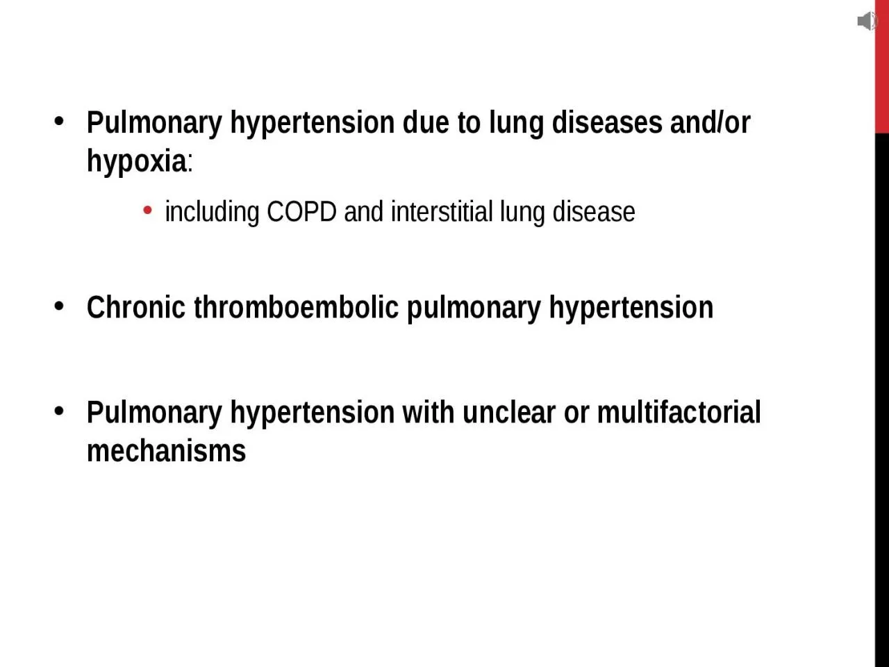
hypoxia including COPD and interstitial lung disease Chronic thromboembolic pulmonary hypertension Pulmonary hypertension with unclear or multifactorial mechanisms Pathogenesis Chronic obstructive or interstitial lung diseases ID: 1035973

disease pulmonary basis lung pulmonary disease lung basis edition 10h pathologic cotran robbins alveolar interstitial hypertrophy granulomatosis necrotizing respiratory

Share:

Link:

Embed:

Download Presentation from below link

Download Presentation The PPT/PDF document "Pulmonary hypertension due to lung disea..." is the property of its rightful owner. Permission is granted to download and print the materials on this web site for personal, non-commercial use only, and to display it on your personal computer provided you do not modify the materials and that you retain all copyright notices contained in the materials. By downloading content from our website, you accept the terms of this agreement.


Presentation Transcript

1. Pulmonary hypertension due to lung diseases and/or hypoxia:including COPD and interstitial lung diseaseChronic thromboembolic pulmonary hypertensionPulmonary hypertension with unclear or multifactorial mechanisms

2. PathogenesisChronic obstructive or interstitial lung diseasescongenital or acquired heart disease:Recurrent thromboemboli Autoimmune diseases

3. Obstructive sleep apnea idiopathic pulmonary arterial hypertensionAlso called primary pulmonary hypertensionUncommonall known causes are excludedUp to 80% has a genetic basis:inherited in families as an autosomal dominant trait. incomplete penetrance

4. Morphology:Medial hypertrophy of the pulmonary muscular and elastic arteries arterioles and small arteriesPulmonary arterial atherosclerosispulmonary artery and its major branchesRight ventricular hypertrophy

5. Plexiform lesion:uncommon a tuft of capillary formations producing a network, or web, that spans the lumens of dilated thin-walled, small arteries and may extend outside the vessel.

6. Robbins and Cotran pathologic basis of disease, 10h edition

7. Robbins and Cotran pathologic basis of disease, 10h edition

8. Clinical Features:Asymptomaticwomen 20-40 & young childrendyspnea and fatigue, anginal chest pain80% of patients within 2-5 years respiratory distress, cyanosis, and right ventricular hypertrophy and death from decompensated cor pulmonale

9. Diffuse Alveolar Hemorrhage SyndromesComplication of some interstitial lung disorders. Includes:Goodpasture syndromeIdiopathic pulmonary hemosiderosis Granulomatosis with polyangiitis

10. Goodpasture Syndrome:Is an uncommon autoimmune disease in which lung and kidney injury are caused by circulating autoantibodies against certain domains of type IV collagen.type IV collagen is intrinsic to the basement membranes of renal glomeruli and pulmonary alveoliResults in necrotizing hemorrhagic interstitial pneumonitis and rapidly progressive glomerulonephritis.

11. Morphology:Grossly, red-brown consolidation due to diffuse alveolar hemorrhageMicroscopically:Focal necrosis of alveolar wall with intraalveolar hemorrhage, Fibrous thickening of septa, Hypertrophic type II pneumocytes. Abundant hemosiderin Linear pattern of immunoglobulin deposition (IgG, sometimes IgA or IgM) seen along the alveolar septa.

12. Robbins and Cotran pathologic basis of disease, 10h edition

13. Robbins and Cotran pathologic basis of disease, 10h edition

14. Clinical features:Teens and twentiesMales > femalesActive smokersPlasmapheresis and immunosuppressive therapy , renal transplantation

15. Granulomatosis and PolyangiitisFormerly called Wegener granulomatosis>80% of patients develop upper-respiratory or pulmonary manifestations. The lung lesions are characterized by a combination of necrotizing vasculitis (“angiitis”) and parenchymal necrotizing granulomatous inflammation.

16. The signs and symptoms stem from involvement of the upper-respiratory tract (chronic sinusitis, epistaxis, nasal perforation) and the lungs (cough, hemoptysis, chest pain). Anti-neutrophil cytoplasmic antibodies (PR3- ANCAs) are present in close to 95% of cases

17. M.abdaljaleel@ju.edu.jo

18. THANK YOU!