/
Characterization of Extracellular Matrix in Abdominal Aorta Sections Characterization of Extracellular Matrix in Abdominal Aorta Sections

Characterization of Extracellular Matrix in Abdominal Aorta Sections - PowerPoint Presentation

singh
singh . @singh
Follow
66 views
Uploaded On 2023-07-09

Characterization of Extracellular Matrix in Abdominal Aorta Sections - PPT Presentation

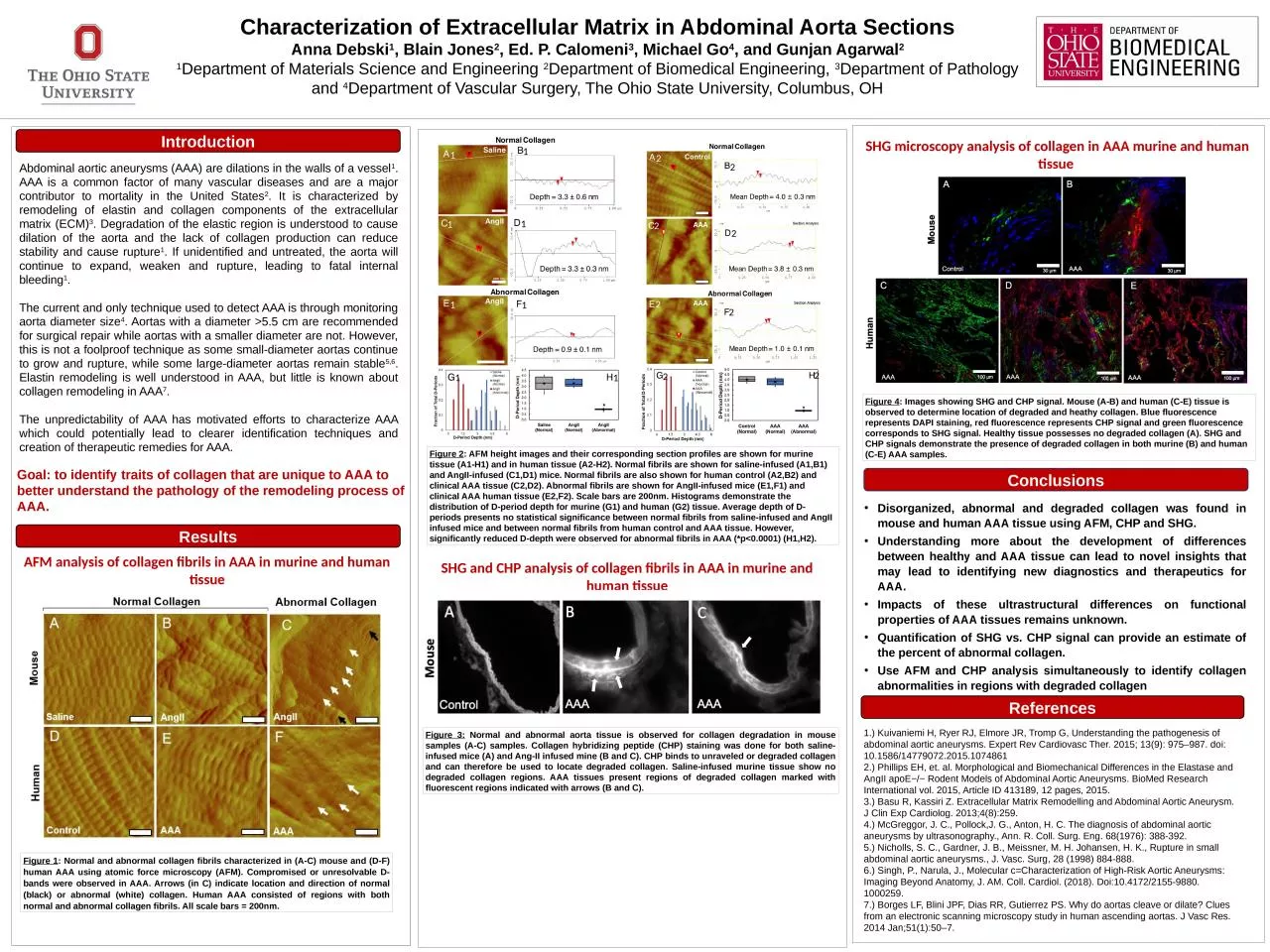
Anna Debski 1 Blain Jones 2 Ed P Calomeni 3 Michael Go 4 and Gunjan Agarwal 2 1 Department of Materials Science and Engineering 2 Department of Biomedical Engineering 3 Department of Pathology and ID: 1007565

collagen aaa tissue human aaa collagen human tissue fibrils chp degraded infused normal abnormal murine aortic aneurysms abdominal shg

Share:

Link:

Embed:

Download Presentation from below link

Download Presentation The PPT/PDF document "Characterization of Extracellular Matrix..." is the property of its rightful owner. Permission is granted to download and print the materials on this web site for personal, non-commercial use only, and to display it on your personal computer provided you do not modify the materials and that you retain all copyright notices contained in the materials. By downloading content from our website, you accept the terms of this agreement.


Presentation Transcript

1. Characterization of Extracellular Matrix in Abdominal Aorta SectionsAnna Debski1, Blain Jones2, Ed. P. Calomeni3, Michael Go4, and Gunjan Agarwal21Department of Materials Science and Engineering 2Department of Biomedical Engineering, 3Department of Pathology and 4Department of Vascular Surgery, The Ohio State University, Columbus, OHAbdominal aortic aneurysms (AAA) are dilations in the walls of a vessel1. AAA is a common factor of many vascular diseases and are a major contributor to mortality in the United States2. It is characterized by remodeling of elastin and collagen components of the extracellular matrix (ECM)3. Degradation of the elastic region is understood to cause dilation of the aorta and the lack of collagen production can reduce stability and cause rupture1. If unidentified and untreated, the aorta will continue to expand, weaken and rupture, leading to fatal internal bleeding1. The current and only technique used to detect AAA is through monitoring aorta diameter size4. Aortas with a diameter >5.5 cm are recommended for surgical repair while aortas with a smaller diameter are not. However, this is not a foolproof technique as some small-diameter aortas continue to grow and rupture, while some large-diameter aortas remain stable5,6. Elastin remodeling is well understood in AAA, but little is known about collagen remodeling in AAA7.The unpredictability of AAA has motivated efforts to characterize AAA which could potentially lead to clearer identification techniques and creation of therapeutic remedies for AAA. ConclusionsDisorganized, abnormal and degraded collagen was found in mouse and human AAA tissue using AFM, CHP and SHG.Understanding more about the development of differences between healthy and AAA tissue can lead to novel insights that may lead to identifying new diagnostics and therapeutics for AAA.Impacts of these ultrastructural differences on functional properties of AAA tissues remains unknown.Quantification of SHG vs. CHP signal can provide an estimate of the percent of abnormal collagen.Use AFM and CHP analysis simultaneously to identify collagen abnormalities in regions with degraded collagen ReferencesFigure 1: Normal and abnormal collagen fibrils characterized in (A-C) mouse and (D-F) human AAA using atomic force microscopy (AFM). Compromised or unresolvable D-bands were observed in AAA. Arrows (in C) indicate location and direction of normal (black) or abnormal (white) collagen. Human AAA consisted of regions with both normal and abnormal collagen fibrils. All scale bars = 200nm.AFM analysis of collagen fibrils in AAA in murine and human tissueFigure 3: Normal and abnormal aorta tissue is observed for collagen degradation in mouse samples (A-C) samples. Collagen hybridizing peptide (CHP) staining was done for both saline-infused mice (A) and Ang-II infused mine (B and C). CHP binds to unraveled or degraded collagen and can therefore be used to locate degraded collagen. Saline-infused murine tissue show no degraded collagen regions. AAA tissues present regions of degraded collagen marked with fluorescent regions indicated with arrows (B and C). 1.) Kuivaniemi H, Ryer RJ, Elmore JR, Tromp G, Understanding the pathogenesis of abdominal aortic aneurysms. Expert Rev Cardiovasc Ther. 2015; 13(9): 975–987. doi: 10.1586/14779072.2015.10748612.) Phillips EH, et. al. Morphological and Biomechanical Differences in the Elastase and AngII apoE−/− Rodent Models of Abdominal Aortic Aneurysms. BioMed Research International vol. 2015, Article ID 413189, 12 pages, 2015.3.) Basu R, Kassiri Z. Extracellular Matrix Remodelling and Abdominal Aortic Aneurysm. J Clin Exp Cardiolog. 2013;4(8):259.4.) McGreggor, J. C., Pollock,J. G., Anton, H. C. The diagnosis of abdominal aortic aneurysms by ultrasonography., Ann. R. Coll. Surg. Eng. 68(1976): 388-392. 5.) Nicholls, S. C., Gardner, J. B., Meissner, M. H. Johansen, H. K., Rupture in small abdominal aortic aneurysms., J. Vasc. Surg, 28 (1998) 884-888. 6.) Singh, P., Narula, J., Molecular c=Characterization of High-Risk Aortic Aneurysms: Imaging Beyond Anatomy, J. AM. Coll. Cardiol. (2018). Doi:10.4172/2155-9880. 1000259. 7.) Borges LF, Blini JPF, Dias RR, Gutierrez PS. Why do aortas cleave or dilate? Clues from an electronic scanning microscopy study in human ascending aortas. J Vasc Res. 2014 Jan;51(1):50–7.400 nmResultsIntroductionSHG microscopy analysis of collagen in AAA murine and human tissue Figure 4: Images showing SHG and CHP signal. Mouse (A-B) and human (C-E) tissue is observed to determine location of degraded and heathy collagen. Blue fluorescence represents DAPI staining, red fluorescence represents CHP signal and green fluorescence corresponds to SHG signal. Healthy tissue possesses no degraded collagen (A). SHG and CHP signals demonstrate the presence of degraded collagen in both murine (B) and human (C-E) AAA samples. Goal: to identify traits of collagen that are unique to AAA to better understand the pathology of the remodeling process of AAA.SHG and CHP analysis of collagen fibrils in AAA in murine and human tissueFigure 2: AFM height images and their corresponding section profiles are shown for murine tissue (A1-H1) and in human tissue (A2-H2). Normal fibrils are shown for saline-infused (A1,B1) and AngII-infused (C1,D1) mice. Normal fibrils are also shown for human control (A2,B2) and clinical AAA tissue (C2,D2). Abnormal fibrils are shown for AngII-infused mice (E1,F1) and clinical AAA human tissue (E2,F2). Scale bars are 200nm. Histograms demonstrate the distribution of D-period depth for murine (G1) and human (G2) tissue. Average depth of D-periods presents no statistical significance between normal fibrils from saline-infused and AngII infused mice and between normal fibrils from human control and AAA tissue. However, significantly reduced D-depth were observed for abnormal fibrils in AAA (*p˂0.0001) (H1,H2).1112222221111122