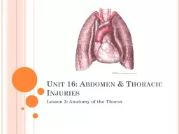
Lesson 2 Anatomy of the Thorax Bell work grab notes and study the functions of the organs Liver Pancreas Small intestine Stomach Spleen Appendix Kidney Large intestine Bladder Gall bladder ID: 779610
Download The PPT/PDF document "Unit 16: Abdomen & Thoracic Injuries" is the property of its rightful owner. Permission is granted to download and print the materials on this web site for personal, non-commercial use only, and to display it on your personal computer provided you do not modify the materials and that you retain all copyright notices contained in the materials. By downloading content from our website, you accept the terms of this agreement.
Slide1
Unit 16: Abdomen & Thoracic Injuries
Lesson 2: Anatomy of the Thorax
Slide2Bell work- grab notes and study the functions of the organs
Liver
Pancreas
Small intestine
StomachSpleenAppendix KidneyLarge intestineBladderGall bladder
Secretes bile/detoxifies
Stores bile
Produces insulin/secrets juice
Filters blood/excretes waste
Food mixes w/secretions & forms
chyme
Stores & regulates RBC
Digestion & absorption takes place
Final
digestion/
chyme
converted to feces
Stores & expels urine
Produces mucus & antibodies
Slide3Objective:
Recognize relevant anatomy of the thorax
Slide4Terminology
Atrium: The upper chambers of the heart
Ventricle
lower chamber of the heart
Intercostal refers to the area between the ribsThorax the chest; the part of the body located between the neck and the abdomen
Slide5Terms in Pictures
Atrium
Intercostal
Thorax
Ventricle
1
2
4
3
Slide6Thoracic Cavity Boarders
Anterior
Sternum, ribs and intercostal muscles
Posterior
Thoracic spine, ribs, scapulae & erector spinae muscle
Superior
Clavicle
Inferior
Diaphragm
Slide7Rib Cage Anatomy
12 pairs of ribs
True ribs (first 7 pairs): attach directly to the sternum by individual cartilage
False ribs (next 3 pairs): attach indirectly to the sternum by shared cartilage
Floating ribs (next 2 pairs): have no attachment to the sternum
Slide8Organs of the Thoracic Cavity
Esophagus
Trachea
Lungs
Heart
Slide9The Esophagus
Location:
In the throat, behind the trachea
Function:
Structure for food to pass from the mouth to the stomach
Slide10The Trachea
Location:A cartilage structure in the throat, directly in front of the esophagus
The trachea divides into 2 branches (bronchi) which enter the lungs
Function:
Allows air to pass into and out of the lungs
Slide11The Lungs
Location:
On either side of the heart
Functions:Exchange carbon dioxide for oxygen Help to remove heat from the body
Slide12The heart
Location:Under the sternum (slightly to the left) and between the lungs
Function:
to pump blood throughout the bodyBlood carries oxygen and nutrients to the cellsBlood carries carbon dioxide and waste products away from the cells
Slide13The Heart
Divided into 4 chambersR and L atrium (upper chambers)
R and L ventricles (lower chambers)
Heart valves separate each chamber and prevent a backflow of the blood
Tricuspid valveBicuspid (mitral) valvePulmonary valveAortic valve
Slide14Direction blood pumps through the heart
Slide15How does the blood flow through the Heart?
The
pulmonary veins empty oxygen-rich blood from the lungs into the left atrium of the heart.
As the atrium contracts, blood flows from your left atrium into your left ventricle through the open mitral valve.
When the ventricle is full, the mitral valve shuts. This prevents blood from flowing backward into the atrium while the ventricle contracts.As the ventricle contracts, blood leaves the heart through the aortic valve, into the aorta and to the body.Blood enters the heart through two large veins, the inferior and superior vena cava, emptying oxygen-poor blood from the body into the right atrium of the heart.As the atrium contracts, blood flows from your right atrium into your right ventricle through the open tricuspid valve.When the ventricle is full, the tricuspid valve shuts. This prevents blood from flowing backward into the atria while the ventricle contracts.As the ventricle contracts, blood leaves the heart through the pulmonic valve, into the pulmonary artery and to the lungs, where it is oxygenated and then returns to the left atrium through the pulmonary veins.
Left Side of Heart
Right Side of Heart
http://www.webmd.com/heart-disease/guide/how-heart-works?page=3
Slide16How blood flows through the lungs
Once blood travels through the pulmonic valve, it enters your lungs. This is called the pulmonary circulation. From your pulmonic valve, blood travels to the pulmonary artery to tiny capillary vessels in the lungs
.
Here, oxygen travels from the tiny air sacs in the lungs, through the walls of the capillaries, into the blood. At the same time, carbon dioxide, a waste product of
metabolism, passes from the blood into the air sacs. Carbon dioxide leaves the body when you exhale. Once the blood is purified and oxygenated, it travels back to the left atrium through the pulmonary veins.
Slide17closure
List the organs of the thoracic cavity.
List all boarders of the thoracic cavity.
The heart sits slightly to the ________.
Name the different segments of the spine; in order from top to bottom. Are atriums on top or bottom?What valve separates the left atrium and left ventricle?