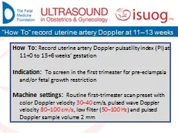
/
Mueller polarimetric imaging for early detection of uterine cervix cancer: from proof Mueller polarimetric imaging for early detection of uterine cervix cancer: from proof

Mueller polarimetric imaging for early detection of uterine cervix cancer: from proof - PowerPoint Presentation

slygrat
slygrat . @slygrat
Follow
345 views
Uploaded On 2020-06-30

Mueller polarimetric imaging for early detection of uterine cervix cancer: from proof - PPT Presentation

in vivo measurements Angelo Pierangelo 1 Jérémy Vizet 1 Jean Rehbinder 1 Stanislas Deby 1 Stéphane Roussel 1 Tatiana Novikova 1 Ranya Soufan 2 Catherine ID: 789918

vivo mueller cancer polarimetric mueller vivo polarimetric cancer cervical imaging polarization retardance application statistical depolarization field tissue healthy samples

Share:

Link:

Embed:

Download Presentation from below link

Download The PPT/PDF document "Mueller polarimetric imaging for early d..." is the property of its rightful owner. Permission is granted to download and print the materials on this web site for personal, non-commercial use only, and to display it on your personal computer provided you do not modify the materials and that you retain all copyright notices contained in the materials. By downloading content from our website, you accept the terms of this agreement.


Presentation Transcript

Slide1

Mueller polarimetric imaging for early detection of uterine cervix cancer: from proof of principle experiments to

in vivo measurementsAngelo Pierangelo1Jérémy Vizet1, Jean Rehbinder1, Stanislas Deby1, Stéphane Roussel1, Tatiana Novikova1, Ranya Soufan2, Catherine Genestie2, Christine Haie-Meder2, Hervé Fernandez3, André Nazac4, François Moreau1(1) Laboratoire de Physique des Interfaces et des Couches Minces (UMR 7647) - Ecole Polytechnique (Palaiseau)(2) Gustave Roussy Institute (Jussieu)(3) Teaching hospital of Kremlin Bicêtre (Kremlin Bicêtre))(4) Teaching hospital of Brugmann (Bruxelles)

Slide2

Epithelial

cancersEpithelial tissueEpitheliumConnectivetissue

Slide3

Cells genetically

alteredHyperplasiaSevere dysplasiaCarcinoma in situInvasive cancerEpitheliumConnective tissue(collagen, elastin…)VesselsTimeEpithelial cancers90% of cancersEpithelial tissueEpitheliumConnectivetissue

Epithelial cancer development

Slide4

Cells genetically

alteredHyperplasiaSevere dysplasiaCarcinoma in situInvasive cancerEpitheliumConnective tissue(collagen, elastin…)VesselsTimeEpithelial cancers90% of cancersEpithelial tissueEpithelium

Connectivetissue

Epithelial cancer development

Detection of precancerous stage

Recovery of patients in 95% of cases

Slide5

Cervical cancer

Major health problem: the second most common female cancer in the world with 275 000 deaths per year, mainly in developing countries (but not only)Slow evolution (10 - 20 years): ideal case for screeningDirect access to cancerModel for other epithelial cancers

Slide6

Cervical cancer screening

ColposcopyConizationSignificant improvement of colposcopy can make the early detectionand surgery both more effective and affordableColposcopy : inefficient technique (generating supplementary costs)(60-70% sensitivity and 50% specificity for high-grade dysplasia detection)Visualization of dysplastic areas for biopsy is very difficult and operator dependentResection margins to achieve a cone biopsy (conization) are poorly definedCurrent practice

Slide7

Electric field (Polarization)

Magnetic field

x

y

x

y

x

y

Linear polarization

Elliptical polarization

Depolarized light

Polarization of light

Slide8

Polarimetric properties

RetardanceDiattenuationDifference of transmission for two polarization eigenstatesPhase difference between two polarization eigenstatesDepolarizationIntroduction of « disorder » in the evolution of the electric fieldBirefringence : refraction index varies with polarizationDichroïsm : Absorption coefficient varies with polarizationStochastic process (multiple-scattering)Biological origins :Single scatteringTransmission anisotropy of some moleculesBiological origins :Collagen, elastin, muscle fibers (myosin, actin…)

Biological origins :

Organelles, cytoplasm, fibers…

slow

fast

Slide9

Mueller polarimetry

Mueller matrix M Complete polarimetric response of a sample (including depolarization)[1] : “Interpretation of Mueller matrices based on polar decomposition” , S.-Y. Lu and R. A. Chipman, JOSA A 13 (5) (2012)Stokes vector4-component vector which describes completely the polarization of lightLu and Chipman [1] : M = Md MR MDReverse : M = MDr MRr MdrSymmetric : M = MD2 MR2

Mds M

R1

M

D1

Decompositions based on hypothesis on

the

physical structure of the

sample

Intensity measurements

Slide10

Mueller polarimetric imaging

PSG : 4 probing polarization statesAnalysis through 4 different configurations of the PSA16 measured intensitiesMueller matrix imageMueller polarimetric imagerProbing polarization statesBackscattered polarization statesPSG (Polarization State Generator): linear polarizer, followed by retarders with changing characteristics over time (rotating waveplates, liquid crystal cells, etc ...)PSA (Polarization State Analyzer): the same elements as PSG, but in the reverse order

Slide11

Quartz plate

MilkTilted glass

Mueller Matrix

Lu-Chipman decomposition

Dichroism

Retardance

Depolarization

2cm

Mueller polarimetric imaging

…on “ad hoc” samples

ex

vivo

Slide12

…on biological samples

ex vivo2cmCancerous zone

5cm

Uterine cervix

(500nm)

Colon (500nm)

Pure Depolarizer

(diagonal matrix)

M22=M33>M44

Rayleigh Scattering

Depolarization

Retardance

Dichroism

(

non-diagonal matrix

)

Mueller polarimetric imaging

Slide13

Mueller polarimetric imaging :is sensitive to the microscopic morphology of the tissue

is realized using simple optical elements in the visible range (450 – 700 nm) : different depths in the tissue can be reached using different wavelengths (due to different light absorption by hemoglobin for different wavelengths, hemoglobin being more absorbing for shorter wavelengths) ;is well suited for « full field » (few cm²) modality (fundamental for in vivo applications) can be easily implemented at low cost Mueller polarimetric imaging

Slide14

Application to cervical cancer

Ex vivo proof of principle measurements1 Full field Mueller imaging polarimeter~10 samples analyzed2013

Slide15

Application to cervical cancer

Ex vivo proof of principle measurementsEx vivo statistical evaluationIn vivo preliminary results1 Full field Mueller imaging polarimeter3 Full field Mueller imaging upgraded polarimeters~10 samples analyzed~100 samples analyzed1 monochromatic Mueller colposcope (550nm)

~15 patients

analyzed

2013

2013-2017

Slide16

Application to cervical cancer

Ex vivo proof of principle measurementsEx vivo statistical evaluationIn vivo preliminary resultsIn vivo statistical evaluation1 Full field Mueller imaging polarimeter3 Full field Mueller imaging upgraded polarimeters~10 samples analyzed~100 samples analyzed

1 monochromatic Mueller colposcope (550nm)

~15 patients

analyzed

Color Mueller colposcope

(450 - 550 - 650nm)

~300 patients

t

o analyze

2013

2013-2017

2018-2020

Slide17

Application to cervical cancer

Mueller matrix imageand polarimetric parameters2cmCharacteristics Field of view ≈ 5 x 4 cm²Wavelengths : 400 nm to 700 nm (50 nm step)Time required to acquire a full Mueller matrix Image : several tens of secondsEx vivo analysis

Slide18

90°Retardance (550nm)

Depolarization (550nm)

Healthy

Glandular

CIN 3

0

1

2cm

Conventional image

A.

Pierangelo

et al.

Opt.Exp

. (2013)

J.

Rehbinder

et

al

. J.

Biomed

.

Opt

. (2015)

Prix de l’innovation de l’Ecole Polytechnique 2012

Application to cervical cancer

Ex vivo

proof of principle measurements

Healthy zones are characterized by a strong

Retardance

(~60°) and

Depolarization

(~1)

Abnormal zones are characterized by a very low

Retardance

(< 10°)

Different degrees of

Depolarization

enable to distinguish malignant lesions from benign transformations of the cervix

Slide19

Application to cervical cancer

12345Cut ACut BCut CCut ECut D

1

2

3

4

5

Automatic reconstruction of the histological mapping

Statistical evaluation of

sensitivity

and

specificity

of the technique

using

Retardance

as a polarimetric diagnostic parameter to distinguish healthy tissue from severe dysplasia (CIN3) on

25

pieces of

conizations

Ex vivo

statistical evaluation

Histology is the gold standard for

the diagnosis

of pre-cancerous lesions

Slide20

Pre-sliced

Polarimetric imageHistological diagnosisApplication to cervical cancerEx vivo statistical evaluation

Slide21

Application to cervical cancer

Uncertainty positioning cut (~1mm) : this introduces an error of 10 – 15% on sensitivity and specificityWe selected subzones between two adjacent lines labeled by the same histological diagnosisEx vivo statistical evaluation

Slide22

Application to cervical cancer

Ex vivo statistical evaluationComparison of polarimetric imaging with the gold-standardPathologicHealthyPositive testTPFPNegative testFNTN  

Conventional colposcopy : Se~60-70%

60-70% of cancers detected

Conventional colposcopy : Sp<50%

Less than 50% of healthy zones are properly identified

Slide23

We fixed a threshold

Rs for RetardanceWe considered that : pixels with R>Rs healthy tissuepixels with R<Rs precancerous stageTrue positive TP are the pixels with R<Rs detected as abnormal zones by pathologists We calculated Se et Sp by varying Rs Application to cervical cancerEx vivo statistical evaluation

Slide24

Application to cervical cancer

Ex vivo statistical evaluationJ. Rehbinder et al. J. Biomed. Opt. (2015)

Slide25

T

he comparison between Mueller polarimetric microscopy and SHG microscopy proved that the strong anisotropy observed in healthy cervix is due to the existence of a highly ordered layer in the connective tissue under the epithelium S. Bancelin et al. Opt. Express 22(19), 22561 (2014)Microscopy and interpretation of the resultsApplication to cervical cancer

Healthy

CIN3

Modeling of anisotropic media in progress

Strong

Retardance

of healthy tissues is the signature of

collagen

Slide26

Mueller polarimetric

macroscope (Ex vivo)Mueller polarimetric colposcope (In vivo)16 images in 20s16 images in 1.6sApplication to cervical cancerIn vivo measurementsLinear polarizersAnalyzed lightIncident light

Slide27

Application to cervical cancer

In vivo measurements on a cohort of patients from January 2018 to January 2020 in hospital setting (CHU Kremlin Bicêtre, CHU Brugmann)Presence of a precancerous lesion (CIN3) confirmed by the biopsyHealthy zones are characterized by a strong Retardance (~80°) and Depolarization (~0.8)Abnormal zones are characterized by a very low Retardance (< 10°)Different degrees of Depolarization enable to distinguish malignant lesions (CIN3) from benign transformations of the cervix (glandular tissue)These results are compatible with ex vivo0°90°01

Algebraic methods (LPICM)

Adaptative polarimetry (SPIM)

Statstical approach (ICUBE, Arizona University)

Retardance

Depolarization

Intensity image

In vivo

measurements

Image obtained combining

550 and 650 nm

Slide28

New version of colposcope that is compact, user friendly, fast (16 images in 0.5s) and enables simultaneous acquisition of images at 450, 550 and 650nm  December

2017Measurements by using Mueller colposcopy in vivo on a cohort of patients in two different hospitals (CHU of the Kremlin Bicêtre and CHU Brugmann of Brussels)  January 2018Modeling of anisotropic tissues (Monte Carlo)  In progressExploration of new applications…Conclusions and perspectives

Slide29

New perspectives and applications

Cancerous pathologiesGastroenterologyPneumologyGynecology (Uterus…)UrologyDermatology…Other non-cancerous pathologiesPreterm delivery (30 patients analyzed at CHU Brugmann of Bruxelles)…

Slide30

Piezoelectric

ScanningOptical fiber0°120°MicroscopyMuscular tissue of colon1mm

Retardance (630nm)

New perspectives and applications

Mueller Microendoscope

(collaboration LPICM – Xlim)

… towards Mueller polarimetric endoscopy

Slide31

Mininvasive surgery

GynecologyChallenge: Mueller polarimetric rigid endoscopeNew perspectives and applications…in progress

Slide32

Thanks to:

Special thanks to:Antonello De Martino