/
Nova Biotechnologica et Chimica 152 2016 Nova Biotechnologica et Chimica 152 2016

Nova Biotechnologica et Chimica 152 2016 - PDF document

sophie
sophie . @sophie
Follow
342 views
Uploaded On 2021-06-07

Nova Biotechnologica et Chimica 152 2016 - PPT Presentation

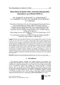
DOI 101515nbec20160017 University of SS Cyril and Methodius in Trnava MOLYBDATEREDUCING AND SDSDEGRADING N GUSMANIZAR13 reasons The aim of this paper is to isolate a bacterium that is ID: 836756

blue molybdenum sds reduction molybdenum blue reduction sds reducing strain shukor molybdate bacteria phosphate bacterium enterobacter bacterial concentrations carbon

Share:

Link:

Embed:

Download Presentation from below link

Download Pdf The PPT/PDF document "Nova Biotechnologica et Chimica 152 2016" is the property of its rightful owner. Permission is granted to download and print the materials on this web site for personal, non-commercial use only, and to display it on your personal computer provided you do not modify the materials and that you retain all copyright notices contained in the materials. By downloading content from our website, you accept the terms of this agreement.


Presentation Transcript

1 Nova Biotechnologica et Chimica 15-2 (20
Nova Biotechnologica et Chimica 15-2 (2016) 166 DOI 10.1515/nbec-2016-0017 University of SS. Cyril and Methodius in Trnava MOLYBDATE-REDUCING AND SDS-DEGRADING *, N. GUSMANIZAR1,3 reasons. The aim of this paper is to isolate a bacterium that is able to reduce molybdenum blue and at the same time can grow on the detergent Sodium Dodecyl Sulfate (SDS). Biochemical analysis resulted in a tentative identification of the bacterium as Enterobacter sp. strain Neni-13. Growth on SDS 1. 167 Rahman, M.F. et al. The Molycorp mine in New Mexico is a molybdenum mine. Its tailing wastes found their way into rivers near the mines including the Red River. Portions of the river exposed to high concentration of mine wastes are declared dead (JACOBS et al., 2014). In Indonesia, the mine tailings from the Batu Hijau, Sumbawa, copper, gold and molybdenum mine are dumped into the ocean with an annual amount close to several million tonnes. The sea is gradually polluted resulting in a reported reduction in fish populations (ANGEL ., 2013). Bacteria are prime candidate for metal including molybdenum remediation. Bacteria have unique enzymes that can convert heavy metals to less toxic forms (AHMAD al., 2013b). In the case of molybdenum, bacteria can convert the soluble molybdenum into insoluble molybdenum disulphide (TUCKER et al., 1997) or to the partially soluble and colloidal molybdenum blue (CAPALDI and PROSKAUER, 1896). Bioremoval columns packed with molybdenum-reducing sulphate-reducing bacteria showed good potential for the removal of molybdenum, but the toxic hydrogen sulphide gas emitted require another removal column packed with hydrogen sulphide-utilizing bacteria (TUCKER et al., 1997). Another promising candidate is the Mo-reducing bacterial system. The system utilizes bacteria that reduce molybdenum into molybdenum blue. The end product can be trapped in membrane (HALMI et al., 2014), and the process does not require strict anaerobic requirements (GHANI et al., 1993). Numerous candidates have been isolated from the genera of (LIM et al., 2012; HALMI et al., 2013b; MASDOR et al., 2015), Bacillus (ABO-SHAKEER et al., 2013; OTHMAN et al., 2013), Pseudomonas (SHUKOR ., 2010; AHMAD et al., 2013a), Acinetobac

2 ter, Serratia Enterobacter (SHUKOR 2009
ter, Serratia Enterobacter (SHUKOR 2009). Aside from heavy metals, detergents are often present as co pollutants, especially in water bodies. Detergents have detrimental effects to aquatic life (AMBILY and JISHA, 2012). The anionic surfactants such as Sodium Dodecylbenzene Sulfonate (SDBS) and Sodium Dodecyl Sulfate (SDS) show toxic effects to Daphnia magnaat concentrations of between 0.0025 and 300 mg/L. Detergents main toxicity have been documented in invertebrates and crustaceans due to the massive amount of these detergents continuously being released into the aquatic environment (CHATURVEDI and KUMAR, 2011). Several microorganisms can degrade SDS and use it for growth. The enzyme alkylsulfatase is the first enzyme of the pathway to degrade and assimilate SDS for microbial growth (CHATURVEDI and KUMAR, 2011). To date numerous SDS-degrading bacteria have been isolated (ABBOUD et al., 2007; HOSSEINI et al., 2007; CHATURVEDI and KUMAR, 2011; AMBILY and JISHA, 2012; HALMI 2013a). The current trend of xenobiotics detoxification research is to isolate multiple degraders or microorganisms showing multiple detoxification ability. This is especially important in polluted sites where multiple contaminants can be found (HUSAINI et al., 2008). The versatility of such multiple-degrading microorganisms are constantly being sought. Previously, two Mo-reducing bacteria have been reported with ability to grow on the detergent SDS (HALMI et al., 2013b; MASDOR et al.,2015). Herein, we report on the isolation of another Mo-reducing bacterium that can Nova Biotechnologica et Chimica 15-2 (2016) 168 grow on SDS as a sole carbon source. We also report on the first utilization of the modified Gompertz model to obtain growth parameters of this bacterium on SDS. All of the chemicals utilized in this work are of analytical grade. The UV-spectrophotometer (Shimadzu 1201) was purchased from Shimadzu Corporation, Kyoto, Japan. The orbital shaker was purchased fromYihder Technology Co., Ltd., Taipei, Taiwan. The microplate reader utilized in this work is a BioRad 680 microplate model and was purchased from BioRad, Richmond, CA, USA. The centrifuge model used was the Beckman GS-15R and was purchased from Beckman Instruments Inc., Fullerton, CA, USA. Bacterial cells were ruptured using a sonicator (Biosonik 111TM, Bronwil

3 l Scientific, Rochester, N.Y) Water sam
l Scientific, Rochester, N.Y) Water samples were taken from the Danau Maninjau Lake in the province of Padang, Sumatera, Indonesia in January 2009. Screening and isolation of potential molybdenum-reducing bacteria were carried in a minimal salts media with phosphate content fixed at 5 mM as most of the Mo-reducing bacteria isolated to date showed optimal molybdenum reduction at this concentration. The media was supplemented with sodium molybdate at 10 mM. About 0.1 mL of the water sample was spread onto a solid low phosphate agar (pH 6.5). The plates were incubated at room temperature (27 C) for 48 hours. The composition (mM) of the reduction media is as follows: glucose anhydrous (55 mM), (NH.SO (22.7 mM), MgSOO (2.03 mM), yeast extract (0.5 %), NaCl (85.56 mM), NaO (10 mM) and NaHPO (5 mM). Solid medium was prepared by adding agar (1.5 %) (MASDOR et al., 2015) This is a low phosphate molybdenum or LPM media. After the incubation period (48 hours), blue colonies were selected, and restreaked on the LPM agar several times to obtain pure culture. The Mo-reducing bacterial isolates were then grown in 20 mL of LPM liquid media to select for the best reducer. The bacterial isolates were grown for 48 hours at room temperature (27 C)on an orbital shaker (120 rpm, Yihder, Taiwan). About 1 mL of the culture media from each bacterial is(Beckman GS-15R, Fullerton, CA, USA) at 10,000 x g for 5 min at room temperature. The amount of molybdenum blue produced in the extracellular liquid was quantified at 865 nm using the specific extinction coefficient of 16.3 mM.A portion of the blue supernatant from culture media for the best isolate was periodically sampled and scanned from 400 to 900 nm (UV-spectrophotometer, Shimadzu 1201) after undergoing the centrifugation step as before. Identification of the best isolate was carried out utilizing biochemical and phenotypical methods in accordance to the Bergey’s Manual of Determinative Bacteriology (HOLT et al., 169 Rahman, M.F. et al. 1994), and followed up by using the ABIS online system software (COSTIN and IONUT 2015) as before (MASDOR 2015). The Mo-reducing bacterium was characterized in a microtiter plate format. The characterization include concentrations of molybdate, carbon sources, phosphate, effect of pH, heavy meta

4 ls, temperature and screening of potenti
ls, temperature and screening of potential xenobiotics as sources of growth or as electron donors supporting molybdenum reduction (SHUKOR and SHUKOR, 2014; MASDOR et al., 2015). A volume of 1 L of high phosphate molybdate media or HPM distributed in several smaller 250 mL conical flasks was utilized for growth. The HPM has the same composition to the LPM but with the phosphate concentration increased to 100 Mm. Briefly, bacterial cells were grown aerobically with shaking at 120 rpm on an orbital shaker (Yihder, Taiwan) at room temperature (27 C). The media utilized is a modification to the LPM in which the phosphate concentration was increased to 100 mM. This was carried out to prevent the formation of bacterial blue aggregates. Cells were harvested by centrifugation at 10,000 g (Beckman at 4 C for 10 min). After rinsing with deionized water twice, the pellets were resuspended in 20 mL of LPM giving an optical density measured at 600 nm of 1.0 absorbance value. In an effort to accommodate the variations in phosphate, molybdate, carbon sources, and pH conditions throughout the characterization works, appropriate modifications to the LPM was carried out. About 180 L of the cellular suspension was transferred into the wells of a sterile microplate, and mixed with 20 L of sterile glucose or other carbon sources from stock solutions. The final concentration of the carbon sources was 1.0 % (w/v). The microplates were covered (Corning® microplate), and then incubated at room temperature. Readings at 750 nm was periodically taken using a BioRad reader (Model No. 680, Richmond, CA). The molar extinction coefficient of 11,687 M.at 750 nm was utilized to quantify molybdenum blue production. This wavelength was chosen as it is the maximum filter available for the microplate unit (SHUKOR and SHUKOR, 2014). The effect of several heavy metals was studied utilizing Atomic Absorption Spectrometry calibration standard solutions from MERCK.2.4. Detergents and hydrocarbons as carbon sources for bacterial Preliminary works showed that none of the detergents and hydrocarbons tested could support molybdenum reduction. Hence, these compounds were then screened for bacterial growth in the microplate format as described by replacing glucose with these xenobiotics at the final concentration of 500 mg/L for detergents. Diesel and crude petroleum was initially added to the final concen

5 tration of 2 % (v/v) in 10 mL of the gr
tration of 2 % (v/v) in 10 mL of the growth media and sonicated at 60 Hz (Biosonik 111TM, Bronwill Scientific, Rochester, N.Y) for 5 minutes. Then 200 L of the media was added into the microplate wells, and mixed with 50 L of bacterial cells (see above). Nova Biotechnologica et Chimica 15-2 (2016) 170 The microplate was incubated at room temperature (27 C) for three days and the amount of molybdenum blue production was measured at 750 nm as before. The ingredients of the growth medium (1 L) were as follows: (NH.SO(22.7 mM), NaNO(23.53 mM), MgSOO (2.03 mM), yeast extract (0.01 %), NaCl (85.56 mM), NaHPO (50 mM) and 1 mL of trace elements solution. The trace elements solution composition (mM) was as follows: CaCl (0.363), FeSO(0.076), MnSOO (0.179), ZnSOO (0.069), CuSOO (0.02), CoClO (0.021), NaMoOO (0.020). The media was adjusted to pH 7.0. Then 200 L of the media was added into the microplate wells and incubated at room temperature (27 C) for 72 hours. Bacterial growth was measured as the increase in optical density at 600 nm. Substances (MBAS) Assay SDS degradation was quantified according to the MBAS assay JURADO et al.(2006). 100 µL of slightly acidic methylene blue reagent (pH of between 5 and 6) was added into 5 mL of the SDS calibration standards or samples (appropriately diluted) in a separating funnel followed by the addition of 200 µL of sodium tetraborate pH 10.5. The mixture was then mixed vigorously and then 4 mL of chloroform was added to the mixture and vigorously stirred for 1 minute. The mixture was left to separate and settle down for several minutes. The chloroform layer was separated and the absorbance was read at 650 nm in a glass cuvette. Values are means ± standard deviation of three replicated experiments. Statistical analyses such as ANOVA and t-test utilized the Graphpad Prism software ver. 5.0 available from www.graphpad.com. 3.1. Detergents as carbon sources for sp. strain Neni-13 Utilization of detergents and hydrocarbons as potential carbon sources for Enterobacter sp. growth was determined after 72 hours of incubation. The data showed that the bacterium was able to grow on the detergent SDS (Fig. 1). However, it was discovered that none of the detergents and hydrocarbons tested in this experiment can support molybdate reduction. SDS degradation studies carried out a

6 t concentrations between 200 and 1400 mg
t concentrations between 200 and 1400 mg/L under aerobic conditions showed that the fastest degradation occurred at 200 mg/L of SDS, with complete degradation occurring at day-4 of incubation. A 100 % of degradation was still observed at the SDS concentration of 800 mg/L. The lowest degradation after 12 days of cultivation occurred at 1400 mg/L showing 19.4 % degradation (Fig. 2). 171 Rahman, M.F. et al. 0.00.20.40.60.8ontrTerito NP9 rgi5S9WitconolthochloridenzoniloriDiesrudAbs 600 nmFig. 1. Growth of Enterobacter sp. strain Neni-13 on xenobiotics as sole source of carbon and energy. Bars signify the standard deviation of triplicates. 024681012Incubation (day)SDS residual (%) 200 400 600 800 1000 1200 1400Fig. 2. SDS degradation by Enterobacter sp. strain Neni-13. The bacterium was grown at various concentrations of SDS in a volume of 50 mL in a 250 mL shake-flask on an orbital shaker at 120 rpm at room temperature (27 C). Bars signify the standard deviation of triplicates. Strain Neni-13 was a Gram-negative, short rod-shaped, motile, and facultative anaerobic bacterium (Table 1). Identification of the bacterium through the ABIS online software suggested that the identity of the bacterium giving the highest similarity or homology score of 90 % and accuracy score of 100 % as Enterobacter sp. strain Neni-13. A more accurate molecular identification technique through phylogenetic analysis of the 16srRNA gene is currently being carried out to identify this species further. However, for now the bacterium is identified as Enterobacter strain Neni-13 in honor of the late Dr. Neni Gusmanizar. Nova Biotechnologica et Chimica 15-2 (2016) 172 Table 1. Overview on the presence (+) or absence (-) of individual biochemical activities for Enterobacter sp. strain Neni-13. Motility + Acid production from: Pigment – Alpha-Methyl-D-Glucoside + Catalase production (24 h) + D-Adonitol + Oxidase (24 h) – L-Arabinose + ONPG (beta-galactosidase) + Cellobiose + Arginine dihydrolase (ADH) + Dulcitol + Lysine decarboxylase (LDC) – Glycerol + Ornithine decarboxylase (ODC) + D-Glucose + Nitrates reduction + myo-Inositol + Methyl red – Lactose + Voges-Proskauer (VP) + Maltose + Indole production – D-Mannitol + Hydrogen sulf

7 ide (HS) – D-Mannose + Acetate utilizati
ide (HS) – D-Mannose + Acetate utilization + Melibiose + Malonate utilization + Mucate + Citrate utilization (Simmons) + Raffinose + Tartrate (Jordans) + L-Rhamnose + Esculin hydrolysis + Salicin + Gelatin hydrolysis – D-Sorbitol + Urea hydrolysis + Sucrose + Deoxyribonuclease – Trehalose + Lipase (corn oil) – D-Xylose + Phenylalanine deaminase – Growth on KCN medium + 3.3. Scanning absorption spectrum of molybdenum blue EnterobacterA scanning absorption spectrum of the molybdenum blue produced from this bacterium from the visible to the near infrared (IR) regions showed a peak with a maximum wavelength of 865 nm and a shoulder at approximately 710 nm (Fig. 3). The molybdenum blue spectra at 24-h and 48-h of incubations were similar. 0.01.02.03.0600650700750800850900950Wavelength (nm) 24 hFig. 3. Scanning absorption spectrum of molybdenum blue from Enterobacter sp. strain Neni-13 at different time intervals. 173 Rahman, M.F. et al. Glucose was the best electron donor for supporting molybdenum reduction sp. strain Neni-13. This is followed in descending order of efficiency by sucrose, l-rhamnose, maltose, lactose, cellobiose, d-mannose, raffinose, mucate, d-mannitol, d-adonitol, melibiose, glycerol, d-sorbitol and l-arabinose (Fig. 4). The optimal concentration of glucose was 1 % (w/v) (data not shown). D-AdelloblcitcerolLacaltD-ManitMelibiosecatoseL-RamnosSalicinD-SorbitolSucroseAbs 750 n m Fig. 4. The effect of various carbon sources on molybdenum blue production by Enterobacter Neni-13. Bars signify the standard deviation of triplicates. 3.5. Molybdenum reduction at various phosphate and molybdate The optimum concentration of phosphate occurred between 2.5 and 5.0 mM with higher concentrations were strongly inhibitory to reduction (Fig. 5). 0.00.51.01.5 0 01020304050Phosphate (mM)Abs 750 n m Fig. 5. The effect of phosphate concentrations on molybdenum blue production by sp. strain Neni-13. Bars signify the standard deviation of triplicates. Nova Biotechnologica et Chimica 15-2 (2016) 174 Molybdenum blue production at various sodium molybdate concentrations was studied by varying molybdate concentrations from 0 to 70 mM. The result shows that a lag period

8 of about 10 hours was observed before m
of about 10 hours was observed before molybdenum blue production started, and a maximum molybdenum blue production of 14.8 nmol molybdenum blue after 52 hours of incubation using 15 mM sodium molybdate was observed. An inhibition to molybdenum reduction was observed at molybdate concentrations higher than 30 mM (Fig. 6). 01122334455Incubation (h)nmole Mo-Blue 0 mM 5 mM 10 mM 15 mM 20 mM 25 mM 30 mM 35 mM 40 mM 50 mM 60 mM 70 mMFig. 6. The effect of molybdate on molybdenum blue production by Enterobacter sp. strain Neni-13. Bars signify the standard deviation of triplicates. ConrseniCad C hromppeercuryLeadMolybdate-reducing Activity (%)Fig. 7. The effect of heavy metals (2 ppm) on molybdenum blue production by Enterobacter sp. strain Neni-13. Bars signify the standard deviation of triplicates. 175 Rahman, M.F. et al. Molybdate reduction was inhibited by the heavy metals mercury, silver, copper and chromium with both mercury and silver showing a similar strong inhibition as judged by ANOVA analysis (Fig. 7). Discussion Although the phenomenon of molybdate reduction to molybdenum blue has been reported in E. coli more than a century ago (CAPALDI and PROSKAUER, 1896), the first detailed study on this phenomenon was carried out in E. coli K12 (CAMPBELL et al., 1985). In 1993, the same phenomenon was reported in Enterobacter cloacae strain 48 (GHANI et al., 1993), without citing previous works, indicating the rarity of similar studies. The potential for utilization in the bioremediation of molybdenum has first beent recognized by GHANI et al.(1993). To date many more Mo-reducing bacteria have been isolated (Table 2), including two strains that can grow on the detergent SDS (HALMI et al., 2013b; MASDOR et al., 2015). In addition, a psychrophilic Mo-reducing bacterium is also isolated from Antarctica. Previously, two molybdenum-reducing bacterium from this genus; Enterobacter cloacae strain 48 (GHANI et al., 1993) and Enterobacter sp. strain Dr.Y13 (SHUKOR 2009) have been isolated. Studies on the optimal conditions for molybdenum reduction utilizes the microtiter plate format with the use of whole or resting cells allowing a high throughput characterization to be carried out (SHUKOR and SHUKOR, 2014; MASDOR et al.,2015). Usage of resting cells or whole cells was

9 first started in the bacterium Enteroba
first started in the bacterium Enterobacter cloacae strain 48 (GHANI et al., 1993). There are considerable reports on the utilization of resting cells in a microtiter plate format indicating the favorable use of this method. This include a study on polyalcohol ethoxylate biodegradation (SHARVELLE et al., 2008). One of the advantages of resting cells is it can give cells tolerance to toxic xenobiotics especially in the beginning of the growth process. The inhibition by heavy metals on molybdenum reduction was observed in nearly all of the molybdenum-reducing bacteria isolate to date (Table 2). The presence of these heavy metals at potential bioremediation site for molybdenum could present a problem if the concentrations of the metals are above 2 ppm. The inhibitory effects of heavy metals such as mercury and copper have been reported in bacterial chromate reduction, with the target of inhibition by these metal ions suggested as the sulfhydryl group of the chromate reductase (AHMAD et al., 2013b). Several chemicals such as calcium carbonate, manganese oxide, and magnesium hydroxide can be added to bioremediation sites to alleviate the effect of toxic cations above as these chemicals can bind and immobilize the metal ions (HETTIARACHCHI et al., 2000). However, the effect of the addition of these sequestering agents to molybdenum reduction remains to be studied. Another option to lessen the toxicity of these cationic heavy metals to bioremediation of molybdenum, especially in aquatic bodies is to entrap the molybdenum-reducing bacterium in dialysis tubing or membrane (HALMI 2014). Nova Biotechnologica et Chimica 15-2 (2016) 176 Table 2. Overview of Mo-reducing bacteria isolated to date. Optimal Optimal (mM) Optimal (mM) Heavy metal inhibitors Reference glucose 5-20 5-7.5 Cu MASDOR et al., 2015 glucose 40 2.5-5 AsABO-SHAKEER et al., 2013 A.rzi glucose 50 4 Cd,AgOTHMAN et al., 2013 Pseudomonas DRY2 glucose 15-20 5 Cr, Hg SHUKOR et al., 2010 Pseudomonas sp. strain DRY1 glucose 30-50 5 Cd,AgAHMAD et al., 2013a Enterobacter strain Dr.Y13glucose 25-50 5 CrSHUKOR et al., 2009 Enterobacter strain 48 sucrose 20 2.9 Cr GHANI et al., 1993 glucose 80 5 Cr CAMPBELL et al., 1985 strain hkeemfructose 80 4.5 Cu LIM et al., Literature search showed a variation in the efficiency of bacterial isolates on SDS degradati

10 on. SDS-degrading bacteria are potential
on. SDS-degrading bacteria are potential agents for aquatic and soil bioremediation of SDS. To date, several SDS-degrading bacteria have been isolated and characterized including cold-adapted, psychrophilic or psychrotolerant SDS-degrading (MARGESIN and SCHINNER, 1998; HALMI et al., 2013a). A bacterial consortium of Pantoea agglomerans and Acinetobacter calcoaceticus is able to degrade 4000 mg/L of SDS in approximately 5 days (ABBOUD et al., 2007). The SDS-degrading bacteria Pseudomonas betelli and Acinetobacter johnsoniidegrades 500 mg/L SDS within 5 days of incubation (HOSSEINI et al. 2007). Klebsiella oxytoca srain DRY14, degrades 80 % of 2 000 mg/L of SDS within 4 days of incubation (SHUKOR et al., 2009), while Pseudomonas aeruginosa sp. degrades 100 % of 1 000 mg/L of SDS within 2 days of incubations (SYED et al., 2010). One of the most efficient SDS-degrading bacterium isolated is a mutated strain of Pseudomonas aeruginosa MTCC 10311 that degrades 1 500 mg/L of SDS within two days of incubation (AMBILY and JISHA, 2012). At high concentrations, SDS disrupts cellular membrane integrity. This leads to disturbances to the ion gradients resulting in the leakage of bacterial cytosolic contents (CHATURVEDI and KUMAR, 2011). Another mechanism of SDS toxicity is through surface protein and enzymes denaturation (AMBILY and JISHA, 2012). The identity of the molybdenum blue produced from bacterial reduction of molybdenum has never been identified since it was first mentioned in 1896. et al. (1985) were the first to suggest that the molybdenum blue found in E. coli K12 is likely a reduced form of phosphomolybdate. They primarily based this evaluation on the molybdenum blue spectrum acquired. Phoshomolybdate (molybdophosphate) was not mentioned in the works on Enterobacter cloacae strain 48 by GHANI et al. (1993). Instead, they proposed that the Mo-reducing enzyme to 177 Rahman, M.F. et al. molybdenum (Mo) reduces molybdenum in the form of molybdate (Moenzymatically to Mo before the reaction with phosphate anion formed molybdenum blue. Nonetheless, there are a few problems with this mechanism. Firstly, molybdenum exist as [MoOor molybdate anions, and does not exist in solution. Secondly, an acidified solution of molybdate above 1 mM will be instantaneously conve

11 rted to polyions such as Mo, and MoThird
rted to polyions such as Mo, and MoThirdly, these polyions combine with phosphate and form the heteropolymolybdate molybdophosphate or phosphomolybdate. Fourthly, enzymes such as aldehyde and xantine oxidases can catalytically reduce phosphomolybdate to molybdenum blue but not molybdate to molybdenum blue (GLENN and CRANE, 1956). Lastly, almost all of the bacterially created molybdenum blue display spectra that demonstrate near resemblance of the molybdenum blue generated from the phosphate determination method (PDM) (SHUKOR et al., 2007; MASDOR et al., 2015). The heteropolymolybdate species of molybdenum blue produced in the PDM is phosphomolybdate, the latter displays a scanning spectrum with a characteristics shoulder of from 700 to 720 nm, and a peak maximum from 870 nm to 890 nm et al., 1989). In line with all of this, we suggested a new hypothesis on the standard mechanism of molybdenum blue production in bacteria. In this new mechanism, we propose that initially, a phoshomolybdate intermediate is formed from molybdate. Phosphomolybdate is then reduced by the Mo-reducing enzyme to molybdenum blue in bacteria (SHUKOR et al., 2007). When we use phosphomolybdate as the electron acceptor substrate instead of the original molybdate in the Mo-reducing enzyme assay, we obtain an increase in enzyme activity of about two hundred times. Utilizing this compound, the Mo-reducing enzyme is purified for the first time after the phenomenon was first documented in 1896 (SHUKOR et al., 2014). The existence of an intermediate species in molybdenum blue production is likewise noticed during heavy metal reduction of chromate. In the bacteriaum Shewanella putrefaciens (now known as S. oneidensis) (MYERS et al., 2000), biological reduction of chromate from the 6+ to 3+ shows the occurrence of the intermediate species Cr. This has been proven via spectroscopic and paramagnetic resonance works. This indicates that the presence of an intermediate species is not unique to molybdenum. Spectroscopic analysis of the resultant molybdenum blue as a method to distinguish between existing heteropolymolybdates including silicomolybdate, sulphomolybdate and phosphomolybdate is a simple and acceptable method, and is routinely used to characterize phoshomolybdate or to differentiate between the numerous heteropolymolybdate (SIMS 1961). However, further investigations making use of nucle

12 ar magnetic resonance and electron spin
ar magnetic resonance and electron spin resonance are needed for detail identification of the possible lacunary species of phosphomolybdate observed in bacterial reduction of molybdenum to molybdenum blue (SIMS, 1961). Carbon sources such as glucose and sucrenergy or electron donors for the reduction of heavy metals. As bioremediation sites are often electron donor deficient, studies on the best electron donor are very useful as an important additive in bioremediation sites. At very low concentration of cells, Nova Biotechnologica et Chimica 15-2 (2016) 178 carbon sources are often utilized as carbon sources for energy, as the cell concentration increases, bacterial cells begin to produce excess reducing equivalents such as NADPH and NADH that are true electron donors for metal reduction. In resting or whole cells preparation, carbon sources are converted to reducing equivalents in a much greater amount, which makes metal reduction increases in efficiency. Glucose is also one of the best electron donors in many metal reductions such as chromate (LLOVERA et al., 1993) and selenate (LOSI and FRANKENBERGER JR., 1997). Mo-reducing bacteria isolated so far either prefer glucose or sucrose as efficient electron donor for supporting molybdenum reduction to molybdenum blue (Table 2). Metabolic pathways such as glycolysis, Kreb’s cycle and the electron transport system are the sites where these carbon sources are converted to the reducing equivalents NADH and NADPH. Both of these reducing equivalents are substrates for the molybdenum reducing-enzyme (SHUKOR et al.,2014). Molybdenum reduction is impacted by phosphate concentrations, with high phosphate concentrations significantly decreasing its production in the course of bacterial reduction of molybdenum. As an example, GHANI et al. revealed that phosphate concentrations greater than 2.9 mM inhibited reduction (GHANI et al.,1993), whilst levels greater than 5 mM inhibited most of the Mo-reducing bacteria isolated up to now (Table 1). Previous studies (GLENN and CRANE, 1956) have demonstrated that high phosphate concentrations negatively affecting phosphomolybdate structure by means of sustaining the pH at neutral. The phosphomolybdate complex is extremely unstable at this pH, and it is solely stable under acidic environments (GLENN and CRANE, 1956). Mo-redu

13 cing bacteria isolated so far can tolera
cing bacteria isolated so far can tolerate and reduce molybdate at the optimal concentrations ranging from 5 to 80 mM (Table 2). As molybdenum concentrations may achieve up to 900 mg/L in waters and 6,500 mg/Kg in soils (STONE and STETLER, 2008), Mo-reducing bacteria represent a meaningful tool for remediation processes. Another essential prerequisite is the soil phosphate concentrations should be about 5 mM but not exceeding 20 mM for reduction to occur. Fortunately, phosphate concentrations are hardly ever observed exceeding beyond these range in regular type of soils (CHAI 2011). A molybdate-reducing bacterium with the ability to grow on SDS as a sole carbon source has been isolated. Molybdenum blue characterization results indicate a good tolerance to high concentration of molybdenum, a requirement for low concentration of phosphate. Glucose, one of the most assimilable carbon sources was the best electron donor for molybdenum reduction. The absorption spectrum of molybdenum blue indicates that molybdenum blue is likely a reduced phosphomolybdate. Molybdenum reduction was inhibited by several toxic heavy metals indicating future bioremediation. Acknowledgment: The company Snoc International Sdn Bhd. financed a portion of this project. 179 Rahman, M.F. et al. References ABBOUD, M.M., KHLEIFAT, K.M., BATARSEH, M., TARAWNEH, K.A., AL-MUSTAFA, A., AL-MADADHAH, M.: Different optimization conditions required for enhancing the biodegradation of linear alkylbenzosulfonate and sodium dodecyl sulfate surfactants by novel consortium of Acinetobacter calcoaceticusPantoea agglomerans. Enzyme Microb. Technol., 41, 2007, 432-439. ABO-SHAKEER, L.K.A., AHMAD, S.A., SHUKOR, M.Y., SHAMAAN, N.A., SYED, M.A.: Isolation and characterization of a molybdenum-reducing Bacillus pumilus strain lbna. J. Environ. Microbiol. Toxicol., 1, 2013, 9-14. AHMAD, S.A., SHUKOR, M.Y., SHAMAAN, N.A., MAC CORMACK, W.P., SYED, M.A.: Molybdate reduction to molybdenum blue by an Antarctic bacterium. BioMed Res. Int., 2013, 2013a, 10 p. AHMAD, W.A., WAN AHMAD, W.H., KARIM, N.A., SANTHANA RAJ, A.S., ZAKARIA, Z.A.: Cr(VI) reduction in naturally rich growth medium and sugarcane bagasse by Acinetobacter haemolyticus. Int. Biodeterior. Biodegrad., 85, 2013b, 571-576. AMBILY, P.S., JISHA, M.S.: Biodegr

14 adation of anionic surfactant, sodium do
adation of anionic surfactant, sodium dodecyl sulphate by Pseudomonas aeruginosa MTCC 10311. J. Environ. Biol., 33, 2012, 717-720. ANGEL, B.M., SIMPSON, S.L., JAROLIMEK, C.V., JUNG, R., WAWORUNTU, J., BATTERHAM, G.: Trace metals associated with deep-sea tailings placement at the Batu Hijau copper-gold mine, Sumbawa, Indonesia. Mar. Pollut. Bull., 73, 2013, 306-313. CAMPBELL, A.M., DEL CAMPILLO-CAMPBELL, A., VILLARET, D.B.: Molybdate reduction by Escherichia coli K-12 and its chl mutants. Proc. Natl. Acad. Sci. U.S.A., 82, 1985, 227-231. CAPALDI, A., PROSKAUER, B.: Beiträge zur Kenntniss der Säurebildung bei Typhus-bacillen und Bacterium coli - Eine differential-diagnostische Studie. Z. Für Hyg. Infect., 23, 1896, 452-474. CHAI, B., WU, Y., LIU, P., LIU, B., GAO, M.: Isolation and phosphate-solubilizing ability of a fungus, Penicillium sp. from soil of an alum mine. J. Basic Microbiol., 51, 2011, 5-14. CHATURVEDI, V., KUMAR, A.: Isolation of a strain of Pseudomonas putidacapable of metabolizing anionic detergent sodium dodecyl sulfate (SDS). Iran. J. Microbiol., 3, 2011, 47-53. CLESCERI, L.S., GREENBERG, A.E., TRUSSELL, R.R.: Standard methods for the examination of water and wastewater, Port City Press, Baltimore, 1989. COSTIN, S., IONUT, S.: ABIS online - bacterial identification software, http://www.tgw1916.net/bacteria_logare.html, database version: Bacillus 022012-2.10, accessed on Mar 2015. GHANI, B., TAKAI, M., HISHAM, N.Z., KISHIMOTO, N., ISMAIL, A.K.M., TANO, T., SUGIO, T.: Isolation and characterization of a Mo-reducing bacterium. Appl. Environ. Microbiol., 59, 1993, 1176-1180. Nova Biotechnologica et Chimica 15-2 (2016) 180 GLENN, J.L., CRANE, F.L.: Studies on metalloflavoproteins. V. The action of silicomolybdate in the reduction of cytochrome c by aldehyde oxidase. Biochim. Biophys. Acta, 22, 1956, 111-115. HALMI, M.I.E., HUSSIN, W.S.W., AQLIMA, A., SYED, M.A., RUBERTO, L., MACCORMACK, W.P., SHUKOR, M.Y.: Characterization of a sodium dodecyl sulphate-degrading Pseudomonas sp. strain DRY15 from Antarctic soil. J. Environ. Biol., 34, 2013, 1077-1082. HALMI, M.I.E., WASOH, H., SUKOR, S., AHMAD, S.A., YUSOF, M.T., SHUKOR, M.Y.: Bioremoval of molybdenum from aqueous solution. Int. J. Agric. Biol., 16, 2014, 848-850. HALMI, M.I.E., ZUHAINIS, S.W., YUSOF, M.T., SHAHARUDDIN, N.A., HELMI, W., SHU

15 KOR, Y., SYED, M.A., AHMAD, S.A.: Hexava
KOR, Y., SYED, M.A., AHMAD, S.A.: Hexavalent molybdenum reduction to Mo-blue by a sodium-dodecyl-sulfate- degrading Klebsiella oxytocastrain dry14. BioMed Res. Int., 2013, 2013, 8 p. HETTIARACHCHI, G.M., PIERZYNSKI, G.M., RANSOM, M.D.: In situstabilization of soil lead using phosphorus and manganese oxide. Environ. Sci. Technol., 34, 2000, 4614-4619. HOLT, J.G., KRIEG, N.R., SNEATH, P.H.A., STALEY, J.T., WILLIAMS, S.T.: Bergey’s Manual of Determinative Bacteriology, 9 Auflage, Lippincott Williams & Wilkins, 1994. HOSSEINI, F., MALEKZADEH, F., AMIRMOZAFARI, N., GHAEMI, N.: Biodegradation of anionic surfactants by isolated bacteria from activated sludge. Int. J. Environ. Sci. Technol., 4, 2007, 127-132. HUSAINI, A., ROSLAN, H.A., HII, K.S.Y., ANG, C.H.: Biodegradation of aliphatic hydrocarbon by indigenous fungi isolated from used motor oil contaminated sites. World J. Microbiol. Biotechnol., 24, 2008, 2789-2797. JACOBS, J.A., LEHR, J.H., TESTA, S.M.: Acid mine drainage, rock drainage, and acid sulfate soils: causes, assessment, prediction, prevention, and remediation, John Wiley & Sons, Inc., Hoboken, New Jersey 2014. JURADO, E., FERNÁNDEZ-SERRANO, M., NÚÑEZ-OLEA, J., LUZÓN, G., LECHUGA, M.: Simplified spectrophotometric method using methylene blue for determining anionic surfactants: Applications to the study of primary biodegradation in aerobic screening tests. Chemosphere, 65, 2006, 278-285. LIM, H.K., SYED, M.A., SHUKOR, M.Y.: Reduction of molybdate to molybdenum blue by Klebsiella sp. strain hkeem. J. Basic Microbiol., 52, 2012, 296-305. LLOVERA, S., BONET, R., SIMON-PUJOL, M.D., CONGREGADO, F.: Chromate reduction by resting cells of Agrobacterium radiobacter EPS-916. Appl. Environ. Microbiol., 59, 1993, 3516-3518. LOSI, M.E., FRANKENBERGER JR., W.T.: Reduction of selenium oxyanions by Enterobacter cloacae strain SLD1a-1: Reduction of selenate to selenite. Environ. Toxicol. Chem., 16, 1997, 1851-1858. MARGESIN, R., SCHINNER, F.: Biodegradation of the anionic surfactant sodium dodecyl sulfate at low temperatures. Int. Biodeterior. Biodegrad., 41, 1998, 139-143. 181 Rahman, M.F. et al. MASDOR, N., ABD SHUKOR, M.S., KHAN, A., BIN HALMI, M.I.E., ABDULLAH, S.R.S., SHAMAAN, N.A., SHUKOR, M.Y.: Isolation and characterization of a molybdenum-reducing and SDS- deg

16 rading Klebsiella oxytoca strain Aft-7 a
rading Klebsiella oxytoca strain Aft-7 and its bioremediation application in the environment. Biodiversitas, 16, 2015, 238-246. MYERS, C.R., CARSTENS, B.P., ANTHOLINE, W.E., MYERS, J.M.: Chromium(VI) reductase activity is associated with the cytoplasmic membrane of anaerobically grown Shewanella putrefaciens MR-1. J. Appl. Microbiol., 88, 2000, 98-106. NEUNHÄUSERER, C., BERRECK, M., INSAM, H.: Remediation of soils contaminated with molybdenum using soil amendments and phytoremediation. Water. Air. Soil Pollut., 128, 2001, 85-96. OTHMAN, A.R., BAKAR, N.A., HALMI, M.I.E., JOHARI, W.L.W., AHMAD, S.A., JIRANGON, H., SYED, M.A., SHUKOR, M.Y.: Kinetics of molybdenum reduction to molybdenum blue by Bacillus sp. strain A.rzi. BioMed Res. Int., 2013, 2013, 9 p. SHARVELLE, S.E., GARLAND, J., BANKS, M.K.: Biodegradation of polyalcohol ethoxylate by a wastewater microbial consortium. Biodegradation, 19, 2008, 215-221. SHUKOR, M.S., SHUKOR, M.Y.: A microplate format for characterizing the growth of molybdenum-reducing bacteria. J. Environ. Microbiol. Toxicol., 2, 2014, 42-44. SHUKOR, M.Y., AHMAD, S.A., NADZIR, M.M.M., ABDULLAH, M.P., SHAMAAN, N.A., SYED, M.A.: Molybdate reduction by Pseudomonas sp. strain DRY2. J. Appl. Microbiol., 108, 2010, 2050-2058. SHUKOR, M.Y., HALMI, M. I.E., RAHMAN, M.F.A., SHAMAAN, N.A., SYED, M.A.: Molybdenum reduction to molybdenum blue in Serratia sp. strain DRY5 is catalyzed by a novel molybdenum-reducing enzyme. BioMed Res. Int., 2014, 2014, 8 p. SHUKOR, M.Y., RAHMAN, M.F., SHAMAAN, N.A., SYED, M.S.: Reduction of molybdate to molybdenum blue by Enterobacter sp. strain Dr.Y13. J. Basic Microbiol., 49, 2009, S43-S54. SIMS, R.P.A.: Formation of heteropoly blue by some reduction procedures used in the micro-determination of phosphorus. The Analyst, 86, 1961, 584-590. STONE, J., STETLER, L.: Environmental impacts from the North Cave Hills abandoned uranium mines, South Dakota. Merkel, B., Hasche-Berger, A. (Eds.): Uranium, mining and hydrogeology, Springer Berlin Heidelberg, 2008, 371-380. SYED, M.A., MAHAMOOD, M., SHUKOR, M.Y., SHAMAAN, N.A.: Isolation and characterization of SDS-degrading Pseudomonas aeruginosa sp. strain D1. Aust. J. Basic Appl. Sci., 4, 2010, 5000-5011. TUCKER, M.D., BARTON, L.L., THOMSON, B.M.: Reduction and immobilization of molybdenum by Desulfovibrio desulfuricans. J. Environ. Qual., 26, 1997, 1146-1152. 15 December 20