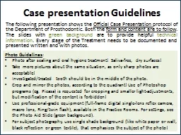
Photo Guidelines Photo after scaling and oral hygiene tr e atment Saliva free dry surfaces Take m ultiple pictures about the same situation as only sharp photos are acceptable ID: 815639
Download The PPT/PDF document "Case presentation Guidelines" is the property of its rightful owner. Permission is granted to download and print the materials on this web site for personal, non-commercial use only, and to display it on your personal computer provided you do not modify the materials and that you retain all copyright notices contained in the materials. By downloading content from our website, you accept the terms of this agreement.
Slide1
Case presentation Guidelines
Photo Guidelines: Photo after scaling and oral hygiene treatment! Saliva-free, dry surfaces!Take multiple pictures about the same situation, as only sharp photos are acceptable!Investigated/treated tooth should be in the middle of the photo. Crop and mirror the photos, according to the quadrant! Use of Photoshop programs (eg. Picasa) is requested for cropping and smaller light-adjustments, but modification of the content is forbidden!Use professional-grade equipment (full-frame digital single-lens reflex camera, macro lens, Ring/twin flash), available in the Practice Rooms. For settings, see the Photo Aid Slide (green background).For subject photography, use single shade background (like white paper or wall, black reflection or green textile), that emphasizes the subject of the photo!
The following presentation shows the Official Case Presentation protocol of the Department of Prosthodontics. Both the form and content are to follow. The slides with the green background are to provide you with helpful technical information. Each stage of the Treatment needs to be documented and presented both written and with photos.
Slide2Prosthodontic Final Exam
Case Presentation TITLE (Type of Denture)Eg. Complete Denture, FPD, Combined Fixed-Removable Partial Denture etc. Student:Consultant: Starting and finishing Date of Treatment:
SEMMELWEIS UNIVERSITYFaculty of DentistryDepartment of ProsthodonticsHead of Dept.: Prof. Dr. Péter HERMANN
Slide3Introduction of the Patient
Age, gender(previous) occupationThe reason for Dental Investigations’s Needs
eg. Pain, Aesthetic Complaints, Instability of Dentures etc. Student
Date
Prosthodontic
Final
Exam
Case
Presentation
Title
(
Type of Denture)
SEMMELWEIS UNIVERSITYDepartment Of Prosthodontics
Slide4Medical History
Factors which would affect the dental treatment General diseases – Medication – relevant adverse effects!Eg. High blood pressure: Amlodipin (amlodipine, Calcium
channel blocker) – AE: gingiva hyperplasia Eg. Osteoporosis: Bonviva (Ibandronate: Bisphosphonate) – AE: MRONJ osteonecrosis of the jawAllergy (eg. medicaments, metals)Bad habits, Addiction: Smoking, Alcohol etc. (how much?)
Student
Date
Prosthodontic
Final
Exam
Case
Presentation
Title
(Type of Denture)
SEMMELWEIS UNIVERSITYDepartment Of
Prosthodontics
Slide5Dental History
(Previously) worn Prosthodontic Appliances (when they were made) Extractions (when?)Other dental treatments, eg. orthodontics, dental surgery etc. Student DateProsthodontic Final Exam Case
Presentation Title (Type of Denture)SEMMELWEIS UNIVERSITYDepartment Of Prosthodontics
Slide6Dental Status
Portrait- and Intraoral Photos
Portrait – with natural smileFull smile
Maximal
Intercuspal
Position
Maxillary
arch
Mouth
slightly
open
Mandibular
arch
Student Date
Prosthodontic
Final
Exam
Case
Presentation
Title
(
Type
of
Denture
)
SEMMELWEIS UNIVERSITY
Department
Of
Prosthodontics
Slide71.
2.
4.
3.
5.
6.
- - - - - - - - - - - - - - - - - - - - - - - - - - - - - - - - - - - - - - - - -
- - - - - - - - - - - - - - - - - - - - - - - - - - - - - - - - - - - - - - - - -
- - - - - - - - - - - - - - - - - - - - - - - - - - - - - - - - - - - - - - - - -
- - - - - - - - - - - - - - - - - - - - - - - - - - - -
- - - - - - - - - - - - - - - - - - - - - - - - - - - - -
- - - - - - - - - - - - - - - - - - - - - - - - - - - - -
- - - - - - - - - - - - - - - - - - - - - - - - - - - - -
- - - - - - - - - - - - - - - - - - - - - - - - - - - - -
- - - - - - - - - - - - - - - - - - - - - - - - - - - - -
f 8
- - - - - - - - - - - - - - - - - - - - - - - - - - - - - - - - - - - - - - - - -
Manual
mode
: f 22, ISO 200, 1/200;
Flash
: M 1/2
Occlusal
mirror
-
Focus
Cheek
retractors
(
big
size
)
Photo-
Aid
: Camera
settings
Portrait
–
with
natural
smile
Maximal
Intercuspal
Position
Maxillary
arch
Full
smile
Mandibular
arch
Mouth
open
Slide8Camera –
Difference in Quality
If you use cellphone for photography, the lighting is poorer and the quality will be lower. The settings (picture composition, focus, patients position etc.) however
remain
the
same
.
Slide9Common
errors in PhotographyA fényképek minőségét jelentősen meghatározza, hogy a kép témája megfelelően helyezkedik-e el a képen. Az alábbiakra kell figyelni tehát:Fényképezőgép megfelelő beállítása (világosság, fókusz)Páciens megfelelő pozicionálása (fejtartás, mosoly, szájnyitás mértéke)Saját pozíciónk, a segédeszközök megfelelő alkalmazása (fénykép szemből, nem oldalról és alulról készüljön, tükör ne legyen párás stb.) !
Slide10Too
bright / too
dark picture (overexposed / underexposed)Image blurry (lack of focus and/or improper depth of field)Bad positioning: image taken from an improper angle (inferior/superior/canted), improper framing with mirror Meaningless content (keep in mind what we want to show!)No uniform backdrop, fogging of mirrorCommon errors in Photography
Slide11NO
PATIENT’S DATA,Just DATE of The X-ray05. 06. 2016.Please indicate, if the actual status differs from what is shown on the radiograph! Eg. Extractions – „X” mark on the tooth!Dental Status
Panoramic Radiograph Student DateProsthodontic Final Exam Case Presentation Title (Type of Denture)SEMMELWEIS UNIVERSITYDepartment Of Prosthodontics
Slide12P
(H
HH
H
H
E,
X
H)
8
7
6
5
4
3
2
1
1
2
3
4
5
6
7
8
8
7
6
5
4
3
2
1
1
2
3
4
5
6
7
8
E,
K
(H
H
H)
Vizsgázó neve
Dátum
Fogpótlástan Szigorlati Esetbemutatás
Cím
(Fogpótlás
Típusa)
SEMMELWEIS EGYETEM
Fogpótlástani Klinika
Dental
Status
Intraoral
radiographs
and
findings
Take
a
periapical
x-
ray
of
each
abutment
!
grey
colour
= missing tooth
X
= hopeless tooth (to be extracted
)
)( =
closed
extraction
gap
C
= crown, bridge – )( dimensions
U
= telescope
I
= implant
E
= root canal treated tooth (
e
ndo
)
L =
periapical
lesion
P
=
p
ost and core
F
= filling
D
=
decay
* =
to
be
replaced
Inspection
of Head and Neck RegionIntraoral Investigation I.Stomato-oncological screening: eg. no findings, red/white lesion (where?), precancerous lesions or cancer etc.TMJ examination: complaints, diagnosis
eg. Reciprocal clicking – diagnosis: Anterior Disc Displacement Static Occlusion - Bite form: eugnathic or dysgnathic Dynamic Occlusion - Type of Guidance: canine/group guidance etc. Student DateProsthodontic Final Exam
Case
Presentation
Title
(
Type
of
Denture
)
SEMMELWEIS UNIVERSITY
Department
Of
Prosthodontics
ANOMALIES
Dental
Skeletal
Transversal
Crossbite
Asymmetry
Crossbite
Asymmetry
Saggital
Angle
Class
II.
Angle
Class
III.
Prognathia
Progenia
Vertical
Open
bite
Deep
bite
Hyperdivergent
Hypodivergent
Slide14Inspection of Head and Neck Region
Intraoral Investigation II.
Oral hygiene eg. Good, poorOral mucosal abnormalities: eg. Denture stomatitis Periodontal health: pl. gingivitis (type) generalized/localized acut/chronic periodontitis etc.
→ in case of periodontal disease, periodontal charting is needed!
www.periodontalchart-online.com
Student
Date
Prosthodontic
Final
Exam
Case
Presentation
Title (
Type
of
Denture
)
SEMMELWEIS UNIVERSITY
Department
Of
Prosthodontics
Slide15Dental status
Upper JawPhoto of Upper Jaw (with and without RPD) and Study castIntraoral x-raysFábián and Fejérdy Classification of Partially Edentulous Arch: 0 / 1A / 1B / 2A / 2B / 2A/1 / 3Status (Zsigmondy cross, with indication of Findings)
Student DateProsthodontic Final Exam Case Presentation Title (Type of Denture)SEMMELWEIS UNIVERSITYDepartment Of Prosthodontics
(B
B
B
B
B
X
E
B)
8
7
6
5
4
3
2
1
1
2
3
4
5
6
7
8
grey
colour
= missing tooth
X
= hopeless tooth (to be extracted)
)( =
closed
extraction
gap
C
= crown, bridge – )( dimensions
U
= telescope
I
= implant
E
= root canal treated tooth (
e
ndo)
L =
periapical
lesion
P
=
p
ost and core
F
= filling
D
= decay
* =
to
be
replaced
Dental
statusLower JawPhoto of Upper Jaw (with and without RPD) and Study modelIntraoral x-
raysFábián and Fejérdy Classification of Partially Edentulous Arch: 0 / 1A / 1B / 2A / 2B / 2A/1 / 3Status (Zsigmondy cross, with indication of Findings) Student DateProsthodontic Final Exam Case
Presentation
Title
(
Type
of
Denture
)
SEMMELWEIS UNIVERSITY
Department
Of
Prosthodontics
8
7
6
5
4
3
2
1
1
2
3
4
5
6
7
8
E
(H
H
H)
grey
colour
= missing tooth
X
= hopeless tooth (to be extracted)
)( =
closed
extraction
gap
C
= crown, bridge – )( dimensions
U
= telescope
I
= implant
E
= root canal treated tooth (
e
ndo)
L =
periapical
lesion
P
=
p
ost and core
F
= filling
D
= decay
* =
to
be
replaced
Preoperative
TreatmentsDetailed description (text, drawings and photos!) of Surgical, Periodontal, Conservative treatment plan and treatments. (with methods and materials used)Detailed description of Prosthodontic Planning phases (eg. Study impressions and casts, diagnostic wax-up and mock-up, smile design etc. Photos!) Student Date
Prosthodontic Final Exam Case Presentation Title (Type of Denture)SEMMELWEIS UNIVERSITYDepartment Of Prosthodontics!
Slide18Prosthodontic Treatment Plan
UPPER JAW: FIXED partial denture (FPD)4-unit porcelain fused to metal (PFM) bridge (CoCr) Retainers: 16, 13 Pontics: 15, 14Cast post and core / Dowel core, PFM (CoCr) crown: 24Lower Jaw: COMBINED FIXED-REMOVABLE PARTIAL DENTUREFixed appliance:2-unit PFM (CoCr) splint: Retainers
: 34, 33 Semi-precision attachment: Preci-Vertix slide attachment patrix (male part) with lingual parallel milled shoulder and interlock (34)2-unit PFM (CoCr) splint: Retainers: 44, 43 Semi-precision attachment: Preci-Vertix slide attachment patrix (male part) with lingual parallel milled shoulder and interlock (34)Removable appliance: attachment-retained removable partial dentureAcrylic base, cast metal mesh framework (CoCr) , lingual bar major connectorSemi-precision attachment: preci
vertix
slide attachment
matrix
(
female part
)
, lingual bracing arm
(
indirect
retainer
)
with interlock 44, 34 Replaced teeth: 47, 46, 45, 35, 36, 37 (6 pcs) acrylic artificial teethDento-mucosal support
Student Date
Prosthodontic
Final
Exam
Case
Presentation
Title
(
Type
of
Denture
)
SEMMELWEIS UNIVERSITY
Department
Of
Prosthodontics
D
escription
and drawings
!
Meaning
of
colours
:
Blue
=
metal,
Green
=
Ceramic
,
Red
=
Acrylic
Yellow
=
precision
attachment (female part)Orange = zirconia
Slide19Prosthodontic Treatment Plan
UPPER JAW: CLASP-RETAINED Removable Partial Denture (RPD)Dento-mucosal supportAcrylic base, cast metal (CoCr) mesh framework and (butterfly) palatal strap major connector14: mesial occlusal rest
Clasp-retention14: back-action clasp23: Roach C clasp13, 22: bracing arms5 pcs acrylic artificial teeth (15, 16, 24, 25, 26)Lower Jaw: telescopic OverdentureMuco-dental supportAcrylic base Telescopic retentionCylindro-conical primary
coping
(
CoCr
): 33, 45
Cylindro-conical
secondary
coping
(
CoCr
): 33, 45
12
pcs
acrylic artificial teeth (36-46)
D
escription and drawings
!
Student
Date
Prosthodontic
Final
Exam
Case
Presentation
Title
(
Type
of
Denture
)
SEMMELWEIS UNIVERSITY
Department
Of
Prosthodontics
Meaning
of
colours
:
Blue
=
metal,
Green
=
Ceramic
,
Red
=
Acrylic
Yellow
=
precision attachment (female part)Orange = zirconia
Slide20Vizsgázó neve
DátumFogpótlástan Szigorlati EsetbemutatásCím (Fogpótlás Típusa)SEMMELWEIS EGYETEMFogpótlástani KlinikaFelső állcsont: RÖGZÍTETT fogpótlás
11, 21 lithium-diszilikát kerámia héj (préskerámia, e.max®)12, 22 lithium-diszilikát kerámia korona (préskerámia, e.max®)[17-x-15] 3-tagú, cirkonium-dioxid vázas kerámia híd Leplezett horgonykoronák: 17, 15 Leplezett hézagfog: 16[27-x-75] 3-tagú, cirkonium-dioxid vázas kerámia híd Leplezett horgonykoronák: 27, 25 Leplezett hézagfog: 26Alsó állcsont: Implantációs rögzített FOGPÓTLÁS34 cementtel rögzített fémkerámia korona (CoCr)gyári titán protetikai fej (pl. TiDesign®) (pl. Astra TX) bone level implantátumon36 cementtel rögzített fémkerámia korona (CoCr)gyári titán protetikai fej (pl. TiDesign®) (pl. Astra TX) bone level implantátumon[36-x-34] 3-tagú, csavaros rögzítésű fémkerámia híd (CoCr)2db gyári titán protetikai fej (pl. Uni Abutment®) (pl. Astra TX) 36 és 34 pozícióban lévő bone level implantátumon
!
Prosthodontic Treatment Plan
D
escription and drawings
!
Meaning
of
colours
:
Blue
=
metal,
Green
=
Ceramic
,
Red
=
Acrylic
Yellow
=
precision
attachment
(
female
part)
Orange
=
zirconia
Slide21Vizsgázó neve
DátumFogpótlástan Szigorlati EsetbemutatásCím (Fogpótlás Típusa)SEMMELWEIS EGYETEMFogpótlástani Klinika
Felső állcsont: Implantációs FEDŐLEMEZES FOGPÓTLÁSMegtámasztás: muko-implantálisPatkó alakban redukált akrilát alaplemez, fémhálós merevítésselElhorgonyzás: gömbretenciós fej 14, 12, 22, 24-es pozícióban14 db keményakrilát műfog (17-27)Alsó állcsont: Implantációs FEDŐLEMEZES FOGPÓTLÁSMegtámasztás: implanto-mukozálisRedukált akrilát alaplemez, fémhálós merevítésselElhorgonyzás: merevítőrúd-rendszerrelPreci horix® típusú merevítőrúd a 45-43-33-35 pozícióban lévő implantátumok között 14 db keményakrilát műfog (37-47)
Prosthodontic Treatment Plan
D
escription and drawings
!
Meaning
of
colours
:
Blue
=
metal,
Green
=
Ceramic
,
Red
=
Acrylic
Yellow
=
precision
attachment
(
female
part)
Orange
=
zirconia
!
Slide22Prosthodontic
ProceduresStep by stepIndicate the different treatment phases on new slides, create a logical structure! The title of the Slides of Prosthodontic treatment Phases should be the name of
these Phases. SEE the Study-Aid (following slides)Indicate the methods used, as well as the type, name and producer of the materials!precisional-situational impression, putty-wash technique; C-Silicone impression material; Zetaplus+Oranwash (Zhermack®)Eg. glass ionomer luting cement, Ketac Cem (3M ESPE®) Student DateProsthodontic Final Exam Case Presentation Title
(
Type
of
Denture
)
SEMMELWEIS UNIVERSITY
Department
Of
Prosthodontics
Slide23Short term control
Long term control Student DateProsthodontic Final Exam Case Presentation
Title (Type of Denture)SEMMELWEIS UNIVERSITYDepartment Of ProsthodonticsProfessional oral hygiene treatment, repeated instruction and motivation - if necessary.Recheck Status.
Slide24CONTENT-AID
Slide25Description of Conservative Treatments
Before photo, periapical X-rayFilling procedure:Prepared, Ready CavityRubber dam, matrix, wedge in situFinished, polished restorationEndodontic treatment:Access cavity with rubber damPhoto with apex locator, X-ray for working length Control radiographInlay/onlay/overlay:Prepared cavity
Impressions, modelsTemporary restorationFinal restoration on the castRubber dam, matrix, wedge in situFinished, polished restoration (occlusal view, ICP)Lsd. Konzerváló Fogászati Klinika esetbemutatási protokollját!
Slide26Complete
dentureClinical phasesAdditional informationPrimary/anatomical impression(impression material type and name)Primary/anatomical castOutline of special tray on the castSecondary/Functional impression
Special tray (on the cast) , Adjusted functional impression traySecondary/Functional impression (from mucosal side)Antagonistic impression(type and material of the tray)(type and name of material used for adjustments)(impression material type and name)Determination of COOcclusal rim on the functional cast Adjusted occlusal rims intraorally and extraorally (with the casts)Shade selection
Using
VITA
Classical
shade
guide
Try-in denture
I
n
the
articulator (ICP, occlusal view)
and
in the mouth (ICP, occlusal view, smile)
Final
denture
+
education
on
hygiene
In
the
articulator
(
ICP,
occlusal
view
,
polished and non-polished surface
s
)
In the mouth (ICP, occlusal view, smile)
Slide27Clasp-retained
RPD with metal baseplateClinical phasesAdditional informationImpressionsSituational impressionAntagonistic impressionBite registration
(if possible)(method, material type and name)(intraorally and extraorally)Bite registration (if it’s necessary for making the metal baseplate)Occlusal rim / Intraoral gothic arch tracer (intraorally and extraorally with the casts)
Outline of the metal baseplate on the master cast
Metal baseplate trial
Metal
baseplate
on the cast
and
in the mouth
ICP
+
occlusal
view
Determination of CO/ICP
(this time at
the latest)
Shade
selection
(
intraorally
and
extraoral
ly
with
the
casts
)
Using
VITA
Classical
shade
guide
Try
-in
denture
In
the
articulator and in
the
mouth
Occlusal
view
+
lateral
view
+ ICP +
smile
Final
denture
+
education
on hygieneIn the articulator (ICP, occlusal view, lateral view, polished and non-polished surfaces)In the mouth (ICP, occlusal view, lateral view, smile)
Slide28Clinical
phasesAdditional informationPreparationSituational impression for the temporary restorationPrepared abutment teethTemporary restorations
(method, material type and name)ICP + occlusal view(material type and name)ImpressionsPrecisional-situational impressionAntagonistic impressionBite registrationMegtartott harapás: wax biteNem megtartott harapás: occlusal rim / intraoral gothic arch tracer(method
,
material type and name)
(method
,
material type and name)
(
intraorally
and
extraorally
with
the
antagonistic cast)
Framework trialSectioned castIn the articulator and in
the mouth Shade
selection
(material type)
(ICP +
occlusal
view
+
lateral
view
)
Using
VITA
3D Master
shade
guide
Bisque
trial
In
the
articulator and in
the
mouth
(ICP
+
occlusal
view
+
lateral
view
+
smile
)
Final restoration, cementation
In
the
articulator and in
the mouth(material type and name)(ICP + occlusal view + lateral view
+ smile)Fixed prosthodontic appliances (FPD)
Slide29Combined
fixed-removable partial dentureClinical phasesAdditional information
Fixed partSee „Fixed prosthodontic appliances (FPD)”!Situational (pick-up) impression with the fixed partRemovable partTry-in of RPD’s metal framework with the FPDDetermination of ICP
(
in the mouth, on the cast
)
(
intraorally
and
extraorally
with
the
casts
)
Try-in denture
In the articulator and in the mouth
ICP + occlusal
view
+
smile
Final
denture
In
the
articulator and in
the
mouth
Cementation of
the
fix
ed
part
(
with
the
removable
part)
Only
the
fix
ed
part
then
the
fix
ed
+
the
removable
part
together. ICP + occlusal view + smilePrecision attachments, parallel milled shoulders, interlock should be visible!(material type and name)
Slide30Telescopic
overdentureClinical phasesAdditional informationPreparationSituational impression for the temporary restoration
Prepared abutment teethTemporary restorations Diagnostic teeth set-up – in case of telescopes in front region.ImpressionsPrecisional-situational impressionAntagonistic impressionPrimary telescopes: on the cast, in the mouth.Special tray, functional tray.Functional-situational impression with the primary telescopes.(on the cast, mucosal surface) Determination
of CO
with
occlusal
rims
Ready primary telescopes
(
on the cast, in the mouth
)
Adjustment
of
the
o
cclusal rim with the secondary telescopes in itShade selection
(intraorally and extraorally with the casts)Try-in
dentureIn the articulator and in
the
mouth
(ICP
+
occlusal
view
+
smile
)
Final
denture
,
cementation of primary telescopes
.
Final denture and telescopes on the cast
and in
the
mouth
.
Cementation
of
primary
telescopes
.
Primary telescopes after cementation
.
Final
denture
in
the
mouth
after
the
cementation
.
(polished and non-polished surfaces)(ICP + occlusal view) (ICP + occlusal view + smile)
Slide31Implant-retained
crowns and bridgesClinical phases and additional information General informationDate of implant placementName, type and description of the implant and its
prosthetic components (material, type, custom/cast etc.)Healing component (healing abutment/cap) in the mouth, peri-implant soft tissuesImpressionsImplant-level impression: impression components in the mouth, x-ray control.Felépítményszintű lenyomatvétel esetén (csavaros rögzítésű fogpótláshoz): behelyezett implantátum fejek, majd az ezekre behelyezett lenyomatvételi fejek + röntgenfelvételClosed-tray impression: impression caps in mouth, precisional-situational impression with implant analogs (material + method).Open-tray impression: open tray on the cast, impression posts in mouth (with tray), precisional-situational impression with implant analogs (material + method).Antagonistic impression (material + method)
.
Wax
bite
(
extraorally
,
material
type
and
name
),
occlusal
rim (intraorally and extraorally with the antagonistic cast)
Framework trial: in the articulator and in the
mouth.
Shade
selection
:
using
VITA 3D Master
shade
guide
.
Bisque
trial
:
in
the
articulator and in
the
mouth
.
Final insertion of restoration
In
the
articulator and in
the
mouth
(ICP +
occlusal
view
)
Control
x-
ray
Cement-retained: cement type
Screw-retained: torque, covering of the screw hole (
teflon
tape, filling)