PDF-4MORPHOLOGYfilamentous, uniseriate protonema. Cell specializationgree
Author : stefany-barnette | Published Date : 2015-09-09
6MORPHOLOGYbecome starchfilled and hyaline toward the stem interiorassimilates The central strand absent in some taxa isalways a solid core of nonlignified waterconductinghydroids
Presentation Embed Code
Download Presentation
Download Presentation The PPT/PDF document "4MORPHOLOGYfilamentous, uniseriate proto..." is the property of its rightful owner. Permission is granted to download and print the materials on this website for personal, non-commercial use only, and to display it on your personal computer provided you do not modify the materials and that you retain all copyright notices contained in the materials. By downloading content from our website, you accept the terms of this agreement.
4MORPHOLOGYfilamentous, uniseriate protonema. Cell specializationgree: Transcript
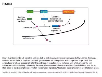
6MORPHOLOGYbecome starchfilled and hyaline toward the stem interiorassimilates The central strand absent in some taxa isalways a solid core of nonlignified waterconductinghydroids Hydroid st. Grace Denton & Stephanie . Tristram. G.E.Denton@warwick.ac.uk. S.C.Tristram@warwick.ac.uk. What are some of the causes of cell injury and cell death?. Causes of cell injury . . CHIPING: C. Lecture 4 . Assays . Based . on Cell Proliferation. Cell counts. can be used to determine the effect of various compounds on cell proliferation, but at least in the early stages of testing, a complete growth curve is required. . Introductory 100 level biology class. Non-majors. Christine Andrews, Katie Morrison-Graham, Bianca . Breland. , Marilyn Thomas, . Sarah Cabbage, Kat Milligan-. Myhre. Goals. Understand how cells communicate and pass on information. A Chromosome and Sister Chromatids. Key Points About Chromosome Structure. A . chromosome consists of DNA that is wrapped around proteins (. histones. ) and condensed. Each . histone. and the DNA wrapped around it make up a . Why do cells need to divide?. Growth and development. You started as 1 cell and now you’re made up of roughly 40 TRILLION cells. Repair and regeneration of tissue. Reproduction. What happens when cells divide? . Cell-to-cell . signaling. : . Important . to both multicellular . & unicellular . organisms. . H. elps coordinate activities & . events necessary for . multicellular . organism to develop from . Learning . Goal . Describe . the composition and function of the lipid bilayer in cell membranes.. Cell Membranes. Cell membranes. . are semipermeable so that nutrients can enter the cell and waste products can leave.. Means “to move the cell”. Approximately 5% of cell’s life. Process differs in . Heterotrophs. (Animals) . Autotrophs. (Plants) due to presence of cell wall. HETEROTROPHS. AUTOTROPHS. Cell. membrane . 1. 2 . 4. 8 . How many chromosomes are in the middle cell?. 1. 2 . 4. 8 . At what part of the cell cycle would you see a chromosome that looks like this. ?. G. 1. G. 2. . M. S . At what part of the cell cycle would you see a chromosome that looks like this? . Two Basic Types. Remember….cells are the basic unit of life for ALL living things.. There are two basic types of cells:. Prokaryotic cells . – found in bacteria. Eukaryotic cells . – found in . Zhang . Junqiu. Paper . presentation. Nature 2004, 428:868-871. Introduction. We want to build a quorum-sensing system which can control cell density at a constant amount autonomously.. Mechanism similar to natural circuit (Streptococcus . Car T Cell Therapy Market report provides the future growth trend of the market based on in-depth research by industry experts.The global and regional market share along with market drivers and restraints are covered in the report. View More @ https://www.valuemarketresearch.com/report/car-t-cell-therapy-market Tissue culture is the general name for the removal of cells, tissues or organs from an animal or plant and their subsequent placement into artificial environment conductive to growth. This environment usually consists of a suitable glass or plastic culture vessel containing a liquid or semi-solid support medium that supplies the nutrients essential for survival and growth. Van Delden C, Iglewski BH. Cell-to-Cell Signaling and Pseudomonas aeruginosa Infections. Emerg Infect Dis. 1998;4(4):551-560. https://doi.org/10.3201/eid0404.980405.
Download Document
Here is the link to download the presentation.
"4MORPHOLOGYfilamentous, uniseriate protonema. Cell specializationgree"The content belongs to its owner. You may download and print it for personal use, without modification, and keep all copyright notices. By downloading, you agree to these terms.
Related Documents