/
Obstetrical Ultrasound  Part I Obstetrical Ultrasound  Part I

Obstetrical Ultrasound Part I - PowerPoint Presentation

stefany-barnette
stefany-barnette . @stefany-barnette
Follow
349 views
Uploaded On 2018-10-30

Obstetrical Ultrasound Part I - PPT Presentation

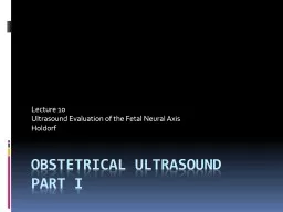
Lecture 10 Ultrasound Evaluation of the Fetal Neural Axis Holdorf Takeaways The Banana Sign Bent Cerebellum The part of the brain at the back of the skull in vertebrates Its function is to coordinate and regulate muscular activity ID: 703775

ventriculomegaly brain spina bifida brain ventriculomegaly bifida spina ventricle findings posterior spinal cerebral sonographic neural lateral disorder sign choroid intracranial hydrocephalus defect

Share:

Link:

Embed:

Download Presentation from below link

Download Presentation The PPT/PDF document "Obstetrical Ultrasound Part I" is the property of its rightful owner. Permission is granted to download and print the materials on this web site for personal, non-commercial use only, and to display it on your personal computer provided you do not modify the materials and that you retain all copyright notices contained in the materials. By downloading content from our website, you accept the terms of this agreement.


Presentation Transcript

Slide1

Obstetrical Ultrasound Part I

Lecture 10

Ultrasound Evaluation of the Fetal Neural Axis

HoldorfSlide2

Take-a-ways…Slide3

The Banana Sign

“Bent” Cerebellum

The part of the brain at the back of the skull in vertebrates. Its function is to coordinate and regulate muscular activity.

Absent Cisterna Magna

One of three principal openings in the subarachnoid space between the arachnoid and pia mater layers of the meninges surrounding the brain. The openings are collectively referred to as cisterns.

The Lemon Sign

Frontal bossing of the cranium

Temporal bonesSlide4

The Banana Sign explained:

Seen on axial imaging through the posterior fossa and is associated with Chiari II Malformation. The Banana sign describes the way the cerebellum is wrapped tightly around the brain stem as a result of spinal cord tethering and downward migration of posterior fossa content. The cisterna magna gets obliterated and the shape of the cerebellum takes on the appearance of a banana. This sign persists into the second and third trimesters. Slide5

The Lemon sign explained:

The lemon sign is the medial inward displacement and scalloping of the frontal bones of the fetal calvarium. This is often associated with an open spina bifida (seen under ultrasound as the widening and flattening of the posterior elements of the spine.Slide6

The formation of the Neural AxisSlide7

The stuff that is always on the boards…Slide8

The lateral, 3rd, and 4th

ventriclesSlide9

Ventriculomegaly

Ventriculomegaly is a brain condition that occurs when the lateral

ventricles become dilated. The most common definition uses a width of the atrium of the lateral ventricle of greater than 10 mm. This occurs in around 1% of pregnancies. When this measurement is between 10 and 15 mm, the ventriculomegaly may be described as mild to moderate. Slide10

VentriculomegalySlide11

When the measurement is greater than 15mm, ventriculomegaly may be classified as more severe.

Enlargement of the ventricles may occur for a number of reasons, for example due to loss of brain volume (perhaps due to infection or infarction), or due to impaired outflow or absorption of cerebrospinal fluid from the ventricles. Slide12

Often, however, there is no identifiable cause. This diagnosis is generally found in routine fetal anomaly scans at 18–22 weeks gestation. It is one of the commonest abnormal brain findings on prenatal ultrasound, occurring in around 1-2 per 1000 pregnancies.

In many cases of mild ventriculomegaly, however, there is resolution of ventriculomegaly during the pregnancy.Slide13

Ventriculomegaly is also known to be associated with other malformations such as agenesis of the corpus callosum (a broad band of nerve fibers joining the two hemispheres of the brain),

spina bifida, and

heart defects.

Fetuses with both isolated

ventriculomegaly and with other anomalies have an increased risk of having a

chromosomal abnormality, including that of

Down Syndrome .Slide14

Anencephaly

Cephalic

disorder that results from a

neural tube defect that occurs when the cephalic (head) end of the neural tube fails to close, usually between the 23rd and 26th day of

pregnancy, resulting in the absence of a major portion of the brain, skull, and

scalp.

Children with this disorder are born without a

forebrain, the largest part of the brain consisting mainly of the

cerebral hemispheres (which include the

isocortex, which is responsible for higher level

cognition, i.e., thinking). The remaining brain tissue is often exposed - not covered by bone or skin.Slide15
Slide16

AnencephalySlide17

Spina bifida

(

Latin: "split spine") is a developmental

birth defect involving the

neural tube: incomplete closure of the

embryonic neural tube results in an incompletely formed spinal cord.

In addition, the

vertebrae overlying the open portion of the spinal cord do not fully form and remain unfused and open

.Slide18

Spina BifidaSlide19

The spinal opening allows the abnormal portion of the spinal cord to protrude through the opening in the bones.

There may or may not be a fluid filled sac surrounding the open spinal cord.

Other neural tube defects include

anencephaly, a condition in which the portion of the neural tube which will become the

cerebrum does not close, and

encephalocele, which results when other parts of the brain remain unfusedSlide20

Spina bifida malformations fall into three categories:

spina bifida occulta, spina bifida cystica (myelomeningocele), and meningocele.

The most common location of the malformations is the

lumbar and

sacral areas of the spinal cord. Myelomeningocele is the most significant form and is that which leads to disability in most affected individuals. The terms spina bifida and myelomeningocele are usually used interchangeably.

Spina bifida can be surgically closed after birth, but this does not restore normal function to the affected part of the spinal cord and an individual with this condition will have dysfunction of the spinal cord and associated nerves from the point of the open defect and below. Intrauterine surgery for spina bifida has also been performed and the safety and efficacy of this procedure is currently being investigated. Slide21

Holoprosencephaly

A type of

cephalic disorder. This is a disorder characterized by the failure of the

prosencephalon (the forebrain of the embryo) to develop.

During normal development the forebrain is formed and the face begins to develop in the fifth and sixth weeks of human pregnancy. (The condition also occurs in other species, as with Cy, the Cyclops kitten.)

Holoprosencephaly is caused by a failure of the embryo's forebrain to divide to form bilateral cerebral hemispheres (the left and right halves of the brain), causing defects in the development of the

face and in brain structure and function.Slide22

HoloprosencephalySlide23

Dandy-Walker syndrome

DWS, or Dandy-Walker complex, is a congenital

brain malformation involving the

cerebellum and the fluid filled spaces around it.

The Dandy-Walker complex is a

genetically sporadic disorder that occurs one in every 25,000 live births, mostly in females.Slide24

Dandy-Walker Syndrome/absent pons and cerebellum in 7y/oSlide25

The key features of DWS are an enlargement of the

fourth ventricle, the space containing cerebrospinal fluid between the medulla and the cerebellum, a partial or complete absence of the

cerebellar vermis, the posterior midline area of cerebellar cortex responsible for coordination of the axial musculature, and cyst formation near the internal base of the

skull.

An increase in the size of the fluid spaces surrounding the brain as well as an increase in pressure may also be present. The syndrome can appear dramatically or develop unnoticed.Slide26

And now…the other stuff Slide27

Chapter 10 outline

Neural Tube Defects (NTD)

Spina Bifida

Anencephaly

Encephalocele

IniencephalySlide28

Intracranial Abnormalities

Hydrocephaly (Ventriculomegaly)

Hydranencephaly

Holoprosencephaly

Porencephaly

Dandy-Walker Syndrome

Agenesis of the Corpus Callosum

Schizencephaly

Lissencephaly

Choroid Plexus Cysts

Choroid Plexus Papillomas

Microcephaly

Intracranial TumorsSlide29

Terminology

Rachischisis: Completely open spine with Herniation and or destruction of the spinal cord

Spina Bifida occulta: Failure of closure of the vertebral column without any associated external abnormalities. Rarely associated with neurologic complications.

Spina Bifida Apperta: Failure of closure of the vertebral column with apparent external abnormalities and neurologic complications

Meningocele: Herniation of the meninges through a schisis defect

Myelomeningocele: Herniation of the mengenges and neural tissue through a schisis defect.Slide30

Neural Tube Defects

On the boards since 1991. There are MANY questions regarding NTDs…study this!!!!

A gamut of CNS anomalies may result from a failure of the normal process of Neurulation (Embryology).

The lesions are frequently open.

This allows CSF to pour into the amniotic cavity, causing elevation of the maternal serum and amniotic fluid levels of AFP.

Routine prenatal testing that reveals elevated MSAFP levels is and indication for a level II sonogram.

When the sonogram does not find the cause of elevated AFP, an amniocentesis is recommended.Slide31

Spina Bifida

Is a general term for lack of closure of the vertebral column.

Prognosis depends on the severity of the lesion.

Pathology

Presence of a Myelomeningocele, a protruding sac which may contain meninges and spinal cord.

May occur anywhere along the spinal column but lumbar region is most common.

Variable in size and content.

Sonographic findings in Transverse view:

Splaying of posterior ossification centers into a U or V shape.

When sac is intact, a cystic structure may be seen extending from the back. Appearances include a small cystic structure, a cyst with septations and or solid matter. Slide32

Sonographic Findings on Sagittal View:Splaying of parallel lateral ossification centers

Soft tissue defect or discontinuity of skin and muscle of posterior backSlide33

An image of a Myelomeningocele in Transverse viewSlide34

Splayed ossification centers in transverse view.Slide35

Soft tissue defect or discontinuity of skin and muscle of posterior back on Sagittal view.Slide36

Spina Bifida Continued…

Intracranial

Lemon sign-frontal bossing of cranium

Banana sign-dislocated cerebellum and obliteration of the cistern magna

Chiari type II malformation-variable displacement of cerebellum, 4

th

ventricle and medulla oblongata through the foramen magnumSlide37

The lemon and Banana signsSlide38

The lemon signSlide39

ANENCEPHALY

Anencephaly occurs at 2-3 weeks when the anterior neuropore fails to fuse.

Pathology

Absence of cranial vault and cerebral hemispheres (functioning brain)

Portions of midbrain and brain stem may be present

Other features include bulging eyes, macroglossia and a very short neck.

Sonographic findings

Fetal head should be identifiable by 12 weeks, definitively by 15 weeks.

Major portions of cranium and intracranial structures are absent: orbits and face are usually present

Associated Polyhydramnios in half the casesSlide40

AnencephalySlide41

Encephalocele

Protrusion of the intracranial contents through a bony defect of the skull can also be called a cephalocele.

Occipital encephaloceles are most frequent. There is a defect in the cranium.Slide42

Pathology

Boney defect in calvarium with herniation of brain and or meninges outside of cranium

Classified as occipital, frontal, or parietal

Hydrocephalus is present 80 % of the time in occipital lesions.

Sonographic findings

Mass extending from calvarium, may be totally cystic, have some meninges (cranial Meningocele, or may contain brain (encephalocele)

Cranial disruption is occasionally seen

Associated with hydrocephalus, PolyhydramniosSlide43

EncephaloceleSlide44

Iniencephaly

Iniencephaly is a dramatic NTD characterized by an exaggerated spinal Lordosis, cervical Rachischisis and an occipital encephalocele

Sonographic findings

Exaggerated, hyperextension of the fetal head

Demonstration of cervical spina bifidaSlide45

IniencephalySlide46

Iniencephaly

A type of

cephalic disorder. It is a rare neural tube defect that combines extreme retroflexion (backward bending) of the head with severe defects of the

spine.

The affected infant tends to be short, with a disproportionately large head. Diagnosis can be made immediately after birth because the head is so severely retroflexed that the face looks upward. The

skin of the face is connected directly to the skin of the

chest and the scalp is directly connected to the skin of the

back. Generally, the

neck is absent.

Most individuals with iniencephaly have other associated anomalies such as

anencephaly,

encephalocele (a disorder in which part of the cranial contents protrudes from the

skull),

hydrocephalus,

cyclopia, absence of the mandible (lower jaw bone), cleft lip and palate,

cardiovascular

disorders,

diaphragmatic hernia, and

gastrointestinal malformation. The disorder is more common among

females.Slide47

IniencephalySlide48

Intracranial Abnormalities

Hydrocephaly AKA Ventriculomegaly

Dilatation of the ventricular system secondary to the increase in the amount of CSF.

Classifications

Aqueductal Stenosis is the cause 43% of the time

Hydrocephalus caused by narrowing of the aqueduct of Sylvius (3

rd

ventricle outflow)

Communicating hydrocephalus is the cause 38% of the time

Dilatation of all ventricles and subarachnoid space caused by an obstruction to CSF follow outside the ventricular system.

Dandy-Walker Syndrome is the cause 12% of the time 

Idiopathic in 7% of the time Cause unknownSlide49

Bilateral ventriculomegalySlide50

diagram of the ventricular system, showing the lateral ventricle, the third ventricle, and the fourth ventricle.Slide51

Ventriculomegaly cont…

Sonographic findings

In the first trimester

CSF seen in the lateral ventricle and possibly compressing choroid plexus


In the 2

nd

and 3

rd

trimester

Normal ventricular configuration…just dilated

Presence of excess fluid in lateral ventricles, with an atrial measurement exceeding 10mm

Dangling choroid plexus

Observe brain echogenicity-echogenic foci in parenchyma may suggest intrauterine infection

Can have Polyhydramnios, abnormal fetal lie, fetal ascites, encephalocele, and Dandy-Walker.Slide52

An image measuring the atrial measurement exceeding 10mmSlide53

a “Dangling” choroid plexus.Slide54
Slide55

DEFINE FILLY’s RULE

Ventriculomegaly

is a brain condition that occurs when the lateral

ventricles

become dilated. The most common definition uses a width of the atrium of the lateral ventricle of greater than 10 mm. This occurs in around 1% of pregnancies. When this measurement is between 10 and 15 mm, the ventriculomegaly may be described as mild to moderate. When the measurement is greater than 15mm, the ventriculomegaly may be classified as more severe. Enlargement of the ventricles may occur for a number of reasons, such as loss of brain volume (perhaps due to infection or infarction), or impaired outflow or absorption of

cerebrospinal fluid

from the ventricles. Often, however, there is no identifiable cause. The interventricular foramen may be congenitally malformed, or may have become obstructed by infection, hemorrhage, or rarely tumor, which may impair the drainage of cerebrospinal fluid, and thus accumulation in the ventricles. This diagnosis is generally found in routine fetal anomaly scans at 18–22 weeks gestation. It is one of the more common abnormal brain findings on prenatal ultrasound, occurring in around 1–2 per 1000 pregnancies. In many cases of mild ventriculomegaly, however, there is resolution of ventriculomegaly during the pregnancySlide56

DEFINE FILLY’s RULESlide57

Hydranencephaly

Means “water, no brain”. Total or near total absence of the cerebral hemispheres. Cause could be intrauterine infarction secondary to bilateral ICA occlusion or malformation.


Sonographic findings

Microcephaly

Large anechoic areas in cranial vault

Variable presence of 3

rd

ventricle

PolyhydramniosSlide58

HydranenephalySlide59

HOLOPROSENCEPHALY (this is always on the boards)

Three classifications

1. Alobar (Most severe)

Large single cavity with minimal amount of cerebral tissue surrounding it, fused thalami, and facial syndrome with hypotelorism and other craniofacial anomalies

2. Semilobar

More cerebral tissue is present. Occipital lobe is present. Associated cleft palate and cleft lip

3. Lobar

Interhemispheric fusion is developed, but there is variable degree of fusion and CSP is absentSlide60
Slide61

Sonographic findings

Fetal cranium appears as a large cystic space with some peripheral cerebral tissue

Presence of a single midline ventricle

Craniofacial abnormalities including a proboscis, severe hypotelorism, Cycolpia

Appearance varies with severity of malformation.Slide62

. Alobar (Most severe)Slide63
Slide64
Slide65

In Holoprosencephaly, remember this hint…”THE FACE PREDICTS THE BRAIN”.Slide66

Porencephaly

The presence of cystic areas within the cerebral parenchyma. The etiology is believed to be the result of lysis of in-utero intracranial hemorrhage or cystic encephalomalacia.


Sonographic findings

Simple cystic structures within the cerebral parenchyma.Slide67

Porencephaly

A type of cephalic disorder involving an extremely rare disorder of the central nervous system involving a cyst or cavity in a cerebral hemisphere. The cysts or cavities are usually the remnants of destructive lesions, but are sometimes the result of abnormal development.

Porencephaly most likely has a number of different, often unknown causes, including absence of brain development and destruction of brain tissue.

More severely affected infants show symptoms of the disorder shortly after birth, and the diagnosis is usually made before age 1. Signs may include delayed growth and development, slight or incomplete paralysis, decreased muscle tone, seizures and macrocephaly or microcephaly.

Individuals with porencephaly may have poor or absent speech development, epilepsy, hydrocephalus, spastic contractures (shrinkage or shortening of muscles), and mental retardation.Slide68

PorencephalySlide69

Dandy-Walker Syndrome

Indicates the association of the following:

Large Cistern Magna

Ventriculomegaly of varying degree

Cerebellar Vermain Agenesis (complete or partial), through which the 4

th

ventricle communicates with the cystic structure in the posterior fossa.

There is obstruction of the 4

th

ventricle outflow and subsequent cystic enlargement of the posterior fossa.

Sonographic findings

Large midline cystic structure in posterior fossa

Enlargement of posterior fossa

Assoc. with ventriculomegaly and PolyhydramniosSlide70

Dandy-Walker Syndrome/fluid filled spaces in the posterior fossaSlide71
Slide72

Agenesis of the Corpus Callosum

Absence of the corpus callosum results from a chromosomal abnormality. May be complete or partial. Is frequently associated with other syndromes and or anomalies.


Sonographic findings

Absent Cavum septum Pellucidum

Lateral ventricles displaced upward and outward with a teardrop shape.

Enlarged occipital horn

3

rd

ventricle enlarged and displaced superiorly.Slide73

The corpus Callosum

The

corpus callosum

(from

Latin

: "tough body"), is a wide, flat bundle of neural fibers beneath the

cortex

at the longitudinal fissure. It connects the left and right

cerebral hemispheres

and facilitates inter-hemispheric communication. It is the largest

white matter

structure in the brain.Slide74
Slide75
Slide76

Teardrop ventricle with Agenesis of the Corpus CallosumSlide77

Schizencephaly

Abnormal clefts in the cerebral hemispheres.

The brain appears split into anterior and posterior partsSlide78
Slide79

Schizencephaly

A rare developmental disorder characterized by abnormal slits, or clefts, in the cerebral hemispheres.

Schizencephaly can be distinguished from

porencephaly by the fact that in schizencephaly the

cyst is lined by

heterotopic

grey matter while a porencephalic cyst is lined by

white matter. Individuals with clefts in both hemispheres, or bilateral clefts, are often developmentally delayed and have delayed speech and

language.

Individuals with smaller, unilateral clefts (clefts in one hemisphere) may be weak on one side of the body and may have average or near-average intelligence.

Patients with schizencephaly may also have varying degrees of

microcephaly,

mental retardation,

hemiparesis (weakness or paralysis affecting one side of the body), or

quadriparesis (weakness or paralysis affecting all four extremities), and may have reduced muscle tone (hypotonia). Most patients have seizures and some may have

hydrocephalus.Slide80

Schizencephaly/destructive brain lesionSlide81
Slide82

Lissencephaly

The surface of the brain lacks the normal sulci and gyri and appears smooth. Diagnosis is not made until the 3

rd

trimester.


Choroid Plexus cyst

The significance is an infrequent association with aneuploidy (Trisomy 18). Can be lateral, bilateral, small, large…

Choroid plexus Papillomas

Are benign tumors of the choroid plexus. Most frequently are unilateral and occur at the level of the atrium of the lateral ventricle. They are frequently associated with hydrocephalusSlide83

LissencephalySlide84

Choroid Plexus cystSlide85

Microcephaly

Considered a symptom of many etiologic disturbances. Defined as a small head circumference below 3 standard deviations of normal.


Sonographic findings

Small HC (other parameters are normal)

Sloping forehead, and disproportion between size of face and skullSlide86

MicrocephalySlide87
Slide88

Intracranial tumors

Rare.

Teratomas are the most common.

Prognosis is dismal

Sonographic findings

Loss of normal intracranial architecture

Presence of space-occupying lesionSlide89

Intracranial TumorSlide90

Homework

1. Rhombencephalon: what is it, and how is it visualized by ultrasound?

2. The ventricular system: Draw and label the ventricle system

3. Choroid plexus: what are they, what do they do, and where are they located in the brain?

4. Ventriculomegaly: how is it defined? Show this with an image. What is measured? What are the upper limits of normal? What measurement is considered abnormal?

5. Define hydrocephalusSlide91

6. Is hydrocephalus usually isolated? Explain

7. Are there any surgical options available for the parents of the fetus diagnosed with intrauterine hydrocephalus?

8. Spina Bifida: Define occulta and aperta.

9. What is myeolmeningocele?

10.What is Arnold-Chiari or Chiari Type II malformation?Slide92

11. What is the banana sign?

12. What is the lemon sign?

13. Define a Cephalocele.

14. What is a cranial Meningocele?

15. What is an encephalocele?Slide93

16. Define Holoprosencephaly.

17. What is agenesis of the Corpus Callosum?

18. What is Dandy-Walker Complex?

19. What is porencephaly?

20. What is Hydranencepahly?

21. What is Microcephaly?

22. Is there such a thing as intracranial calcification? Explain.

23. Please discuss the occurrence of choroid plexus cysts.