/
Scholars BIO  315 Instructor: Dr Scholars BIO  315 Instructor: Dr

Scholars BIO 315 Instructor: Dr - PowerPoint Presentation

stefany-barnette
stefany-barnette . @stefany-barnette
Follow
343 views
Uploaded On 2019-11-20

Scholars BIO 315 Instructor: Dr - PPT Presentation

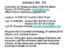
Scholars BIO 315 Instructor Dr Rebecca Kellum THM 319 office Phone 2579741email rkellumukyedu Office Hours anytime by appointment Lecture in THM 116 T and R 200315 pm Lab in THM B03 Section 001 M 9001150 am ID: 766078

protein cell fluorescent light cell protein light fluorescent gfp lab cells discussion questions resolution actin class microscope tag beam

Share:

Link:

Embed:

Download Presentation from below link

Download Presentation The PPT/PDF document "Scholars BIO 315 Instructor: Dr" is the property of its rightful owner. Permission is granted to download and print the materials on this web site for personal, non-commercial use only, and to display it on your personal computer provided you do not modify the materials and that you retain all copyright notices contained in the materials. By downloading content from our website, you accept the terms of this agreement.


Presentation Transcript

Scholars BIO 315 Instructor: Dr . Rebecca Kellum (THM 319 office) Phone: 257-9741/e-mail: rkellum@uky.edu Office Hours: anytime by appointment Lecture : in THM 116, T and R, 2:00-3:15 pm Lab: in THM B03, Section 001: M 9:00-11:50 am Section 002: M 1:00-3:50 pm Lab TA: Brandon Franklin brandon.franklin@uky.edu Required Text: Essential Cell Biology, 4 th edition (2014) Alberts , et al. Garland Science Canvas: Old exams, Echo recordings of lectures, lab exercises, pdf files of lecture slides and discussion articles, on-line homework assignments, and grade postings available on Canvas.

Music lovers: r emind you of something?

The scholars section are designed to give you more stimulation…. not necessarily more rigor. Therefore, the primary differentiating feature of the scholars s ections is a set of research articles for discussion. You will be asked to answer questions about the articles in class discussions and will also be assigned essay format questions on them to prepare you for similar questions on in-class exams (either lecture or lab exam). I will keep a record of your contributions. You must have participation in 4 out of 6 discussion periods for full credit . (Don’t worry. I’ll make sure you all do this.)

Grade Breakdown Lecture 70%; Discussion 15%; Lab 15% Lecture: Lecture Exams: 56.5%Homework: 17.5% Discussion: Discussion Participation: 7.5% Article Questions: 7.5% Lab: Lab Reports: 7.5% Lab Exams: 7.5%

Questions When Reading ANY Research Article: Intro : What was known before the work described in the paper? What new question is the paper addressing? Materials & Methods: Were any new materials or methods developed specifically for this study? Why were they needed? Results : What question is being addressed in each figure, what experiment was used to answer that question, and how were the results interpreted? Discussion : What did we learn from the study? Are there any caveats to the conclusions? What new or remaining questions are there ? Also Posted on Canvas (Discussion Articles link)

On-line Homework on Canvas 10 assignments All but one (HW7) due on Sun. at 11:59 pm. -Feedback given to incorrect responses,but not until entire assignment completed. This is a non-ideal feature of Canvas. -I recommend copying and pasting the entire a ssignment to a Word document and beginning w ork on it before the due date. Questions appear in the order material is covered in class. -You will get 3 attempts to answer each question correctly. But you will receive a grade that is an a verage of all attempts to minimize gaming of the system. Late assignments will receive half credit. -Article questions will also be managed on Canvas.

Note on Class Attendance Class attendance will be taken sporadically to encourage class participation that will be beneficial to you.This will not directly impact your course grade,b ut will factor into my assessment of your s uitability for any post-graduate education. My recommendation letter can only be as good as the data you give me to use. An important piece of data for any recommendation letter is active and regular class participation. Exceptional participation with a B grade often gets a better recommendation than poor participation with an A grade.

Chapter 1 Cells: The Fundamental Units of Life Essential Cell Biology FOURTH EDITION Copyright © Garland Science 2014 Alberts • Bray • Hopkin • Johnson • Lewis • Raff • Roberts • Walter

Cells come in a variety of shapes and sizes mammalian nerve cell Paramecium Chlamydomonas budding yeast Heliobacter pylori Fig. 1-1

Cells form tissues in multi-cellular plants and animals. plant root tip animal kidney tubules Fig. 1-5

The invention of the light microscope in the mid-17 th century allowed discovery of cells byRobert Hooke and Antoni van Leeuwenhoek. Two centuries later, c ells were proposed to form the basis of all life. Cells originate from other c ells only- Louis Pasteur(1860) Panel 1-1

Panel 1-1a - beam of light is focused on, then diffracted off specimen -diffracted light captured a nd re-diffracted through pair o f glass lenses: 1) objective l ens and 2) ocular lens (magnifying image up to 1000-fold) -light from ocular lens captured by lens of eye THE LIGHT MICROSCOPE

Cell visualized by light microscopy What are fibers inside t he cell made of ? Fluorescence microscopy a llows researchers to s ee specific molecules inside cells by labeling them with fluorescent tags. actin

Panel 1-1b Excitation Filter Only light l that excites fluorescent tag allowed to reach specimen Only light l emitted from fluorescent tag allowed to reach eye Emission Filter have light filtersthat are specific for the fluorescent tag used

Nuclear Division: microtubules of mitotic spindle and chromosomes

3) in vitro labeling of protein with fluorescent tag- then inject into living cell 2) indirect labeling of protein with fluorescent tag through an antibody (Immunostaining ) Methods for Labeling Molecules with Fluorescent Tags 1) specific binding of fluorescent stain -limited to only a few molecules (e.g., DAPI-staining of DNA) 4) i n vivo expression of the protein fused to a naturally fluorescing protein (e.g., GFP-tagging) * * * i n lab

Panel 4-2b Cell Biologists Can Raise Antibodies to Specific Proteins They are used in immunostaining and other types of cell biology experiments. B Cells Produce Antibodies Immunostaining requires an antibody

variable n on-variable

Observed in fixed cell Immunofluorescence Anti-actin Antibody ( Ab ) actin fluorescently labeled a ntibody used to indirectly label protein (actin here) in fixed cell DAPI-staining Usually the fluorescent tag is linked to a 2 o antibody that recognizes any 1 o antibody from a given species.

variable n on-variable 2 o antibodies recognize the non-variable portion of the 1 o antibody.

in vitro- labeling fluorescently labeled p rotein ( actin here) injected into living cell Fluorescent tag covalently linked to protein in test tube Observe in a living cell Labeled protein incorporates i nto its normal cellular structure

Expression of GFP-tagged protein for GFP-tagged actin Example: expressed in a living cell

GFP Tagging Green Fluorescent Protein from jellyfish can be fused to any protein and expressed in almost any cell type GFP-tagged skin protein in mice GFP-tagged wing protein in fly Aequoria victoria

ATG GGG AAG GGA CCG TTG…. coding sequence DNA: ATG CCC CGT AAA… coding sequence DNA: fusion gene inserted into genome of cell and protein then expressed in living cell Method 3: GFP Tagging protein protein fused coding sequence DNA ATG GGG AAG GGA CCG TTG CCC CGT AAA… fusion protein actin GFP actin GFP Transcription Translation Transcription Translation Transcription Translation a ctin - GFP a ctin - GFP w ill be used in several of our discussion articles

All 3 Methods for Labeling Proteins Should Produce the Same Result (Will Discuss Further in Lab)

  Earliest Microscopes of 1600’s   R. Hooke (Royal Society o f Science) A. van Leeuwenhoek (microscope h obbyist) double lens (compound) single biconvex lens 270x Mag 1.35 mm Res Modern Microscope: 1500x Mag and 0.2 µm Res

Resolution = distance (d) between two points that can be discriminated Higher Magnification Only Microscopes magnify and resolve. Fig. 18.3 Higher Resolution a nd Magnification d = 0.61 λ n sin α limited by wavelike properties o f light ( λ ) and light gathering a a bility of lens (N.A.) N.A . Theoretical Resolution L imit : 0.2 mm α = angle of c one of light collected New Super-resolution microscopes push that limit (Article #2)

Light Microscope Electron Microscope Electron Microscopy Allow s Higher Resolution Uses an electron beam instead of light beam (allows lower l ) Karp CMB7

ELECTRON MICROSCOPE Panel 1-1f -beam of accelerated electrons i nstead of beam of light focused o n specimen -electromagnet lenses capture a nd diffract electron beam to m agnify specimen up to 250,000-fold -can resolve points 1 nm (10-9 m) apartGold-labeled antibodies can be used to label molecules to provide specificity along with higher resolution.

Immunoelectron Microscopy of Membrane Proteins occludin (large Gold, orange arrowhead) claudin (small Gold, black arrowhead) Karp CMB

Ch 11, 12 Ch 5-8 Ch 15 Ch 13, 14 Ch 15 Ch 13, 14 Fig. 1-7 Ch 1-4 layfoundation Ch 16-18ties all together

X-Ray Diffraction Gives Even Higher Resolution- (level of individual atoms in molecules) Karp CMB7 Maximum Resolution ~ 1 A o (10 -10 m) ( 10 x higher than EM) MyoglobinArticle #1

Model Organisms Have Been Used to Learn About Cells Both genetic and biochemical experimental tools available. u nicellular yeast Drosophila C. elegans mice Arabidopsis Zebra fish

Human Genes Can Substitute f or MutantYeast Genes! How We Know, pg. 30

Human Cells Can Also Be Cultured Outside of Body ( in vitro ) fibroblast cell skeletal muscle cell epithelial cell (found in connective tissue) (found lining tissues) Fig. 1-38