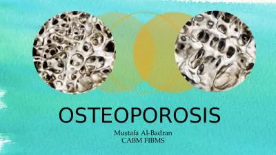
/
OSTEOPOROSIS Osteoporosis is a ‘ systematic skeletal disease characterized by low bone OSTEOPOROSIS Osteoporosis is a ‘ systematic skeletal disease characterized by low bone

OSTEOPOROSIS Osteoporosis is a ‘ systematic skeletal disease characterized by low bone - PowerPoint Presentation

susan
susan . @susan
Follow
27 views
Uploaded On 2024-02-09

OSTEOPOROSIS Osteoporosis is a ‘ systematic skeletal disease characterized by low bone - PPT Presentation

Normally there exists a balance between bone formation amp the bone resorption In osteoporosis The rate of bone resorption exceeds the rate of bone formation rendering the bone weak and brittle with loss of bone mass resulting in the reduction of bone mineral density BMD due to the ID: 1046187

amp bone calcium osteoporosis bone amp osteoporosis calcium exercise fracture decrease secondary activity due mass prolonged fractures years vitamin

Share:

Link:

Embed:

Download Presentation from below link

Download Presentation The PPT/PDF document "OSTEOPOROSIS Osteoporosis is a ‘ syste..." is the property of its rightful owner. Permission is granted to download and print the materials on this web site for personal, non-commercial use only, and to display it on your personal computer provided you do not modify the materials and that you retain all copyright notices contained in the materials. By downloading content from our website, you accept the terms of this agreement.


Presentation Transcript

1. OSTEOPOROSIS

2. Osteoporosis is a ‘ systematic skeletal disease characterized by low bone mass and microarchitectural deterioration of bone tissue leading to enhanced bone fragility & a consequent increase in fracture risk. Normally, there exists a balance between bone formation & the bone resorption. In osteoporosis, The rate of bone resorption exceeds the rate of bone formation rendering the bone weak and brittle with loss of bone mass resulting in the reduction of bone mineral density (BMD) due to the deficiency of Vitamin D & calcium.Osteoporosis

3. Osteoporosis is classified as follows- 1. Primary osteoporosis-, which is further divided into the following-Type-1: Post-osteoporosisType-2: Senile osteoporosis 2. Secondary osteoporosis- developed at any age, with equal distribution among males & females. It is usually secondary to some underlying disease such as endocrine disorders, malignancy or due to prolonged intake of certain medicines. e.g- steroids &Antiepileptic drugs. Classification of Osteoporosis

4. Classification of OsteoporosisCharacteristics Type1(postmenopausal)1. Age 55-75 years 2. Sex (female & 6:1Male) 3. Bone metabolism Pathogenesis increase osteoclast activity Type-270 years (female) 50 years (male)2:1Increase osteoblastic activity

5. Net bone mass mainly tubercularRate of bone rapid/short duration mass Bone density 2SD below normal 4) Clinical signs-Sites Pain & fracture of vertebra(rush). distal forearm.5. Laboratory values-Serum calcium NormalCortical & tubercular.Slowing durationLow or mediumPain & stress fracture of vertebra.Proximal hip & tibia.Normal

6. Serum phosphorus Normal Alk phosphatase Normal Urine calcium increased PTH function decrease Renal conservation secondary of 25 (OH)2 D to 1,25 decrease due(OH) D to decrease PTH. GIT calcium decreasedabsorption NormalNormalNormalIncreasedPrimary decrease due to decrease responsiveness. -

7. Disuse- prolonged bed rest or inactivity. - Prolonged casting or splinting. - Paralysis, space, travel etc. Diet- Calcium, protein, vitamin-C low in diet. - Chronic alcoholism - Anorexia nervosa Drugs- whose prolonged use causes osteoporosis are heparin, methotrexate, ethanol, glucocorticoids etc. Idiopathic- variety is seen in adolesent & middle aged male population.CAUSES

8. Genetic role is seen in osteogenesis imperfecta. Chronic illness like RA, cirrhosis, sarcoidosis, renal tubular acidosis etc. Neoplasms- like bone marrow tumors.CLINICAL FEATURES- In the early stage, the low and asymptomatic progress of the disease goes unnoticed. In the late stage, the patient develops pain in the dorsolumbar spine. There is development of a kyphotic hump deformity due to osteoporosis collapse of multiple vertebrae and loss of vertebral height.

9.

10.

11. There is generalised rarefaction of bone, making them susceptible to fractures after a trivial injury / fall. There is early fatigue.DIAGNOSIS- Several methods are available- Radiography- loss of bone density, presence of a ballooning of IV disc or compression fracture of the body of vertebra. Dual energy X-ray absorptiometry (Dexa- scan)- for progressive investigation of risk.

12.

13. 3. MRI Scan- details patterns of bone trabecular.4. Neutron activation analysis- to measure the activity of calcium in bone. TREATMENT- Regular check-up of the level of BMD after 50-55 years or menopause in women. Good nutrition diet to control curb bone loss. Vitamin D to facilitate calcium absorption from the gut. Hormone replacement therapy (HRT) in post-menopausal women.

14. Selective oestrogen receptor modulation (SERM) raloxifene is a drug to choice. Calcitonin secreted by thyroid gland- improves bone mass, preventing fracture. Associated symptoms- Backache (kyphosis)- is treated by Taylor brace or anteroposterior hyperextension brace (ASHE Brace).Fractures of the femoral neck or distal radius may require surgical intervention with intramedullary nails or plates.

15. Physiotherapy play a significant role in both primary & secondary prevention-For primary prevention- there are two different systems of exercise to be incorporated depending upon patient’s age and physical fitness. This is done by only after testing exercise tolerance and cardiac clearance.1. A system of structured exercise is ideal for young adults and middle aged group individuals and physically and medically fit elderly people.PHYSIOTHERAPEUTIC MANAGEMENT-

16. 2. Lifestyles exercise a) It is simply means not to avoid a single opportunity to perform some physical activity by increasing the speed, frequency and vigorousity of each physical activity one performs in a daily routine.b) Those who are unable to perform vigorous exercise should try and increase the duration of specific exercise at a slower speed.Secondary prevention- once the diagnosis is confirmed, physiotherapy is directed to prevent or control the expected complications:

17. Tendency to develop postural deformity of kyphosis leading to vertebral compression fracture. Incidences of hip fractures are controlled by applying the methods of safe ambulation.Incidences of colles fractures.Avoid hurry, be watch always.Get BMD level checked at regular intervals.Be regular on calcium and vitamin D suppliments and HRT in postmenopausal women.

18. Exercises-

19. THANK YOU