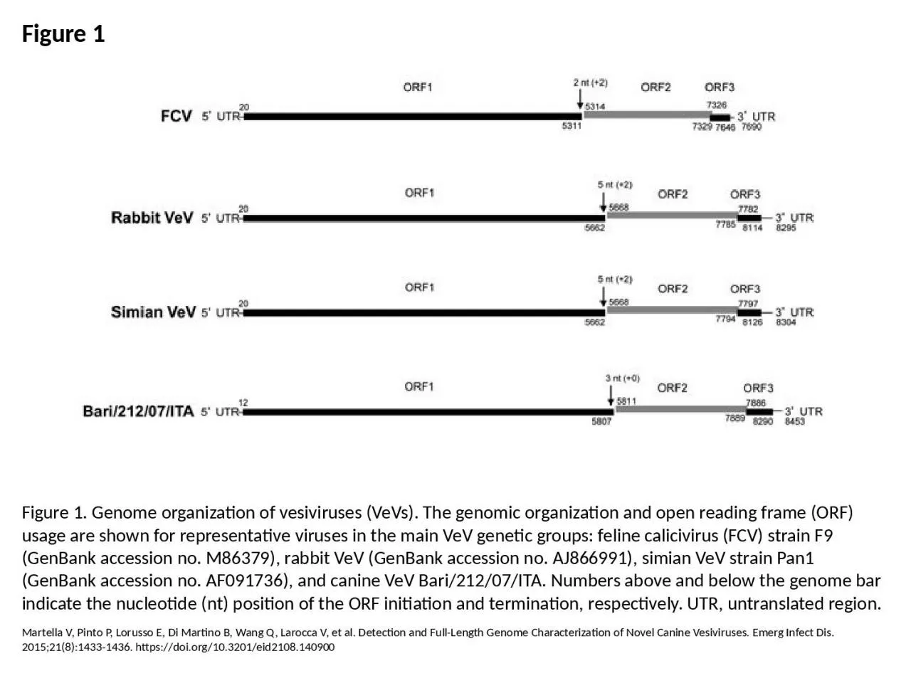
PPT-Figure 1 Figure 1. Genome organization of vesiviruses (VeVs). The genomic organization

Author : tabitha | Published Date : 2023-06-10

Martella V Pinto P Lorusso E Di Martino B Wang Q Larocca V et al Detection and FullLength Genome Characterization of Novel Canine Vesiviruses Emerg Infect Dis 201521814331436

Presentation Embed Code

Download Presentation

Download Presentation The PPT/PDF document "Figure 1 Figure 1. Genome organization o..." is the property of its rightful owner. Permission is granted to download and print the materials on this website for personal, non-commercial use only, and to display it on your personal computer provided you do not modify the materials and that you retain all copyright notices contained in the materials. By downloading content from our website, you accept the terms of this agreement.

Figure 1 Figure 1. Genome organization of vesiviruses (VeVs). The genomic organization: Transcript


Martella V Pinto P Lorusso E Di Martino B Wang Q Larocca V et al Detection and FullLength Genome Characterization of Novel Canine Vesiviruses Emerg Infect Dis 201521814331436 httpsdoiorg103201eid2108140900. MG2G1G0S Figure 1. Telophase Figure 2. BIO-FAX . . .makes science teaching easier. chapter one. l. ecture 2. McGraw-Hill/Irwin. Copyright © 2011 by The McGraw-Hill Companies, Inc. All rights reserved.. Managerial Roles. Identified by . Mintzberg. Followed managers around to see what they DO. Collaboration. Logistical organization. Development of collaborative relationships. Relationship management. Topics. Development and management of internal logistics relationships. Development and management of supply chain relationships. Copyright © . 2015 . McGraw-Hill Education. . All . rights reserved. No reproduction or distribution without the prior written consent of McGraw-Hill Education.. Learning Objectives. LO 1 . Explain how differentiation and integration influence an organization’s structure.. Figure 8.01. Sequencing—Fragments of All Possible Lengths. During chain termination or . Sanger sequencing. , the target DNA fragments are copied millions of times, but each copy ends at a different nucleotide position. These subsets of fragments end with a fluorescently-labeled nucleotide that reveal the identity of the final base. The final sequencing data are a series of fluorescent peaks that correspond to the original template . Doublet B, Lailler R, Meunier D, Brisabois A, Boyd D, Mulvey MR, et al. Variant Salmonella Genomic Island 1 Antibiotic Resistance Gene Cluster in Salmonella enterica Serovar Albany. Emerg Infect Dis. 2003;9(5):585-591. https://doi.org/10.3201/eid0905.020609. Figure 9.01. PCR Detection of . Sequence Tagged Site. Mapping large genomes requires many . genetic markers. . Those based solely on their unique sequences are known as sequence tagged sites (STS). These unique sequences may be amplified by PCR and mapped relative to each other. In practice, each STS is defined by a pair of specific PCR primers at its ends that generate a PCR fragment of defined length.. the Environment. Figure 16.1 Key Global Trends, 1961-2017. Sources:. World Bank, World Development Indicators; U.S. Energy Information Administration, International Energy Statistics; U.N. Food and Agriculture Organization, FAOSTAT; International Energy Agency, World Energy Balances; Organization of the Petroleum Exporting Countries, Annual Statistical Bulletin, 2018; BP, Statistical Review of World Energy, 2019.. Sonntag M, Mühldorfer K, Speck S, Wibbelt G, Kurth A. New Adenovirus in Bats, Germany. Emerg Infect Dis. 2009;15(12):2052-2055. https://doi.org/10.3201/eid1512.090646. Smits SL, Zijlstra E, van Hellemond JJ, Schapendonk C, Bodewes R, Schürch AC, et al. Novel Cyclovirus in Human Cerebrospinal Fluid, Malawi, 2010–2011. Emerg Infect Dis. 2013;19(9):1511-1513. https://doi.org/10.3201/eid1909.130404. Salzberg SL, Kingsford C, Cattoli G, Spiro DJ, Janies DA, Aly MM, et al. Genome Analysis Linking Recent European and African Influenza (H5N1) Viruses. Emerg Infect Dis. 2007;13(5):713. https://doi.org/10.3201/eid1305.070013. Albariño CG, Foltzer M, Towner JS, Rowe LA, Campbell S, Jaramillo CM, et al. Novel Paramyxovirus Associated with Severe Acute Febrile Disease, South Sudan and Uganda, 2012. Emerg Infect Dis. 2014;20(2):211-216. https://doi.org/10.3201/eid2002.131620. Woo P, Lau S, Teng J, Tsang A, Joseph M, Wong E, et al. New Hepatitis E Virus Genotype in Camels, the Middle East. Emerg Infect Dis. 2014;20(6):1044-1048. https://doi.org/10.3201/eid2006.140140. Li L, McGraw S, Zhu K, Leutenegger CM, Marks SL, Kubiski S, et al. Circovirus in Tissues of Dogs with Vasculitis and Hemorrhage. Emerg Infect Dis. 2013;19(4):534-541. https://doi.org/10.3201/eid1904.121390.

Download Document

Here is the link to download the presentation.
"Figure 1 Figure 1. Genome organization of vesiviruses (VeVs). The genomic organization"The content belongs to its owner. You may download and print it for personal use, without modification, and keep all copyright notices. By downloading, you agree to these terms.

Related Documents